- Animal cages. Plant cells

Содержание
- 2. Animal cells have the following parts A cell membrane (a thin layer that surrounds the cell):
- 3. Animal cells have the following parts
- 4. Plant cells have the following parts A cell wall (a strong structure that surrounds the cell
- 5. Plant cells have the following parts Cytoplasm (a liquid that surrounds the nucleus): carries out some
- 6. Cell division is the basis for the growth of an organism. Organisms are organised into: Cells→Tissues→Organs→Systems
- 7. Activity 2.1: How can we examine animal cells Prepare the slide Scrape the inside of your
- 8. Activity 2.1: How can we examine animal cells Viewing the slide Turn on the light on
- 9. To examine animal or plant cells under a microscope: Make sure there is light passing through
- 10. Turn the coarse focus knob to bring the lens away from the slide. Move the slide
- 11. Activity 2.3 To prepare a slide from plant tissue: Cut an onion and remove a thin
- 12. Draw a few drops of iodine solution across the cells using absorbent paper. MODULE 2 CELLS
- 13. Prokaryotic cells: Do not have a nucleus or membrane-enclosed organelles Are small and more primitive than
- 14. Eukaryotic cells: Have a membrane-enclosed nucleus and cell organelles Are larger and more advanced than prokaryotic
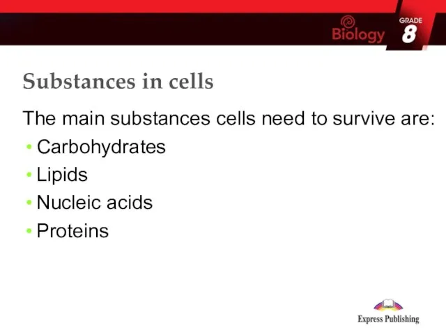
- 15. Substances in cells The main substances found in cells are: Lipids Carbohydrates Nucleic acids Proteins
- 16. The main substances cells need to survive are: Carbohydrates Lipids Nucleic acids Proteins Substances in cells
- 17. Carbohydrates: Made of C, H and O in the ratio Cx(H2O)y, where x = y Types:
- 18. Lipids: Made of C, H and O Fats are solid and oils are liquid at room
- 20. Скачать презентацию
Animal cells have the following parts
A cell membrane (a thin layer
Animal cells have the following parts
A cell membrane (a thin layer
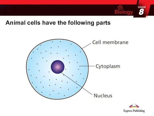
Animal cells have the following parts
Animal cells have the following parts
Plant cells have the following parts
A cell wall (a strong structure
Plant cells have the following parts
A cell wall (a strong structure
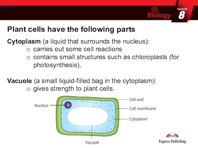
Plant cells have the following parts
Cytoplasm (a liquid that surrounds the
Plant cells have the following parts
Cytoplasm (a liquid that surrounds the
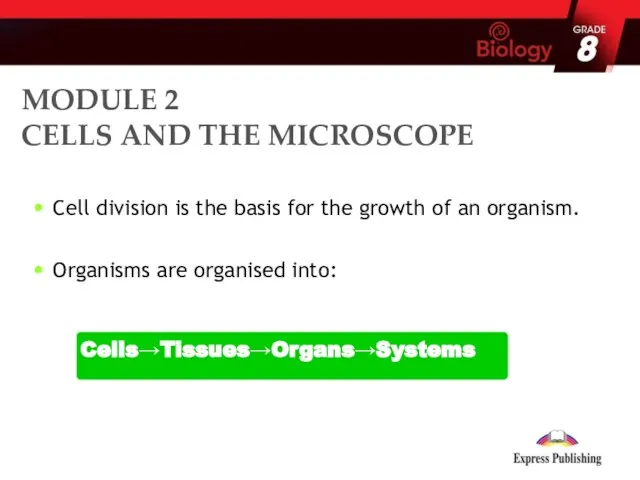
Cell division is the basis for the growth of an organism.
Organisms
Cell division is the basis for the growth of an organism.
Organisms
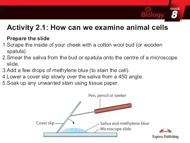
Activity 2.1: How can we examine animal cells
Prepare the slide
Scrape the
Activity 2.1: How can we examine animal cells
Prepare the slide
Scrape the

Activity 2.1: How can we examine animal cells
Viewing the slide
Turn on
Activity 2.1: How can we examine animal cells
Viewing the slide
Turn on
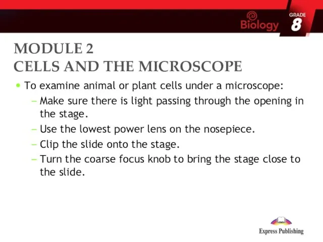
To examine animal or plant cells under a microscope:
Make sure there
To examine animal or plant cells under a microscope:
Make sure there
Turn the coarse focus knob to bring the lens away from
Turn the coarse focus knob to bring the lens away from
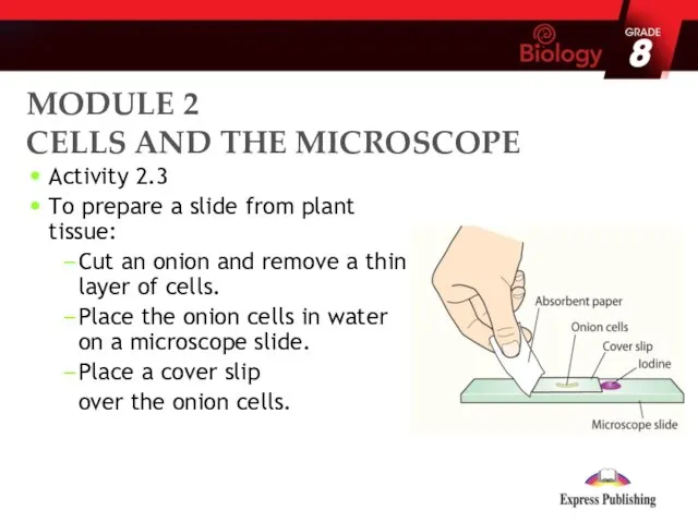
Activity 2.3
To prepare a slide from plant tissue:
Cut an onion
Activity 2.3
To prepare a slide from plant tissue:
Cut an onion

Draw a few drops of iodine solution across the cells using
Draw a few drops of iodine solution across the cells using
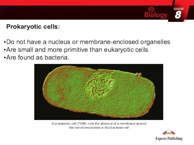
Prokaryotic cells:
Do not have a nucleus or membrane-enclosed organelles
Are small and
Prokaryotic cells:
Do not have a nucleus or membrane-enclosed organelles
Are small and
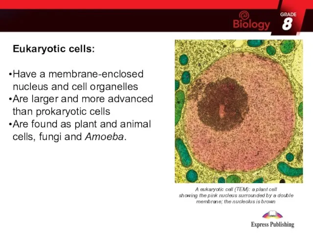
Eukaryotic cells:
Have a membrane-enclosed nucleus and cell organelles
Are larger and more
Eukaryotic cells:
Have a membrane-enclosed nucleus and cell organelles
Are larger and more
Substances in cells
The main substances found in cells are:
Lipids
Carbohydrates
Nucleic acids
Proteins
Substances in cells
The main substances found in cells are:
Lipids
Carbohydrates
Nucleic acids
Proteins
The main substances cells need to survive are:
Carbohydrates
Lipids
Nucleic acids
Proteins
Substances in
Carbohydrates
Lipids
Nucleic acids
Proteins
Substances in
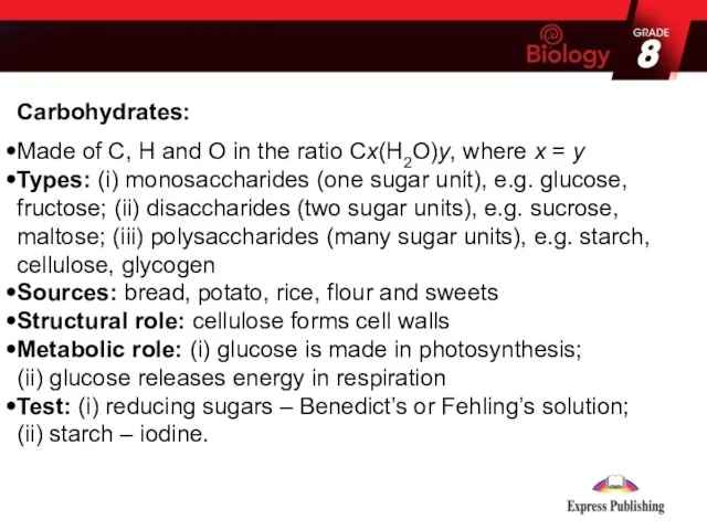
Carbohydrates:
Made of C, H and O in the ratio Cx(H2O)y, where
Made of C, H and O in the ratio Cx(H2O)y, where

Lipids:
Made of C, H and O
Fats are solid and oils are
Lipids:
Made of C, H and O
Fats are solid and oils are
 Регуляция внутренних органов: роль отделов ЦНС, вегетативной нервной системы
 Вирусы: строение, виды, значение. Прионы. Вироиды
 Исчезнувшие животные
 Презентация на тему "Влияние ионизирующих излучений на живые организмы" - скачать презентации по Биологии
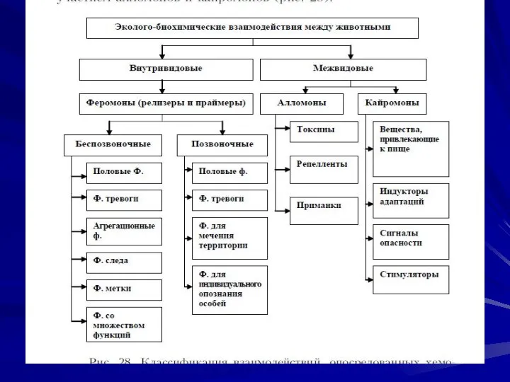 Взаимодействие между животными
 Взаимодействие генов
 Распространение плодов и семян
 Характеристика царства растения
 Водоросли
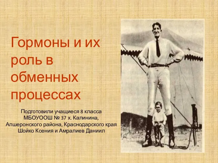 Презентация на тему "Гормоны и их роль в обменных процессах" - скачать презентации по Биологии
 МОУ СОШ п. АЛЯБЬЕВСКИЙ КЛАСС ПАУКООБРАЗНЫЕ – ХЕЛИЦЕРОВЫЕ, особенности строения и жизнедеятельности Презентацию подготовил:
 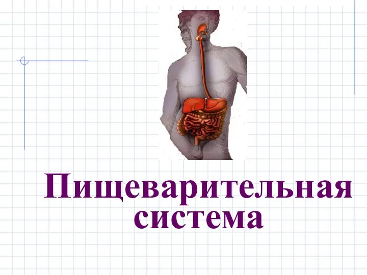 Презентация на тему "Пищеварительная система" - скачать презентации по Биологии
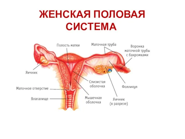 Женская половая система
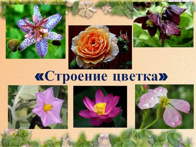 Строение цветка
 Презентация на тему Человек – часть живой природы
 Яблочный спас познавательная игра по биологии, 6 класс Автор: Лаврентьева Снежана Павловна, учитель химии и биологии
 Обмен веществ в организме
  Изучение настоящих тюленей как потенциальных объектов промысла и использования в биотехнических системах
 Семейство кошачьих Презентация к уроку. Выполнила : учитель высшей категории МОУ «СОШ п. Новопушкинское» Энгельсского района Са
 Презентация на тему "Вид- эволюционная единица, его критерии и структура" - скачать презентации по Биологии
 Кто такой павлин?
 СӨЖ Көмірсулар
 Водный и минеральный обмен
 Основные законы наследования признаков
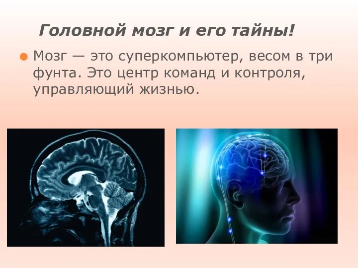 Головной мозг и его тайны!
 Функциональная анатомия мышц головы, шеи и туловища
 Тема: Световая фаза фотосинтеза
 Сільськогосподарські рослини