- Articular system

Содержание
- 2. PLAN: Development of joints. Classification of bone articulations. Continuous bone articulations. Synovial joints. The most typical
- 3. Development of joints Embryonic development of the joints is greatly dependent on the bone development. In
- 4. CLASSIFICATION OF BONE ARTICULATIONS Continuous articulations(synarthroses) are characterized by the presence of an uninterrupted articulating tissue
- 5. CONTINUOUS BONE ARTICULATIONS Depending on the tissue type, which articulates two bones, continuous articulations are divided
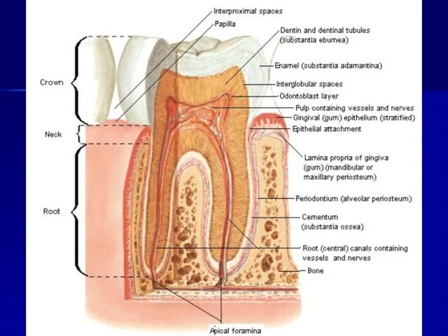
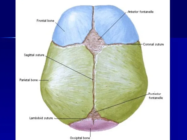
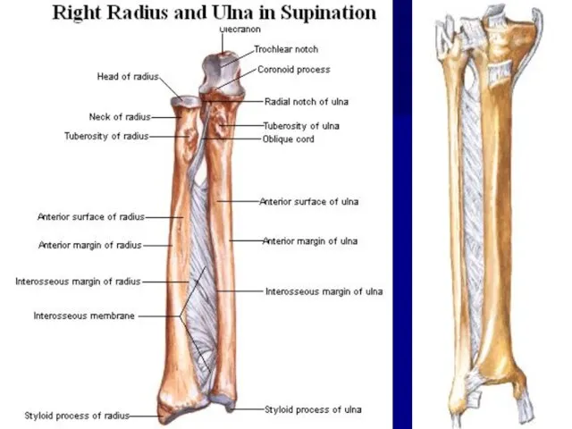
- 7. Syndesmoses, syndesmosis, (juncturae fibrosae) are formed from the fibrous connective tissue and are divided into the
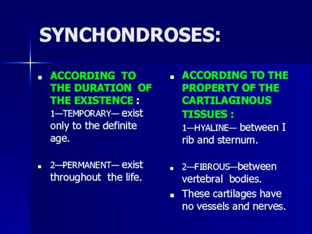
- 12. SYNCHONDROSES: ACCORDING TO THE DURATION OF THE EXISTENCE : 1—TEMPORARY— exist only to the definite age.
- 15. When ossified, synchondrosis can be transformed into the osseous articulation synostosis (synostosis). Synostoses or bone articulations
- 16. SYNOVIAL JOINTS The synovial articulations (juncturae synoviales, articulatio, diarthrosis) are discontinuous joints characterized by the presence
- 17. THE MOST TYPICAL FEATURES OF THE JOINTS Each joint possesses four basic elements: articular surfaces, facies
- 19. Articular surfaces. The articular surfaces of the bones are covered with the hyaline cartilage (sometimes fibrous),
- 20. Synovial folds and bursae. The synovial membrane is well developed and, in the majority of the
- 21. Accessory structures of joints Synovial joints may possess several accessory elements, which facilitate movement to a
- 22. Factors, which aid in the stabilization of articular surfaces: The fibrous capsule, which is reinforced by
- 23. Congruent and incongruent joints In those cases, when articulating surfaces entirely correspond to each other and
- 24. Movements in the joints The frontal axis runs through the joint from right to left. Flexion
- 25. CLASSIFICATION OF JOINTS Classification of the joints based on the number of articulating surfaces: simple -
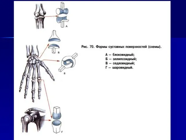
- 26. Classification of the joints based on the shape of articulating surfaces: Uni-axial joints Bi-axial joints Multi-axial
- 27. Uni-axial joints. In these joints, movement is restricted to a single axis. There are two types
- 28. Bi-axial joints. These joints permit movements around two axes, which are perpendicular to each other. The
- 29. Multi-axial joints include spheroidal joints and their variations. Even though, in principle, movements in these joints
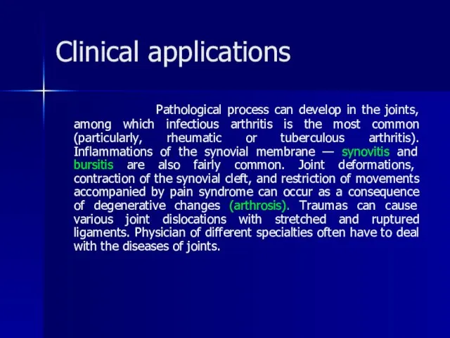
- 31. Clinical applications Pathological process can develop in the joints, among which infectious arthritis is the most
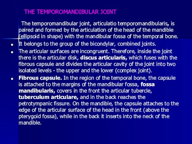
- 53. THE TEMPOROMANDIBULAR JOINT The temporomandibular joint, articulatio temporomandibularis, is paired and formed by the articulation of
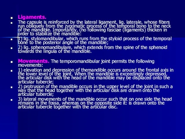
- 54. Ligaments. The capsule is reinforced by the lateral ligament, lig. laterale, whose fibers run obliquely from
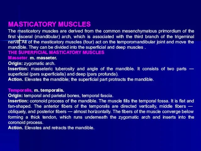
- 55. MASTICATORY MUSCLES The masticatory muscles are derived from the common mesenchymatous primordium of the first visceral
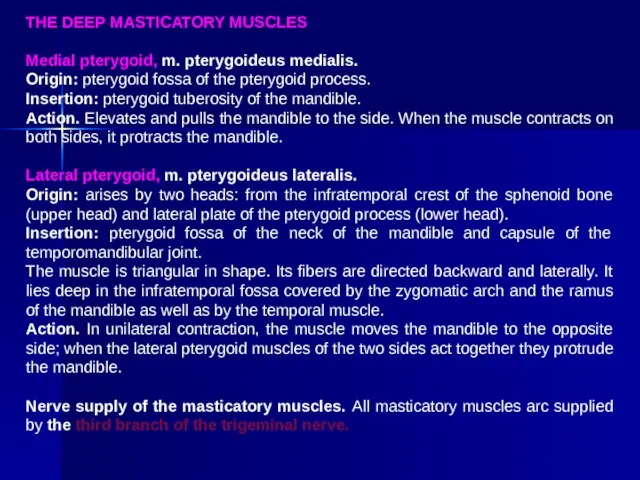
- 56. THE DEEP MASTICATORY MUSCLES Medial pterygoid, m. pterygoideus medialis. Origin: pterygoid fossa of the pterygoid process.
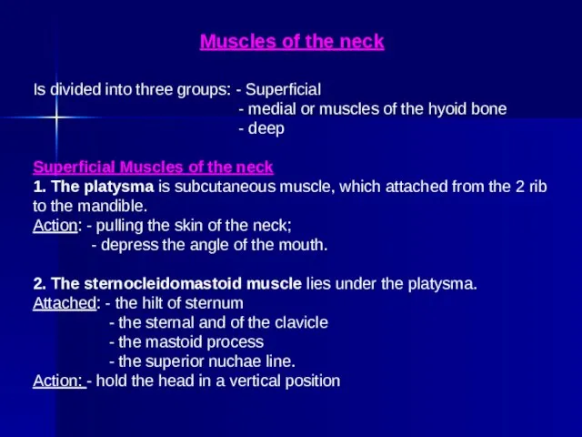
- 57. Muscles of the neck Is divided into three groups: - Superficial - medial or muscles of
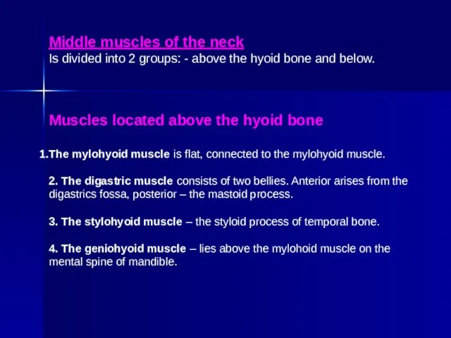
- 58. Middle muscles of the neck Is divided into 2 groups: - above the hyoid bone and
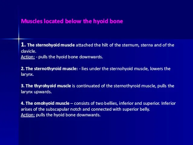
- 59. Muscles located below the hyoid bone 1. The sternohyoid muscle attached the hilt of the sternum,
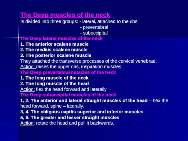
- 60. The Deep muscles of the neck Is divided into three groups: - lateral, attached to the
- 62. Скачать презентацию
PLAN:
Development of joints.
Classification of bone articulations.
Continuous bone articulations.
Synovial joints.
The most typical
PLAN:
Development of joints.
Classification of bone articulations.
Continuous bone articulations.
Synovial joints.
The most typical
Development of joints
Embryonic development of the joints is greatly
Development of joints
Embryonic development of the joints is greatly
CLASSIFICATION OF BONE ARTICULATIONS
Continuous articulations(synarthroses) are characterized by the presence of
CLASSIFICATION OF BONE ARTICULATIONS
Continuous articulations(synarthroses) are characterized by the presence of
CONTINUOUS BONE ARTICULATIONS
Depending on the tissue type, which articulates two
CONTINUOUS BONE ARTICULATIONS
Depending on the tissue type, which articulates two

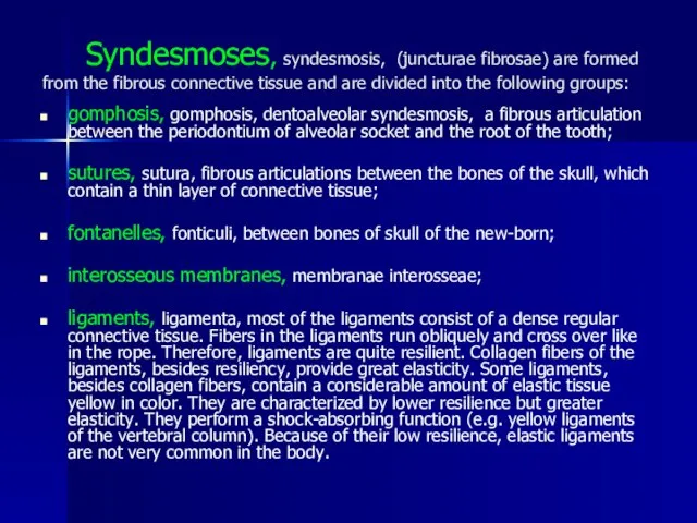
Syndesmoses, syndesmosis, (juncturae fibrosae) are formed from the fibrous connective
Syndesmoses, syndesmosis, (juncturae fibrosae) are formed from the fibrous connective

SYNCHONDROSES:
ACCORDING TO THE DURATION OF THE EXISTENCE : 1—TEMPORARY— exist only
SYNCHONDROSES:
ACCORDING TO THE DURATION OF THE EXISTENCE : 1—TEMPORARY— exist only


When ossified, synchondrosis can be transformed into the osseous articulation
When ossified, synchondrosis can be transformed into the osseous articulation
SYNOVIAL JOINTS
The synovial articulations (juncturae synoviales, articulatio, diarthrosis) are discontinuous
SYNOVIAL JOINTS
The synovial articulations (juncturae synoviales, articulatio, diarthrosis) are discontinuous
THE MOST TYPICAL FEATURES
OF THE JOINTS
Each joint possesses four
THE MOST TYPICAL FEATURES
OF THE JOINTS
Each joint possesses four
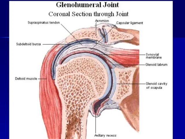
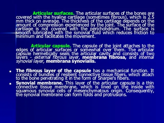
Articular surfaces. The articular surfaces of the bones are covered
Articular surfaces. The articular surfaces of the bones are covered
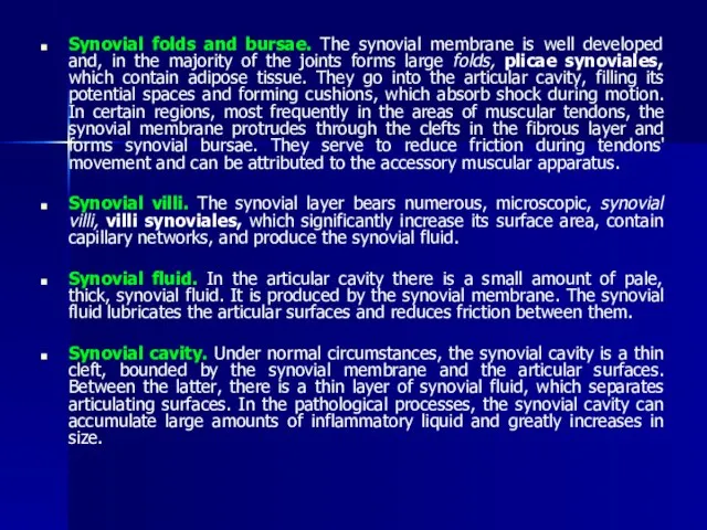
Synovial folds and bursae. The synovial membrane is well developed and,
Synovial folds and bursae. The synovial membrane is well developed and,
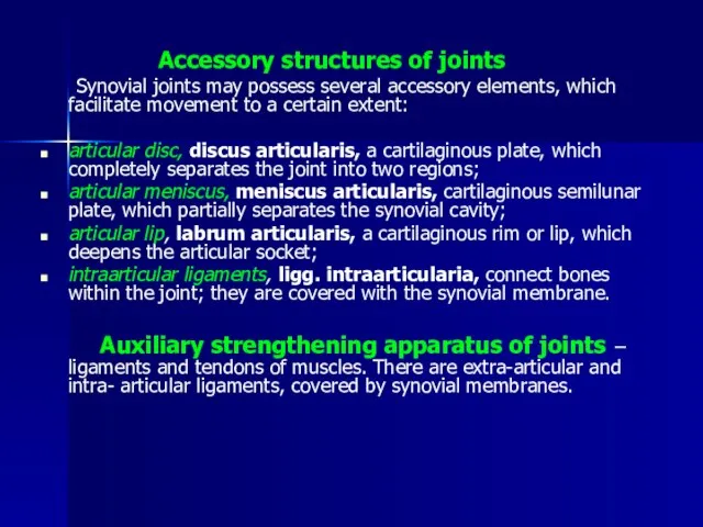
Accessory structures of joints
Synovial joints may possess several
Accessory structures of joints
Synovial joints may possess several
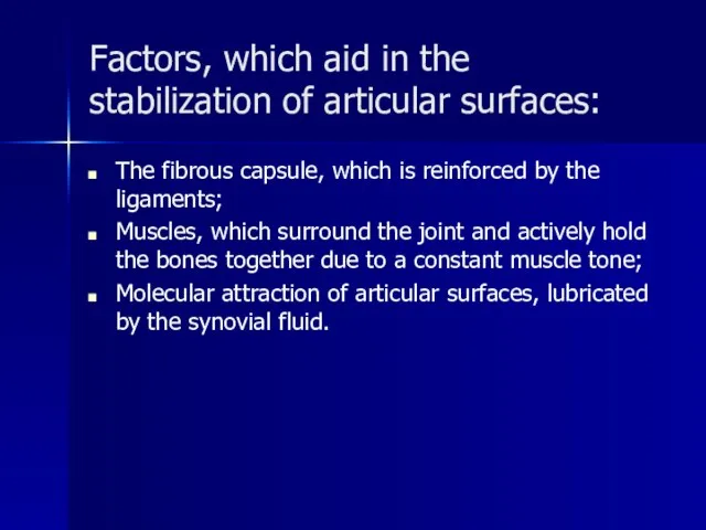
Factors, which aid in the stabilization of articular surfaces:
The fibrous
Factors, which aid in the stabilization of articular surfaces:
The fibrous
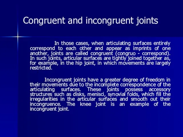
Congruent and incongruent joints
In those cases, when articulating surfaces entirely
Congruent and incongruent joints
In those cases, when articulating surfaces entirely
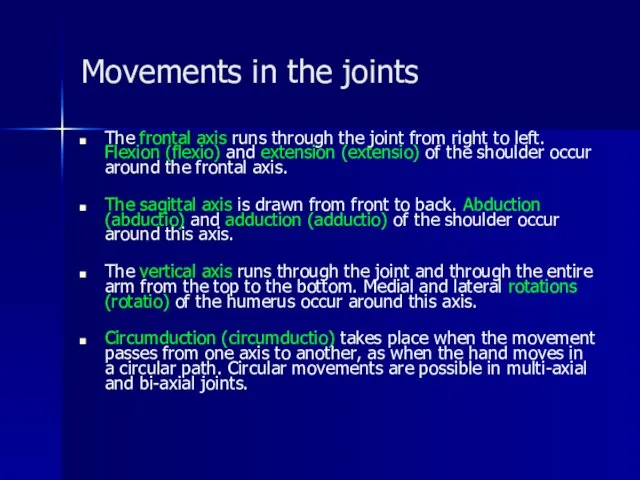
Movements in the joints
The frontal axis runs through the joint from
Movements in the joints
The frontal axis runs through the joint from
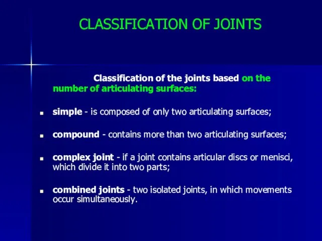
CLASSIFICATION OF JOINTS
Classification of the joints based on the number
CLASSIFICATION OF JOINTS
Classification of the joints based on the number
Classification of the joints based on the shape of articulating surfaces:
Classification of the joints based on the shape of articulating surfaces:
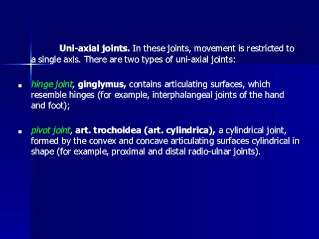
Uni-axial joints. In these joints, movement is restricted to a
Uni-axial joints. In these joints, movement is restricted to a
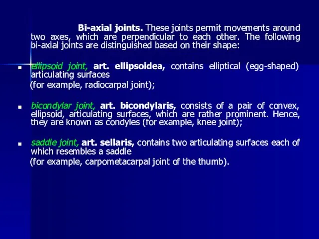
Bi-axial joints. These joints permit movements around two axes, which
Bi-axial joints. These joints permit movements around two axes, which
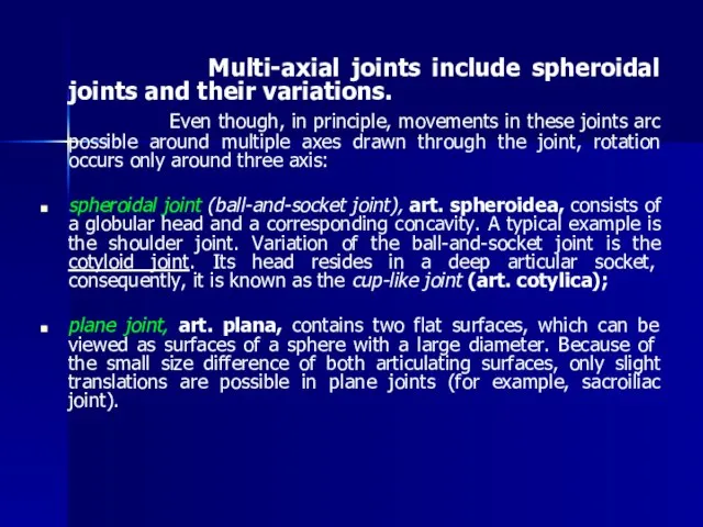
Multi-axial joints include spheroidal joints and their variations.
Even
Multi-axial joints include spheroidal joints and their variations.
Even
Clinical applications
Pathological process can develop in the joints, among which
Clinical applications
Pathological process can develop in the joints, among which


THE TEMPOROMANDIBULAR JOINT
The temporomandibular joint, articulatio temporomandibularis, is paired and
THE TEMPOROMANDIBULAR JOINT
The temporomandibular joint, articulatio temporomandibularis, is paired and
Ligaments.
The capsule is reinforced by the lateral ligament, lig. laterale,
Ligaments.
The capsule is reinforced by the lateral ligament, lig. laterale,
MASTICATORY MUSCLES
The masticatory muscles are derived from the common mesenchymatous primordium
MASTICATORY MUSCLES
The masticatory muscles are derived from the common mesenchymatous primordium
THE DEEP MASTICATORY MUSCLES
Medial pterygoid, m. pterygoideus medialis.
Origin: pterygoid fossa of
THE DEEP MASTICATORY MUSCLES
Medial pterygoid, m. pterygoideus medialis.
Origin: pterygoid fossa of
Muscles of the neck
Is divided into three groups: - Superficial
-
Muscles of the neck
Is divided into three groups: - Superficial
-
Middle muscles of the neck
Is divided into 2 groups: - above
Middle muscles of the neck
Is divided into 2 groups: - above
Muscles located below the hyoid bone
1. The sternohyoid muscle attached the
Muscles located below the hyoid bone
1. The sternohyoid muscle attached the
The Deep muscles of the neck
Is divided into three groups: -
The Deep muscles of the neck
Is divided into three groups: -
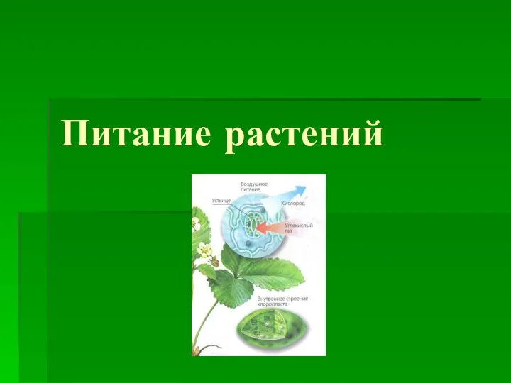 Презентация на тему Питание растений
  Митоз
Митоз Рыба фугу
 Внутреннее строение птицы Презентация к уроку биологии Отряскиной Т.А.
  Многообразие и классификация голосеменных растений. Отдел голосеменные: 1) класс семенные папоротники 2) класс саговниковые (цика
 Генетика человека
 Презентация на тему Классификация углеводов
  Прибрежно-водные растения . Дубинчина Ирина Васильевна учитель биологии МОУ «СОШ имени М. М. Рудченко с. Перелюб Перелюбско
 Транспорт веществ в организме
 Проект. Создание производства гранулированных биоорганических удобрений
 Зелёная аптека. Подорожник
 Забота о потомстве
  Генетическая информация. Удвоение ДНК
 Развитие жизни на земле Мезозойская эра
  Презентация на тему "Царство прокариоты, подцарство бактерии" - скачать презентации по Биологии
 Тема: «Приспособленности живых организмов к условиям окружающей среды.»
 Генетика как наука. Закономерности наследственности установленные Г. Менделем
 Мир вокруг нас. Кто такие птицы
 Грибы
Грибы Формы взаимоотношений организмов
 Содержание паразитологии. Введение в курс
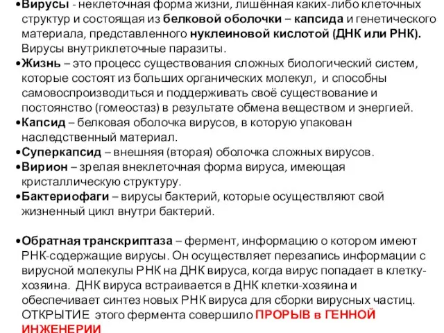 Вирусы. Бактериофаги
 Фотосинтез 6 класс
 Почему на нашей планете сейчас не живут динозавры
 Клітина – одиниця живого. Історія вивчення клітин Розробила вчитель ХЗОШ № 76 Малік Н.А.
  DNA Structure. Nitrogen bases. (Chapter 9.2)
 Класс Ланцетники
 Таламус. Ядра таламуса