- Fertilization and development fertilization

Содержание
- 2. Copyright Pearson Prentice Hall What is fertilization? Fertilization
- 3. Copyright Pearson Prentice Hall Fertilization The process of a sperm joining an egg is called fertilization.
- 4. Copyright Pearson Prentice Hall Fertilization After the two haploid (N) nuclei fuse, a single diploid (2N)
- 5. Copyright Pearson Prentice Hall Early Development Early Development While still in the Fallopian tube, the zygote
- 6. Copyright Pearson Prentice Hall Early Development What are the stages of early development?
- 7. Copyright Pearson Prentice Hall Early Development The stages of early development include implantation, gastrulation, and neurulation.
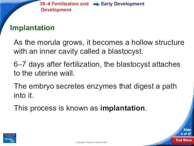
- 8. Copyright Pearson Prentice Hall Early Development Implantation As the morula grows, it becomes a hollow structure
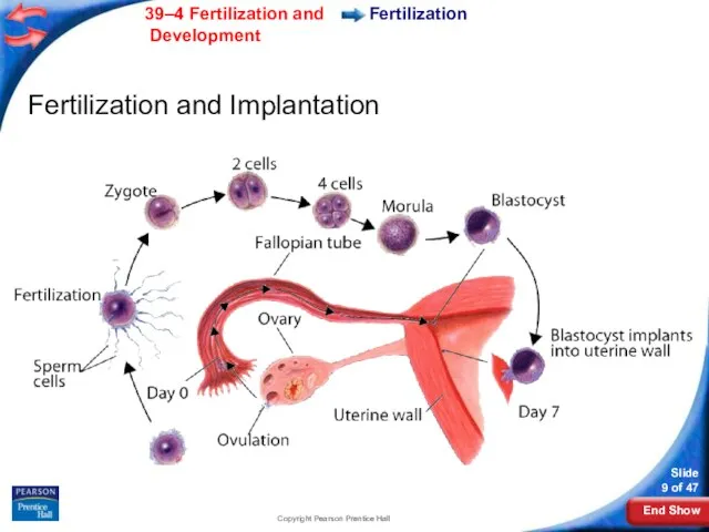
- 9. Copyright Pearson Prentice Hall Fertilization Fertilization and Implantation
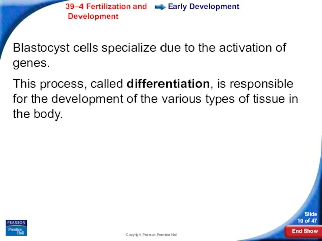
- 10. Copyright Pearson Prentice Hall Early Development Blastocyst cells specialize due to the activation of genes. This
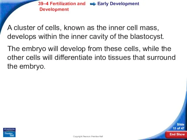
- 11. Copyright Pearson Prentice Hall Early Development A cluster of cells, known as the inner cell mass,
- 12. Copyright Pearson Prentice Hall Early Development Gastrulation The inner cell mass of the blastocyst gradually sorts
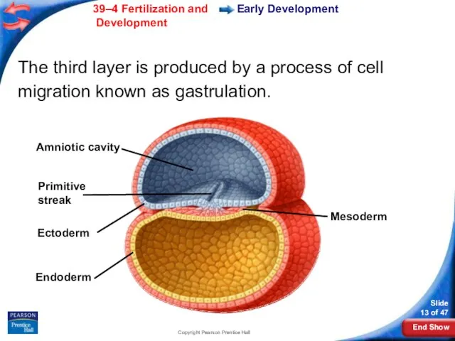
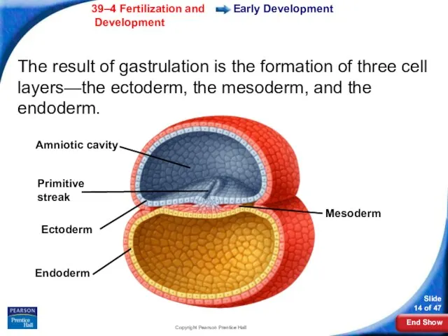
- 13. Copyright Pearson Prentice Hall Mesoderm Amniotic cavity Primitive streak Ectoderm Endoderm Early Development The third layer
- 14. Copyright Pearson Prentice Hall Early Development The result of gastrulation is the formation of three cell
- 15. Copyright Pearson Prentice Hall Early Development The ectoderm develops into the skin and nervous system. The
- 16. Copyright Pearson Prentice Hall Early Development Neurulation Gastrulation is followed by neurulation. Neurulation is the development
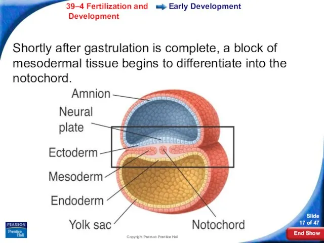
- 17. Copyright Pearson Prentice Hall Early Development Shortly after gastrulation is complete, a block of mesodermal tissue
- 18. Copyright Pearson Prentice Hall Neural crest Neural fold Notochord Early Development As the notochord develops, the
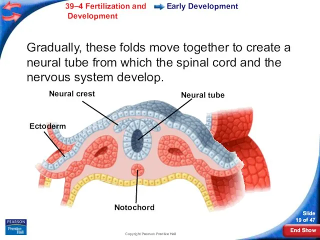
- 19. Copyright Pearson Prentice Hall Neural crest Neural tube Ectoderm Notochord Early Development Gradually, these folds move
- 20. Copyright Pearson Prentice Hall Early Development Extraembryonic Membranes As the embryo develops, membranes form to protect
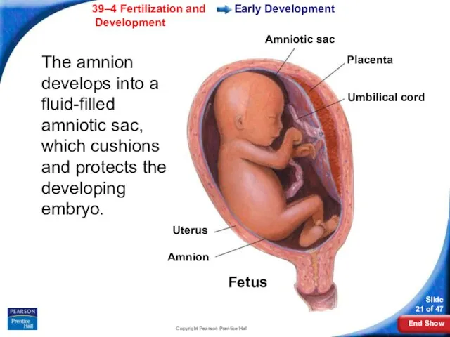
- 21. Copyright Pearson Prentice Hall Early Development The amnion develops into a fluid-filled amniotic sac, which cushions
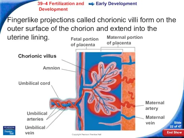
- 22. Copyright Pearson Prentice Hall Fingerlike projections called chorionic villi form on the outer surface of the
- 23. Copyright Pearson Prentice Hall The chorionic villi and uterine lining form the placenta. The placenta connects
- 24. Copyright Pearson Prentice Hall Early Development What is the function of the placenta?
- 25. Copyright Pearson Prentice Hall Early Development The placenta is the embryo's organ of respiration, nourishment, and
- 26. Copyright Pearson Prentice Hall Early Development The placenta acts as a barrier to some harmful or
- 27. Copyright Pearson Prentice Hall Early Development After eight weeks, the embryo is called a fetus. After
- 28. Copyright Pearson Prentice Hall Control of Development Control of Development The fates of many cells in
- 29. Copyright Pearson Prentice Hall Later Development Later Development 4–6 months after fertilization: The heart can be
- 30. Copyright Pearson Prentice Hall Later Development During the last three months, the organ systems mature. The
- 31. Copyright Pearson Prentice Hall Childbirth Childbirth About nine months after fertilization, the fetus is ready for
- 32. Copyright Pearson Prentice Hall Childbirth The mother’s posterior pituitary gland releases the hormone oxytocin, which affects
- 33. Copyright Pearson Prentice Hall Childbirth The opening of the cervix expands until it is large enough
- 34. Copyright Pearson Prentice Hall Childbirth The baby now begins an independent existence. Its systems quickly adapt
- 35. Copyright Pearson Prentice Hall Multiple Births Multiple Births If two eggs are released during the same
- 36. Copyright Pearson Prentice Hall Early Years Early Years The first two years of life are called
- 37. Copyright Pearson Prentice Hall Adulthood Adulthood Development continues during adulthood. Adults reach their highest levels of
- 38. Copyright Pearson Prentice Hall 39–4
- 39. Copyright Pearson Prentice Hall 39–4 Fertilization takes place in the ovary. Fallopian tube. cavity of the
- 40. Copyright Pearson Prentice Hall 39–4 The process in which a blastocyst attaches to the wall of
- 41. Copyright Pearson Prentice Hall 39–4 The central nervous system develops during which phase of early development?
- 42. Copyright Pearson Prentice Hall 39–4 The placenta is a structure that belongs entirely to the mother.
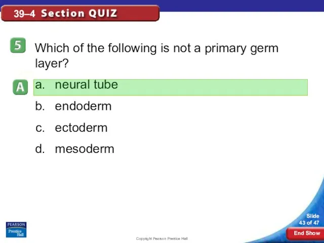
- 43. Copyright Pearson Prentice Hall 39–4 Which of the following is not a primary germ layer? neural
- 45. Скачать презентацию
Copyright Pearson Prentice Hall
What is fertilization?
Fertilization
Copyright Pearson Prentice Hall
What is fertilization?
Fertilization
Copyright Pearson Prentice Hall
Fertilization
The process of a sperm joining an egg
Copyright Pearson Prentice Hall
Fertilization
The process of a sperm joining an egg
Copyright Pearson Prentice Hall
Fertilization
After the two haploid (N) nuclei fuse, a
Copyright Pearson Prentice Hall
Fertilization
After the two haploid (N) nuclei fuse, a
Copyright Pearson Prentice Hall
Early Development
Early Development
While still in the Fallopian tube,
Copyright Pearson Prentice Hall
Early Development
Early Development
While still in the Fallopian tube,
Copyright Pearson Prentice Hall
Early Development
What are the stages of early development?
Copyright Pearson Prentice Hall
Early Development
What are the stages of early development?
Copyright Pearson Prentice Hall
Early Development
The stages of early development include implantation,
Copyright Pearson Prentice Hall
Early Development
The stages of early development include implantation,
Copyright Pearson Prentice Hall
Early Development
Implantation
As the morula grows, it becomes a
Copyright Pearson Prentice Hall
Early Development
Implantation
As the morula grows, it becomes a
Copyright Pearson Prentice Hall
Fertilization
Fertilization and Implantation
Copyright Pearson Prentice Hall
Fertilization
Fertilization and Implantation
Copyright Pearson Prentice Hall
Early Development
Blastocyst cells specialize due to the activation
Copyright Pearson Prentice Hall
Early Development
Blastocyst cells specialize due to the activation
Copyright Pearson Prentice Hall
Early Development
A cluster of cells, known as the
Copyright Pearson Prentice Hall
Early Development
A cluster of cells, known as the
Copyright Pearson Prentice Hall
Early Development
Gastrulation
The inner cell mass of the blastocyst
Copyright Pearson Prentice Hall
Early Development
Gastrulation
The inner cell mass of the blastocyst
Copyright Pearson Prentice Hall
Mesoderm
Amniotic cavity
Primitive streak
Ectoderm
Endoderm
Early Development
The third layer is produced
Copyright Pearson Prentice Hall
Mesoderm
Amniotic cavity
Primitive streak
Ectoderm
Endoderm
Early Development
The third layer is produced
Copyright Pearson Prentice Hall
Early Development
The result of gastrulation is the formation
Copyright Pearson Prentice Hall
Early Development
The result of gastrulation is the formation
Copyright Pearson Prentice Hall
Early Development
The ectoderm develops into the skin and
Copyright Pearson Prentice Hall
Early Development
The ectoderm develops into the skin and
Copyright Pearson Prentice Hall
Early Development
Neurulation
Gastrulation is followed by neurulation.
Neurulation is
Copyright Pearson Prentice Hall
Early Development
Neurulation
Gastrulation is followed by neurulation.
Neurulation is
Copyright Pearson Prentice Hall
Early Development
Shortly after gastrulation is complete, a block
Copyright Pearson Prentice Hall
Early Development
Shortly after gastrulation is complete, a block

Copyright Pearson Prentice Hall
Neural crest
Neural fold
Notochord
Early Development
As the notochord develops, the
Copyright Pearson Prentice Hall
Neural crest
Neural fold
Notochord
Early Development
As the notochord develops, the
Copyright Pearson Prentice Hall
Neural crest
Neural tube
Ectoderm
Notochord
Early Development
Gradually, these folds move together
Copyright Pearson Prentice Hall
Neural crest
Neural tube
Ectoderm
Notochord
Early Development
Gradually, these folds move together
Copyright Pearson Prentice Hall
Early Development
Extraembryonic Membranes
As the embryo develops, membranes form
Copyright Pearson Prentice Hall
Early Development
Extraembryonic Membranes
As the embryo develops, membranes form
Copyright Pearson Prentice Hall
Early Development
The amnion develops into a fluid-filled amniotic
Copyright Pearson Prentice Hall
Early Development
The amnion develops into a fluid-filled amniotic
Copyright Pearson Prentice Hall
Fingerlike projections called chorionic villi form on the
Copyright Pearson Prentice Hall
Fingerlike projections called chorionic villi form on the
Copyright Pearson Prentice Hall
The chorionic villi and uterine lining form the
Copyright Pearson Prentice Hall
The chorionic villi and uterine lining form the
Copyright Pearson Prentice Hall
Early Development
What is the function of the placenta?
Copyright Pearson Prentice Hall
Early Development
What is the function of the placenta?
Copyright Pearson Prentice Hall
Early Development
The placenta is the embryo's organ of
Copyright Pearson Prentice Hall
Early Development
The placenta is the embryo's organ of
Copyright Pearson Prentice Hall
Early Development
The placenta acts as a barrier to
Copyright Pearson Prentice Hall
Early Development
The placenta acts as a barrier to
Copyright Pearson Prentice Hall
Early Development
After eight weeks, the embryo is called
Copyright Pearson Prentice Hall
Early Development
After eight weeks, the embryo is called
Copyright Pearson Prentice Hall
Control of Development
Control of Development
The fates of many
Copyright Pearson Prentice Hall
Control of Development
Control of Development
The fates of many
Copyright Pearson Prentice Hall
Later Development
Later Development
4–6 months after fertilization:
The heart can
Copyright Pearson Prentice Hall
Later Development
Later Development
4–6 months after fertilization:
The heart can
Copyright Pearson Prentice Hall
Later Development
During the last three months, the organ
Copyright Pearson Prentice Hall
Later Development
During the last three months, the organ
Copyright Pearson Prentice Hall
Childbirth
Childbirth
About nine months after fertilization, the fetus is
Copyright Pearson Prentice Hall
Childbirth
Childbirth
About nine months after fertilization, the fetus is
Copyright Pearson Prentice Hall
Childbirth
The mother’s posterior pituitary gland releases the hormone
Copyright Pearson Prentice Hall
Childbirth
The mother’s posterior pituitary gland releases the hormone
Copyright Pearson Prentice Hall
Childbirth
The opening of the cervix expands until it
Copyright Pearson Prentice Hall
Childbirth
The opening of the cervix expands until it
Copyright Pearson Prentice Hall
Childbirth
The baby now begins an independent existence.
Its
Copyright Pearson Prentice Hall
Childbirth
The baby now begins an independent existence.
Its
Copyright Pearson Prentice Hall
Multiple Births
Multiple Births
If two eggs are released during
Copyright Pearson Prentice Hall
Multiple Births
Multiple Births
If two eggs are released during
Copyright Pearson Prentice Hall
Early Years
Early Years
The first two years of life
Copyright Pearson Prentice Hall
Early Years
Early Years
The first two years of life
Copyright Pearson Prentice Hall
Adulthood
Adulthood
Development continues during adulthood.
Adults reach their highest
Copyright Pearson Prentice Hall
Adulthood
Adulthood
Development continues during adulthood.
Adults reach their highest
Copyright Pearson Prentice Hall
39–4
Copyright Pearson Prentice Hall
39–4
Copyright Pearson Prentice Hall
39–4
Fertilization takes place in the
ovary.
Fallopian tube.
cavity of the
Copyright Pearson Prentice Hall
39–4
Fertilization takes place in the
ovary.
Fallopian tube.
cavity of the
Copyright Pearson Prentice Hall
39–4
The process in which a blastocyst attaches to
Copyright Pearson Prentice Hall
39–4
The process in which a blastocyst attaches to
Copyright Pearson Prentice Hall
39–4
The central nervous system develops during which phase
Copyright Pearson Prentice Hall
39–4
The central nervous system develops during which phase
Copyright Pearson Prentice Hall
39–4
The placenta is a structure that
belongs entirely to
Copyright Pearson Prentice Hall
39–4
The placenta is a structure that
belongs entirely to
Copyright Pearson Prentice Hall
39–4
Which of the following is not a primary
Copyright Pearson Prentice Hall
39–4
Which of the following is not a primary
 Биогеоценозы. Экосистемы. Строение и свойства.
  Подцарство Простейшие (Protozoa 7 класс 9 урок Зоология
  Презентация Тема_ _Заяц - _Длинное ухо_. (1)
 Кровь и остальные компоненты внутренней среды организма. Автор учитель биологии МОУ «Гимназия № 23» г. Троицк Че
 Северный и южный полюса. (Окружающий мир, 1 класс)
 Бабочки и цветы (для дошкольников)
 Презентация по биологии Пищеварение в ротовой полости
  Тема: «Наследственная изменчивость»
 Клеточная теория развития организмов
 Презентация на тему "Черты сходства человека и человекообразных обезьян" - скачать презентации по Биологии
 Разнообразие животных
 Учение об экосистеме
 Среднеазиатская черепаха Работа учителя биологии Красносельской школы-интарната Мысковой О.П.
 Презентация Сообщества живых организмов
 Обмен веществ
 Бесполое размножение
 Зоология - наука о животных. Подготовка к ОГЭ и ЕГЭ по биологии
 Обмен аминокислот. Синтез и распад нуклеиновых кислот
 Генетика
 Cell life cycle, cell division, mitotis, meiosis, amitosis and endomitosis
 «Практическая биология, или биология в профессии» курс предпрофильной подготовки
  Физиология дыхания. Зачем мы дышим?
 Деление клетки. Митоз. Мугаллимова Л.А учитель биологии МБОУ «СОШ№1»
 Анатомия и физиология человека
 Интересные факты о водорослях
 Урок на тему : «Положение человека в системе животного мира»
 Тема « Обмен веществ. Обменные процессы в организме»
 Презентация на тему "Краткая история эмбриологии" - скачать презентации по Биологии