- Herpes - infections

Содержание
- 2. CLASSIFICATION Family : Herpesviridae Genus : Simplexvirus Subfamily : Alphaherpesvirinae Species : a) Herpes Simplex Virus
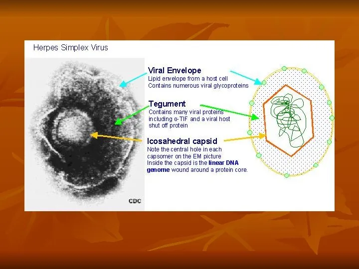
- 3. MORPHOLOGY Large , enveloped viruses containing double stranded DNA Virion : 120-300nm in diameter Icosadeltahedral protein
- 4. RESISTANCE Sensitive to: Acid Solvents Detergents Drying
- 5. ANTIGENIC STRUCTURE HSV 1 & HSV 2 differentiated by serologi typing and by DNA homology Distinguished
- 6. TRANSMISSION Generally transmitted by direct contact of lips or genitals when the sores are present, or
- 7. CELLULAR ENTRY Entry of HSV into the host cell involves interactions of several viral glycoproteins with
- 8. Replication In the case of Herpes virus, initial interactions occur when glycoprotein C, on the surface
- 9. REPLICATION After the viral envelope contents of capsid with tegument proteins has entered the cell via
- 10. REPLICATION
- 11. LATENT INFECTION Especially in neurons, Herpes may persist in a quiescent but persistent form known as
- 12. ENCAPSIDATION AND EGRESS The procapsid proteins (UL18, UL19 and UL38) assemble around scaffolding proteins (UL26 and
- 13. ENVELOPMENT & RELEASE Viral glycoproteins are translated from HSV RNA on the rough endoplasmic reticulum then
- 14. LABORATORY DIAGNOSIS Microscopical Scrapings of lesion: examined microscopically for multinucleated giant cell whose nuclei contain eosinophilic
- 15. Laboratory diagnosis Virological Rapid,definitive determination HSV is made by demonstrating in tissue : a) viral antigen
- 16. Laboratory diagnosis Serological Only useful for diagnosing primary HSV infection & epidemiological studies Not useful for
- 17. TREATMENT Anti HSC drugs like acyclovir, vidarabine, idoxuridine and trifluridine Act as inhibitors of viral DNA
- 20. Vesiculobullous eruption Herpetic Whitlow (HSV 1)
- 21. HSV in situ hybridization
- 23. Скачать презентацию
CLASSIFICATION
Family : Herpesviridae
Genus : Simplexvirus
Subfamily : Alphaherpesvirinae
Species : a) Herpes Simplex
CLASSIFICATION
Family : Herpesviridae
Genus : Simplexvirus
Subfamily : Alphaherpesvirinae
Species : a) Herpes Simplex

MORPHOLOGY
Large , enveloped viruses containing double stranded DNA
Virion : 120-300nm in
MORPHOLOGY
Large , enveloped viruses containing double stranded DNA
Virion : 120-300nm in
RESISTANCE
Sensitive to:
Acid
Solvents
Detergents
Drying
RESISTANCE
Sensitive to:
Acid
Solvents
Detergents
Drying
ANTIGENIC STRUCTURE
HSV 1 & HSV 2 differentiated by serologi typing and
ANTIGENIC STRUCTURE
HSV 1 & HSV 2 differentiated by serologi typing and
TRANSMISSION
Generally transmitted by direct contact of lips or genitals when the
TRANSMISSION
Generally transmitted by direct contact of lips or genitals when the
CELLULAR ENTRY
Entry of HSV into the host cell involves interactions of
CELLULAR ENTRY
Entry of HSV into the host cell involves interactions of
Replication
In the case of Herpes virus, initial interactions occur when glycoprotein
Replication
In the case of Herpes virus, initial interactions occur when glycoprotein
REPLICATION
After the viral envelope contents of capsid with tegument proteins has
REPLICATION
After the viral envelope contents of capsid with tegument proteins has
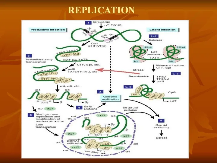
REPLICATION
REPLICATION
LATENT INFECTION
Especially in neurons, Herpes may persist in a quiescent but
LATENT INFECTION
Especially in neurons, Herpes may persist in a quiescent but
ENCAPSIDATION AND EGRESS
The procapsid proteins (UL18, UL19 and UL38) assemble around
ENCAPSIDATION AND EGRESS
The procapsid proteins (UL18, UL19 and UL38) assemble around
ENVELOPMENT & RELEASE
Viral glycoproteins are translated from HSV RNA on the
ENVELOPMENT & RELEASE
Viral glycoproteins are translated from HSV RNA on the
LABORATORY DIAGNOSIS
Microscopical
Scrapings of lesion: examined microscopically for multinucleated giant cell whose
LABORATORY DIAGNOSIS
Microscopical
Scrapings of lesion: examined microscopically for multinucleated giant cell whose
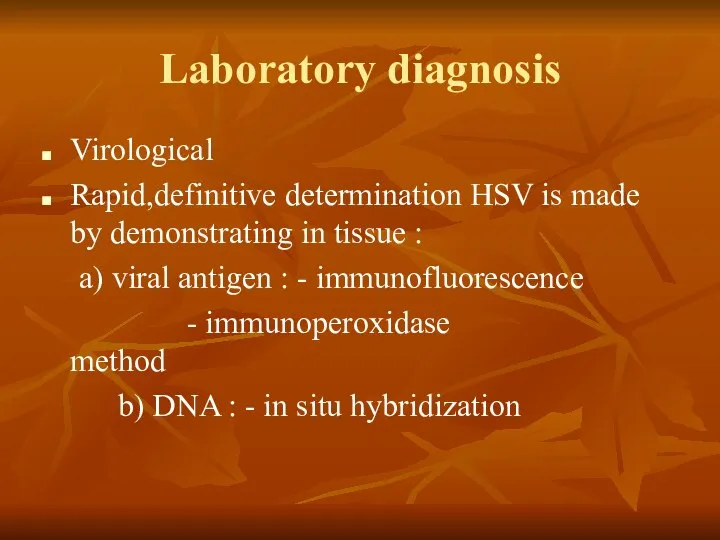
Laboratory diagnosis
Virological
Rapid,definitive determination HSV is made by demonstrating in tissue :
a)
Laboratory diagnosis
Virological
Rapid,definitive determination HSV is made by demonstrating in tissue :
a)
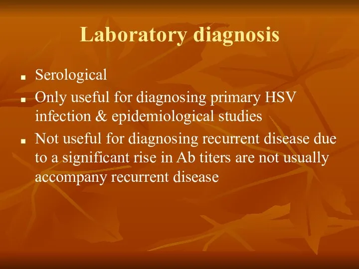
Laboratory diagnosis
Serological
Only useful for diagnosing primary HSV infection & epidemiological studies
Not
Laboratory diagnosis
Serological
Only useful for diagnosing primary HSV infection & epidemiological studies
Not

TREATMENT
Anti HSC drugs like acyclovir, vidarabine, idoxuridine and trifluridine
Act as
TREATMENT
Anti HSC drugs like acyclovir, vidarabine, idoxuridine and trifluridine
Act as
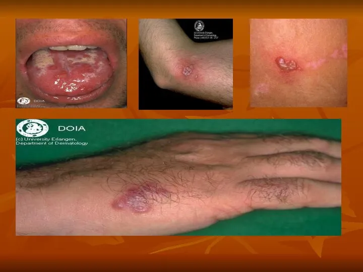

Vesiculobullous eruption
Herpetic Whitlow (HSV 1)
Vesiculobullous eruption
Herpetic Whitlow (HSV 1)
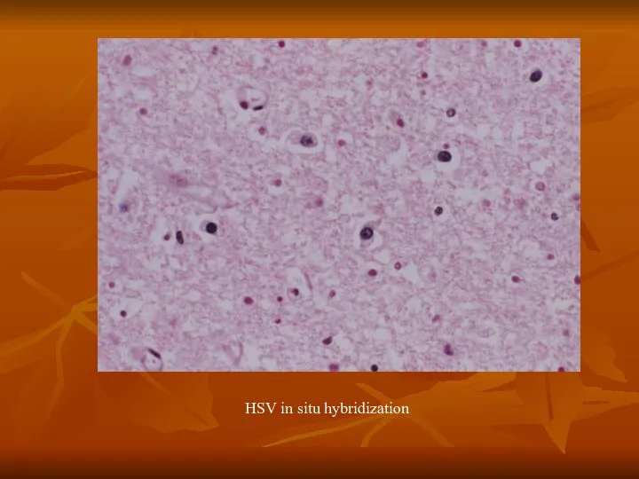
HSV in situ hybridization
HSV in situ hybridization
 «GDP» Склад медикаментов
 Динамикалық SQL
 Нормы литературного языка
 InterSystems Ensemble Преимущества при выполнении SOA-проектов и внедрении SOA-решений
 Автоматизация бизнес-процессов с RPA
 Подача бетонной смеси в конструкции
 Ветхозаветная Троица Призвание Авраама Библейская история встречи Авраама с Богом Изображения Троицы в русской и европейской жи
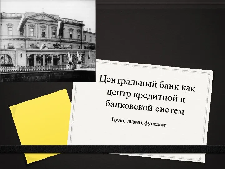 Центральный банк как центр кредитной и банковской систем
 Intermediate 08. Located
 Концепции управления персоналом
 Описание модели приложения с помощью UML
 Физика в архитектуре
 Задания по работе с образовательными интернет-ресурсами
 IT – Скорая. Организация по ремонту компьютеров
 ГПРО ППО
 Микропроцессор дегеніміз
 Проектирование комплекта шкатулок и выполнение в технике лаковой миниатюрной живописи на тему: «Ярмарка»
 Самород сульф
  Значение государственной службы в Российской
 Презентация "Салон красоты" - скачать презентации по Экономике
 Байсаров vs Орбакайте
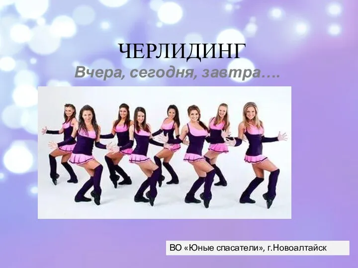 Черлидинг вчера, сегодня, завтра…
 Автор данного шаблона: Ермолаева Ирина Алексеевна учитель информатики и математики МОУ «Павловская сош» с.Павловск Алтайский
  ИНФРАСТРУКТУРА В материале «Услуга» уже были описаны по отдельности основные виды услуг, входящие в более общую категорию «ин
 Схема Горнера. Уильям Джордж Горнер
 Конструкции фильтров
 Автор: к.э.н. доцент каф. «ОД Н и ФР» Дюжов А. В.
 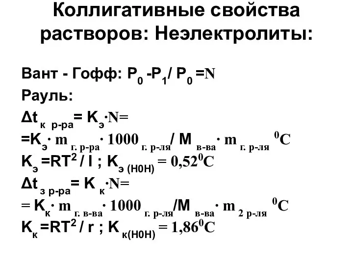 Коллигативные свойства растворов