- Cytogenetic method

Содержание
- 2. Cytogenetics = The study of chromosome number, structure, function, and behavior in relation to gene inheritance,
- 3. Chromosome Chromo = colored in response to dye Some = body Chromosome of Eukaryotes have been
- 4. Why Analyse Chromosomes and Genes? Genetic errors arise from deletions or insertions of genetic material, abnormal
- 5. Cytogenetic methods to detect chromosomal abnormalities underlying human birth defects usually involve analysis of mitotic chromosome
- 6. What tissues are appropriate for chromosome study? • A tissue that can be stimulated to undergo
- 7. The chromosomes are so named as they may be stained by certain dyes Chromosomes are composed
- 8. Chromosome Sister Chromatides
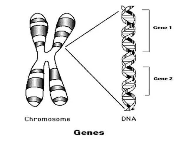
- 9. Chromosomes of different species differ in number and information content Humans and several other species of
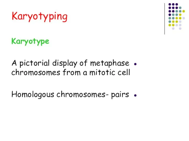
- 10. Karyotyping Karyotype A pictorial display of metaphase chromosomes from a mitotic cell Homologous chromosomes- pairs
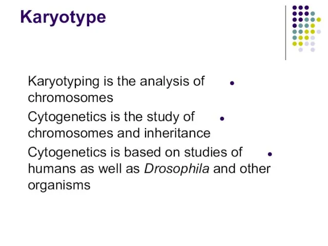
- 11. Karyotype Karyotyping is the analysis of chromosomes Cytogenetics is the study of chromosomes and inheritance Cytogenetics
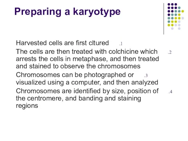
- 12. Preparing a karyotype Harvested cells are first cltured The cells are then treated with colchicine which
- 15. The analysis involves comparing chromosomes for their length, the placement of centromeres (areas where the two
- 17. Metaphase chromosomes
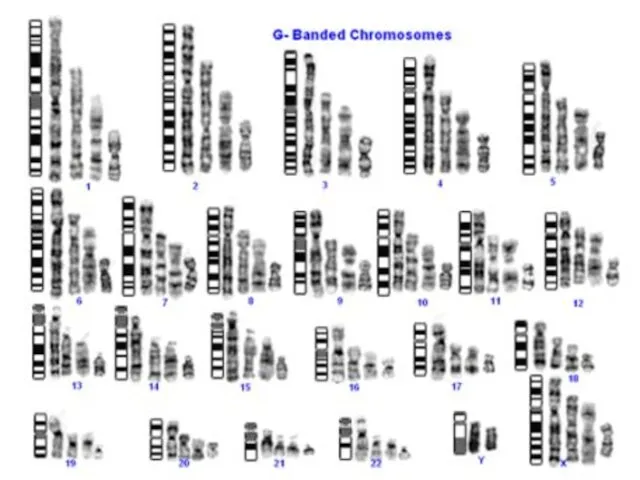
- 18. Karyotyped chromosomes
- 20. Banding patterns on human mitotic chomosomes due to regions of condensed chomatin (darker - G bands)
- 21. Human chromosome number is determined by their length in “mitotic figures"
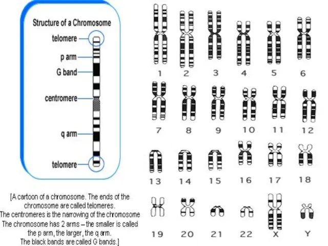
- 23. International System for Cytogenetic Nomenclature, (ISCN,1995) Short arm of the chromosome = p Long arm of
- 24. Hundreds of genes are encompassed within a single G-band. Therefore, most constitutional chromosome abnormalities are associated
- 25. Conclusion The evolution of cytogenetic techniques and the mapping of the human genome have provided scientists
- 27. Скачать презентацию
Cytogenetics = The study of chromosome number, structure, function, and behavior
Cytogenetics = The study of chromosome number, structure, function, and behavior
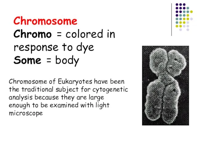
Chromosome
Chromo = colored in response to dye
Some = body
Chromosome of Eukaryotes
Chromosome
Chromo = colored in response to dye
Some = body
Chromosome of Eukaryotes
Why Analyse Chromosomes and Genes?
Genetic errors arise from deletions or insertions
Why Analyse Chromosomes and Genes? Genetic errors arise from deletions or insertions
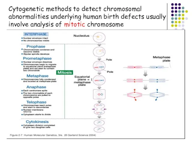
Cytogenetic methods to detect chromosomal
abnormalities underlying human birth defects usually
involve analysis
Cytogenetic methods to detect chromosomal
abnormalities underlying human birth defects usually
involve analysis
What tissues are appropriate for chromosome study?
• A tissue that can
What tissues are appropriate for chromosome study?
• A tissue that can
The chromosomes are so named as they may be stained by
The chromosomes are so named as they may be stained by
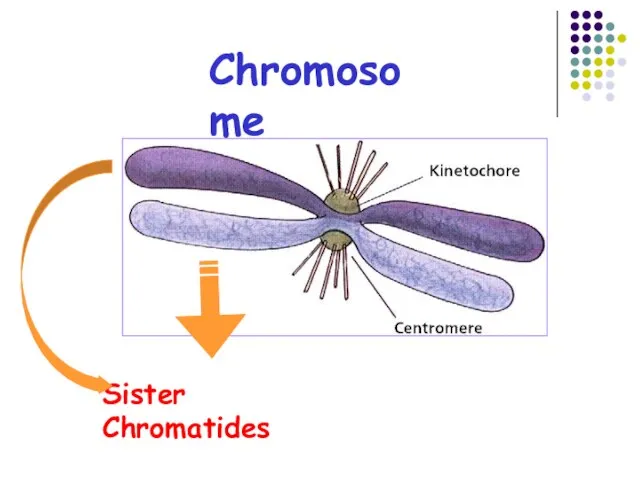
Chromosome
Sister Chromatides
Chromosome
Sister Chromatides
Chromosomes of different species differ in number and information content
Humans
Chromosomes of different species differ in number and information content
Humans
Karyotyping
Karyotype
A pictorial display of metaphase chromosomes from a mitotic cell
Homologous
Karyotyping
Karyotype
A pictorial display of metaphase chromosomes from a mitotic cell
Homologous
Karyotype
Karyotyping is the analysis of chromosomes
Cytogenetics is the study of
Karyotype
Karyotyping is the analysis of chromosomes
Cytogenetics is the study of
Preparing a karyotype
Harvested cells are first cltured
The cells are then
Preparing a karyotype
Harvested cells are first cltured
The cells are then
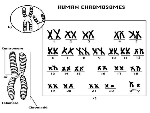
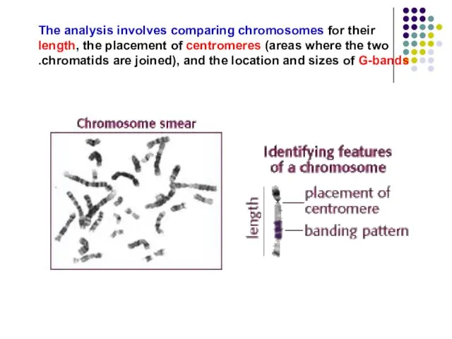
The analysis involves comparing chromosomes for their length, the placement of
The analysis involves comparing chromosomes for their length, the placement of
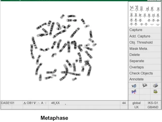
Metaphase chromosomes
Metaphase chromosomes
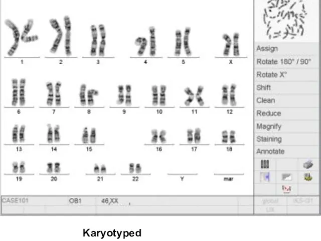
Karyotyped chromosomes
Karyotyped chromosomes
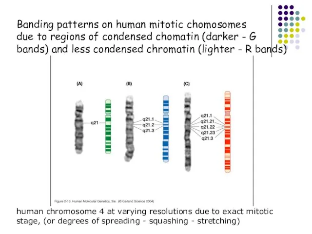
Banding patterns on human mitotic chomosomes
due to regions of condensed chomatin
Banding patterns on human mitotic chomosomes
due to regions of condensed chomatin
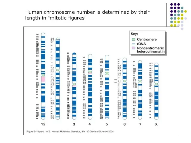
Human chromosome number is determined by their
length in “mitotic figures"
Human chromosome number is determined by their
length in “mitotic figures"
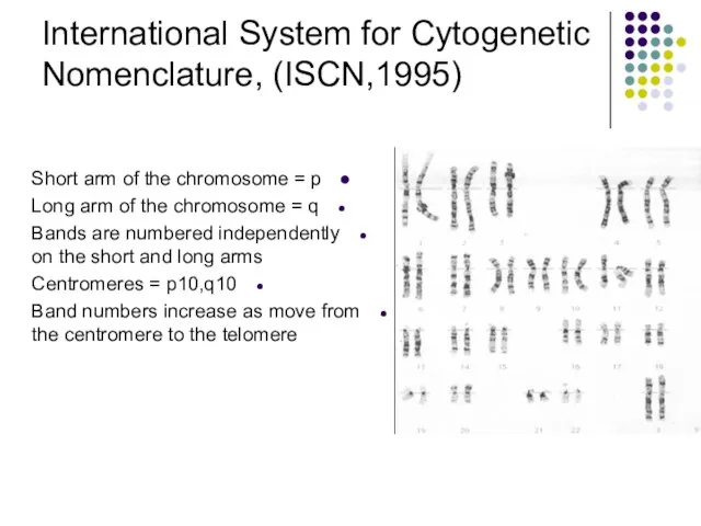
International System for Cytogenetic
Nomenclature, (ISCN,1995)
Short arm of the chromosome = p
Long
International System for Cytogenetic
Nomenclature, (ISCN,1995)
Short arm of the chromosome = p
Long
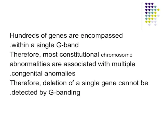
Hundreds of genes are encompassed
within a single G-band.
Therefore, most constitutional chromosome
abnormalities
Hundreds of genes are encompassed
within a single G-band.
Therefore, most constitutional chromosome
abnormalities
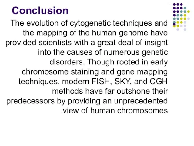
Conclusion
The evolution of cytogenetic techniques and the mapping of the human
Conclusion
The evolution of cytogenetic techniques and the mapping of the human
 Обитатели морей и океанов
 Путешествие по островам "Роботоландии"
 Главные направления эволюции
 Происхождение человека и становление общества
 Брюхоногие моллюски. Виноградная улитка
 Интеллектуальная игра. Птицы
 МОУ Сосновская СОШ №1 Учитель биологии Кинзбурская Татьяна Васильевна
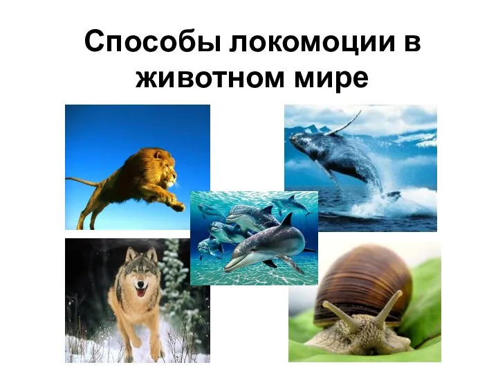 Способы локомоции в животном мире
 Иммунитет Виды иммунитета
  Биосинтез белковой молекулы
 ВЛИЯНИЕ ПОДКОРМКИ НА РОСТ И РАЗВИТИЕ ЛУКА РЕПЧАТОГО Выполнила: Творогова Елизавета Руководитель: Анисимова Светлан
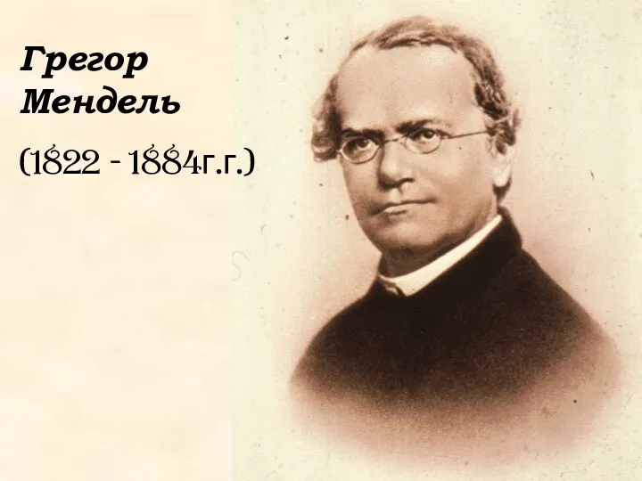 Грегор Мендель (1822 1884г.г.)
 Взаимосвязь между Репродуктивным здоровьем и демографической политикой
 Характеристика биогеоценоза
 Анатомия и физиология репродуктивной системы. Мужские половые органы
 Клетка, как структурно-функциональная единица живого
 Размножение. Органы размножения и их функции у самцов
 Гормоны-2
 Презентация на тему "Движущие силы антропогенеза" - скачать презентации по Биологии
 Презентация на тему "Дикие - дикие кошки" - скачать бесплатно презентации по Биологии
 МБОУ «Михневский районный Детско-Юношеский-Центр Ступинского муниципального района Московской области» Проект «Видовой состав
 Функциональная анатомия вегетативной нервной системы
 Мой домашний питомец Кеша
 Презентация на тему Понятие о систематике растений
 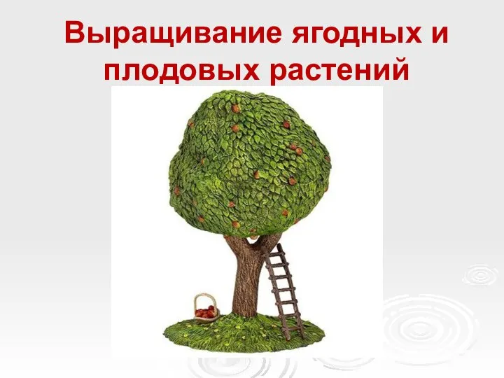 Выращивание ягодных и плодовых растений. Размножение
 Матричные процессы. Этапы транскрипции
 Клетка - элементарная биологическая система
 Водоросли. Мхи