- The biological perspective

Содержание
- 2. Learning Objectives 2.1 What are the nervous system, neurons, and nerves, and how do they relate
- 3. Overview of Nervous System Nervous system an extensive network of specialized cells that carry information to
- 4. Structure of the Neuron Neuron the basic cell that makes up the nervous system and receives
- 5. Structure of the Neuron Parts of a neuron dendrites: branch-like structures that receive messages from other
- 6. Figure 2.2 The Structure of the Neuron The electronmicrograph on the left shows myelinated axons.
- 7. Other Types of Brain Cells Glial cells are grey fatty cells that: provide support for the
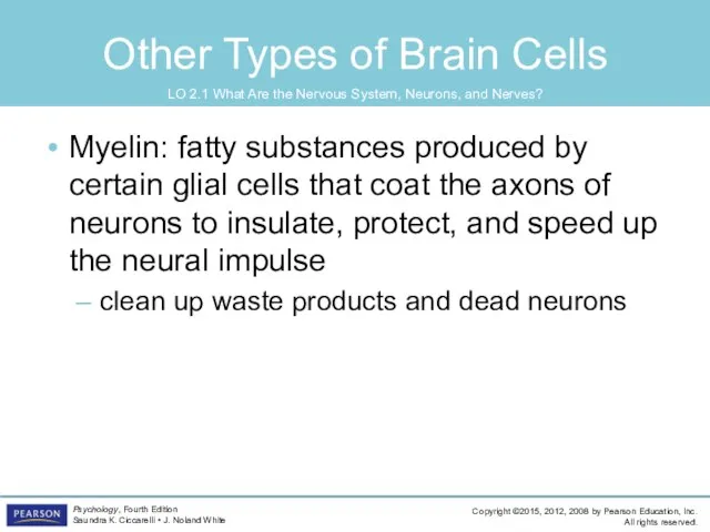
- 8. Other Types of Brain Cells Myelin: fatty substances produced by certain glial cells that coat the
- 9. Generating the Message: Neural Impulse Ions: charged particles inside neuron: negatively charged outside neuron: positively charged
- 10. Generating the Message: Neural Impulse All-or-none: a neuron either fires completely or does not fire at
- 11. Figure 2.3 The Neural Impulse Action Potential In the graph below, voltage readings are shown at
- 12. Figure 2.3 (continued) The Neural Impulse Action Potential In the graph below, voltage readings are shown
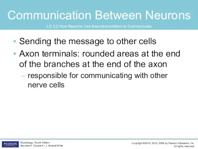
- 13. Communication Between Neurons Sending the message to other cells Axon terminals: rounded areas at the end
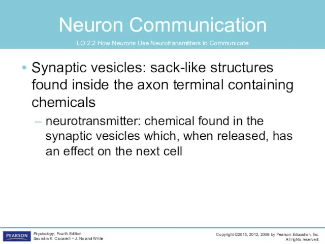
- 14. Neuron Communication Synaptic vesicles: sack-like structures found inside the axon terminal containing chemicals neurotransmitter: chemical found
- 15. Neuron Communication synapse/synaptic gap: microscopic fluid-filled space between the rounded areas on the end of the
- 16. Figure 2.4 Reuptake of Dopamine Dopamine is removed from the synapse by reuptake sites. Cocaine acts
- 17. Neuron Communication Neurons must be turned ON and OFF excitatory neurotransmitter: neurotransmitter that causes the receiving
- 18. Neuron Communication Chemical substances can affect neuronal communication agonists: mimic or enhance the effects of a
- 20. Cleaning up the Synapse reuptake: process by which neurotransmitters are taken back into the synaptic vesicles
- 21. Figure 2.5 An Overview of the Nervous System
- 22. Central Nervous System Central nervous system (CNS): part of the nervous system consisting of the brain
- 23. The Reflex Arc: Three Types of Neurons Sensory neuron: a neuron that carries information from the
- 24. The Reflex Arc: Three Types of Neurons Interneuron: a neuron found in the center of the
- 25. The Reflex Arc: Three Types of Neurons Neuroplasticity: the ability to constantly change both the structure
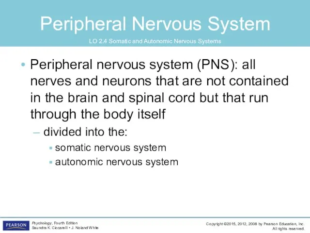
- 26. Peripheral Nervous System Peripheral nervous system (PNS): all nerves and neurons that are not contained in
- 27. Figure 2.7 The Peripheral Nervous System
- 28. Somatic Nervous System Soma = “body” Somatic nervous system: division of the PNS consisting of nerves
- 29. Somatic Nervous System Somatic nervous system (cont’d) motor pathway: nerves coming from the CNS to the
- 30. Autonomic Nervous System Autonomic nervous system (ANS) division of the PNS consisting of nerves that control
- 31. Autonomic Nervous System Autonomic Nervous System (ANS) (cont’d) sympathetic division (fight-or-flight system): part of the ANS
- 32. Figure 2.8 Functions of the Parasympathetic and Sympathetic Divisions of the Nervous System
- 33. The Endocrine Glands Endocrine glands: glands that secrete chemicals called hormones directly into the bloodstream hormones:
- 34. Figure 2.9 The Endocrine Glands
- 35. The Endocrine Glands pituitary gland: gland located in the brain that secretes human growth hormone and
- 36. The Endocrine Glands gonads: the sex glands; secrete hormones that regulate sexual development and behavior as
- 37. Looking inside the Living Brain Clinical Studies deep lesioning: insertion of a thin, insulated wire into
- 38. Looking inside the Living Brain Clinical Studies transcranial magnetic stimulation (TMS), magnetic pulses are applied to
- 39. Mapping Structure computed tomography (CT): brain-imaging method using computer-controlled X-rays of the brain magnetic resonance imaging
- 40. Mapping Structure Mapping Function electroencephalogram (EEG): records electric activity of the brain below specific areas of
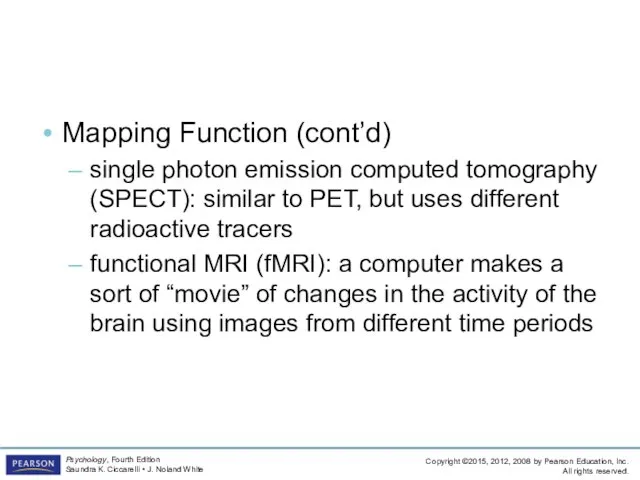
- 41. Mapping Structure Mapping Function (cont’d) single photon emission computed tomography (SPECT): similar to PET, but uses
- 42. Figure 2.12 Major Structures of the Human Brain
- 43. The Hindbrain The Hindbrain medulla: first large swelling at the top of the spinal cord, forming
- 44. The Hindbrain reticular formation (RF): area of neurons running through the middle of the medulla and
- 45. Figure 2.13 The Limbic System
- 46. Structures under the Cortex Limbic system: a group of several brain structures located under the cortex
- 47. Structures under the Cortex Limbic System (cont’d) hypothalamus: small structure in the brain located below the
- 48. Structures under the Cortex Limbic System (cont’d) amygdala: brain structure located near the hippocampus responsible for
- 49. Cortex cortex: outermost covering of the brain consisting of densely packed neurons responsible for higher thought
- 50. Cerebral Hemispheres cerebral hemispheres: the two sections of the cortex on the left and right sides
- 51. Figure 2.14 The Lobes of the Brain
- 52. Four Lobes of the Brain occipital lobe: section of the brain located at the rear and
- 53. Four Lobes of the Brain parietal lobes sections of the brain located at the top and
- 54. Figure 2.15 The Motor and Somatosensory Cortex
- 55. Four Lobes of the Brain temporal lobes: areas of the cortex located just behind the temples
- 56. Four Lobes of the Brain frontal lobes: areas of the cortex located in the front and
- 57. Association Areas of Cortex association areas: areas within each lobe of the cortex responsible for the
- 58. Association Areas of Cortex Broca’s aphasia: condition resulting from damage to Broca’s area (usually in left
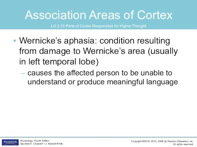
- 59. Association Areas of Cortex Wernicke’s aphasia: condition resulting from damage to Wernicke’s area (usually in left
- 60. Association Areas of Cortex spatial neglect: condition produced by damage to the association areas of the
- 61. Split-Brain Research Cerebrum: the upper part of the brain consisting of the two hemispheres and the
- 62. Split-Brain Research Split-Brain Research study of patients with severed corpus callosum involves sending messages to only
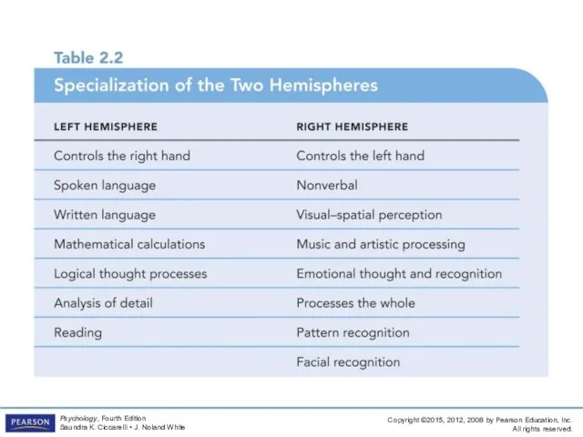
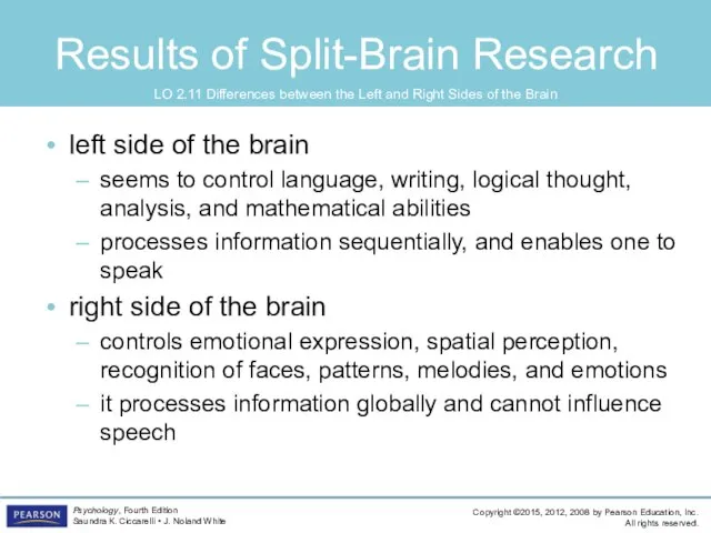
- 64. Results of Split-Brain Research left side of the brain seems to control language, writing, logical thought,
- 66. Скачать презентацию

Learning Objectives
2.1 What are the nervous system, neurons, and nerves, and how
Learning Objectives
2.1 What are the nervous system, neurons, and nerves, and how

Overview of Nervous System
Nervous system
an extensive network of specialized cells that
Overview of Nervous System
Nervous system
an extensive network of specialized cells that
Structure of the Neuron
Neuron
the basic cell that makes up the nervous
Structure of the Neuron
Neuron
the basic cell that makes up the nervous
Structure of the Neuron
Parts of a neuron
dendrites: branch-like structures that receive
Structure of the Neuron
Parts of a neuron
dendrites: branch-like structures that receive
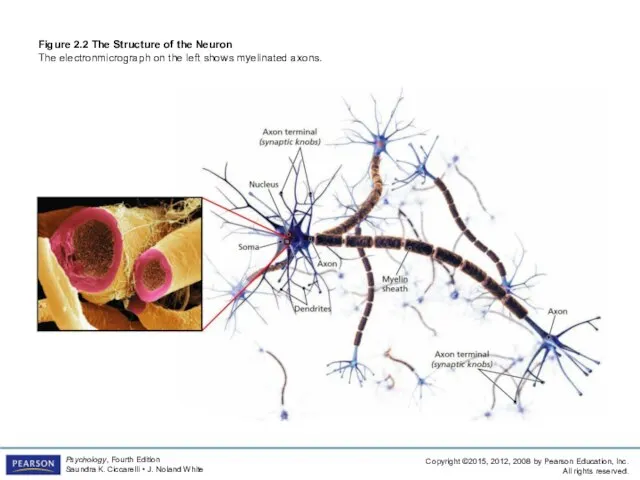
Figure 2.2 The Structure of the Neuron
The electronmicrograph on the left
Figure 2.2 The Structure of the Neuron The electronmicrograph on the left
Other Types of Brain Cells
Glial cells are grey fatty cells that:
Other Types of Brain Cells
Glial cells are grey fatty cells that:
Other Types of Brain Cells
Myelin: fatty substances produced by certain glial
Other Types of Brain Cells
Myelin: fatty substances produced by certain glial

Generating the Message: Neural Impulse
Ions: charged particles
inside neuron: negatively charged
outside neuron:
Generating the Message: Neural Impulse
Ions: charged particles
inside neuron: negatively charged
outside neuron:
Generating the Message: Neural Impulse
All-or-none: a neuron either fires completely or
Generating the Message: Neural Impulse
All-or-none: a neuron either fires completely or

Figure 2.3 The Neural Impulse Action Potential
In the graph below, voltage
Figure 2.3 The Neural Impulse Action Potential In the graph below, voltage
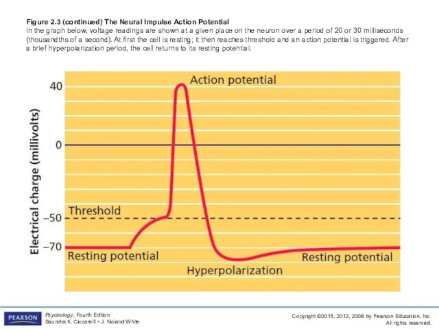
Figure 2.3 (continued) The Neural Impulse Action Potential
In the graph below,
Figure 2.3 (continued) The Neural Impulse Action Potential In the graph below,
Communication Between Neurons
Sending the message to other cells
Axon terminals: rounded areas
Communication Between Neurons
Sending the message to other cells
Axon terminals: rounded areas
Neuron Communication
Synaptic vesicles: sack-like structures found inside the axon terminal containing
Neuron Communication
Synaptic vesicles: sack-like structures found inside the axon terminal containing

Neuron Communication
synapse/synaptic gap: microscopic fluid-filled space between the rounded areas on
Neuron Communication
synapse/synaptic gap: microscopic fluid-filled space between the rounded areas on
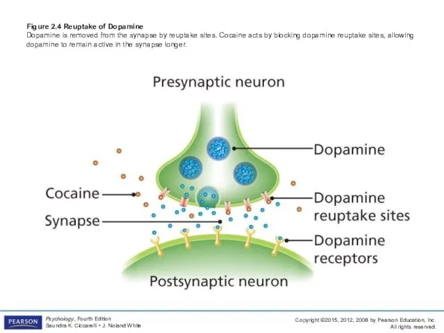
Figure 2.4 Reuptake of Dopamine
Dopamine is removed from the synapse by
Figure 2.4 Reuptake of Dopamine Dopamine is removed from the synapse by
Neuron Communication
Neurons must be turned ON and OFF
excitatory neurotransmitter: neurotransmitter that
Neuron Communication
Neurons must be turned ON and OFF
excitatory neurotransmitter: neurotransmitter that
Neuron Communication
Chemical substances can affect neuronal communication
agonists: mimic or enhance the
Neuron Communication
Chemical substances can affect neuronal communication
agonists: mimic or enhance the


Cleaning up the Synapse
reuptake: process by which neurotransmitters are taken back
Cleaning up the Synapse
reuptake: process by which neurotransmitters are taken back
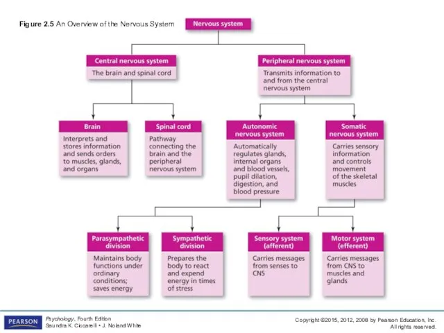
Figure 2.5 An Overview of the Nervous System
Figure 2.5 An Overview of the Nervous System

Central Nervous System
Central nervous system (CNS): part of the nervous system
Central Nervous System
Central nervous system (CNS): part of the nervous system
The Reflex Arc: Three Types of Neurons
Sensory neuron: a neuron that
The Reflex Arc: Three Types of Neurons
Sensory neuron: a neuron that
The Reflex Arc: Three Types of Neurons
Interneuron: a neuron found in
The Reflex Arc: Three Types of Neurons
Interneuron: a neuron found in
The Reflex Arc: Three Types of Neurons
Neuroplasticity: the ability to constantly
The Reflex Arc: Three Types of Neurons
Neuroplasticity: the ability to constantly
Peripheral Nervous System
Peripheral nervous system (PNS): all nerves and neurons that
Peripheral Nervous System
Peripheral nervous system (PNS): all nerves and neurons that
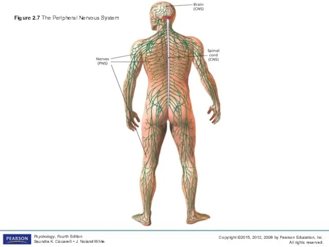
Figure 2.7 The Peripheral Nervous System
Figure 2.7 The Peripheral Nervous System

Somatic Nervous System
Soma = “body”
Somatic nervous system: division of the PNS
Somatic Nervous System
Soma = “body”
Somatic nervous system: division of the PNS

Somatic Nervous System
Somatic nervous system (cont’d)
motor pathway: nerves coming from the
Somatic Nervous System
Somatic nervous system (cont’d)
motor pathway: nerves coming from the
Autonomic Nervous System
Autonomic nervous system (ANS)
division of the PNS consisting of
Autonomic Nervous System
Autonomic nervous system (ANS)
division of the PNS consisting of

Autonomic Nervous System
Autonomic Nervous System (ANS) (cont’d)
sympathetic division (fight-or-flight system): part
Autonomic Nervous System
Autonomic Nervous System (ANS) (cont’d)
sympathetic division (fight-or-flight system): part
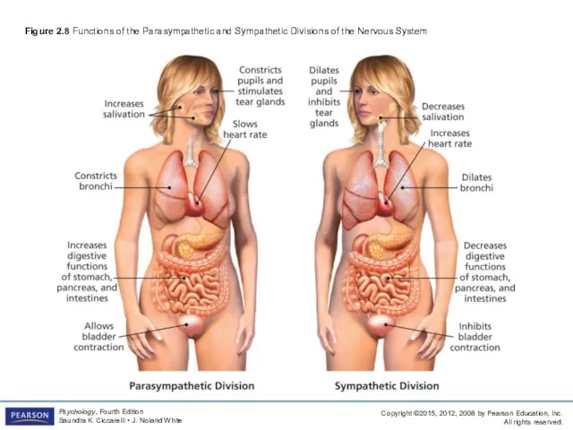
Figure 2.8 Functions of the Parasympathetic and Sympathetic Divisions of the
Figure 2.8 Functions of the Parasympathetic and Sympathetic Divisions of the
The Endocrine Glands
Endocrine glands: glands that secrete chemicals called hormones directly
The Endocrine Glands
Endocrine glands: glands that secrete chemicals called hormones directly

Figure 2.9 The Endocrine Glands
Figure 2.9 The Endocrine Glands

The Endocrine Glands
pituitary gland: gland located in the brain that secretes
The Endocrine Glands
pituitary gland: gland located in the brain that secretes
The Endocrine Glands
gonads: the sex glands; secrete hormones that regulate sexual
The Endocrine Glands
gonads: the sex glands; secrete hormones that regulate sexual
Looking inside the Living Brain
Clinical Studies
deep lesioning: insertion of a thin,
Looking inside the Living Brain
Clinical Studies
deep lesioning: insertion of a thin,
Looking inside the Living Brain
Clinical Studies
transcranial magnetic stimulation (TMS), magnetic pulses
Looking inside the Living Brain
Clinical Studies
transcranial magnetic stimulation (TMS), magnetic pulses

Mapping Structure
computed tomography (CT): brain-imaging method using computer-controlled X-rays of the
Mapping Structure
computed tomography (CT): brain-imaging method using computer-controlled X-rays of the

Mapping Structure
Mapping Function
electroencephalogram (EEG): records electric activity of the brain below
Mapping Structure
Mapping Function
electroencephalogram (EEG): records electric activity of the brain below
Mapping Structure
Mapping Function (cont’d)
single photon emission computed tomography (SPECT): similar to
Mapping Structure
Mapping Function (cont’d)
single photon emission computed tomography (SPECT): similar to
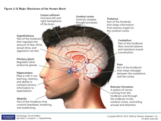
Figure 2.12 Major Structures of the Human Brain
Figure 2.12 Major Structures of the Human Brain
The Hindbrain
The Hindbrain
medulla: first large swelling at the top of the
The Hindbrain
The Hindbrain
medulla: first large swelling at the top of the
The Hindbrain
reticular formation (RF): area of neurons running through the middle
The Hindbrain
reticular formation (RF): area of neurons running through the middle

Figure 2.13 The Limbic System
Figure 2.13 The Limbic System
Structures under the Cortex
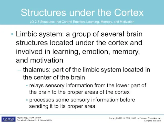
Limbic system: a group of several brain structures
Structures under the Cortex
Limbic system: a group of several brain structures
Structures under the Cortex
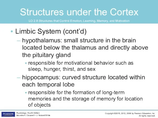
Limbic System (cont’d)
hypothalamus: small structure in the brain
Structures under the Cortex
Limbic System (cont’d)
hypothalamus: small structure in the brain

Structures under the Cortex
Limbic System (cont’d)
amygdala: brain structure located near the
Structures under the Cortex
Limbic System (cont’d)
amygdala: brain structure located near the
Cortex
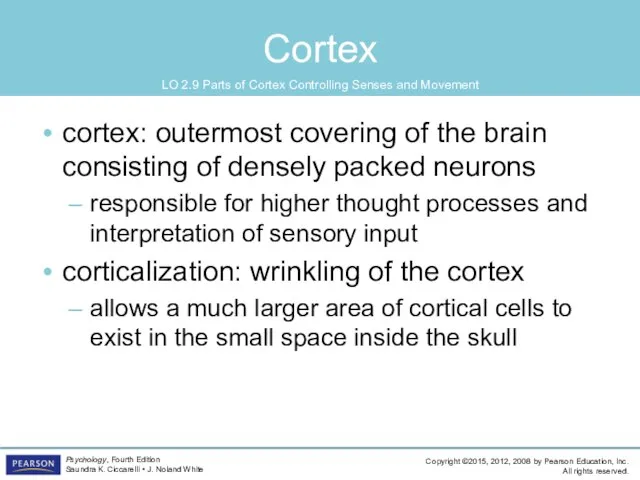
cortex: outermost covering of the brain consisting of densely packed neurons
responsible
Cortex
cortex: outermost covering of the brain consisting of densely packed neurons
responsible
Cerebral Hemispheres
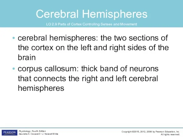
cerebral hemispheres: the two sections of the cortex on the
Cerebral Hemispheres
cerebral hemispheres: the two sections of the cortex on the
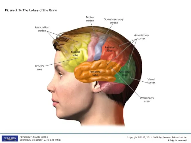
Figure 2.14 The Lobes of the Brain
Figure 2.14 The Lobes of the Brain

Four Lobes of the Brain
occipital lobe: section of the brain located
Four Lobes of the Brain
occipital lobe: section of the brain located

Four Lobes of the Brain
parietal lobes
sections of the brain located at
Four Lobes of the Brain
parietal lobes
sections of the brain located at

Figure 2.15 The Motor and Somatosensory Cortex
Figure 2.15 The Motor and Somatosensory Cortex
Four Lobes of the Brain
temporal lobes: areas of the cortex located
Four Lobes of the Brain
temporal lobes: areas of the cortex located
Four Lobes of the Brain
frontal lobes: areas of the cortex located
Four Lobes of the Brain
frontal lobes: areas of the cortex located

Association Areas of Cortex
association areas: areas within each lobe of the
Association Areas of Cortex
association areas: areas within each lobe of the
Association Areas of Cortex
Broca’s aphasia: condition resulting from damage to Broca’s
Association Areas of Cortex
Broca’s aphasia: condition resulting from damage to Broca’s
Association Areas of Cortex
Wernicke’s aphasia: condition resulting from damage to Wernicke’s
Association Areas of Cortex
Wernicke’s aphasia: condition resulting from damage to Wernicke’s

Association Areas of Cortex
spatial neglect: condition produced by damage to the
Association Areas of Cortex
spatial neglect: condition produced by damage to the
Split-Brain Research
Cerebrum: the upper part of the brain consisting of the
Split-Brain Research
Cerebrum: the upper part of the brain consisting of the

Split-Brain Research
Split-Brain Research
study of patients with severed corpus callosum
involves sending messages
Split-Brain Research
Split-Brain Research
study of patients with severed corpus callosum
involves sending messages
Results of Split-Brain Research
left side of the brain
seems to control language,
Results of Split-Brain Research
left side of the brain
seems to control language,
 Презентация на тему "Хищные птицы" - скачать презентации по Биологии
 Задачи, методы и достижения БИОТЕХНОЛОГИИ
  Харчові ланцюги
 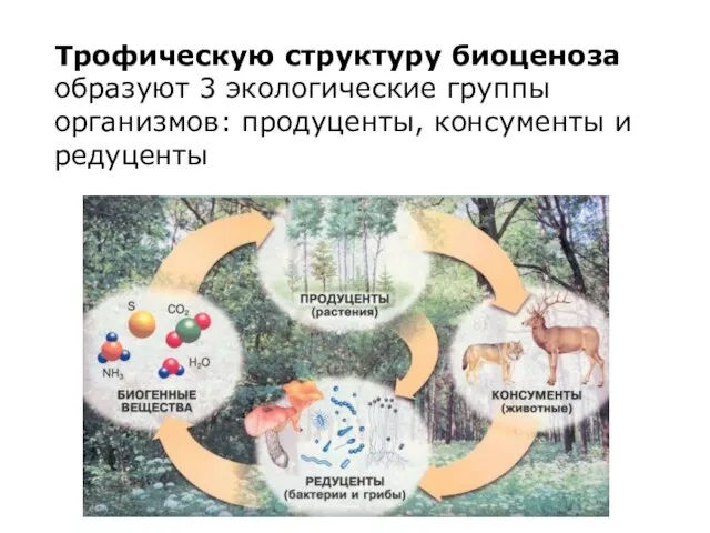 Трофическая структура биоценоза. Продуценты, консументы и редуценты
 Презентация на тему "Смешанные леса " - скачать презентации по Биологии
 Особенности ионообменных реакций с участием биологически активных веществ. Ионообменная сорбция пептидов и белков
 Развитие жизни на Земле в архейскую и протерозойскую эры
 Потребности человека
 Метод фистул И.П.Павлова Го Сергеевна
 Введение. Принцип строения и функции нервной системы. Общая чувствительность и семиотика ее нарушения
 Газообмен в легких и тканях
 Птицы
Птицы Презентация на тему "Железо внутри нас" - скачать презентации по Биологии
 Проект Лапа помощи по защите амурских тигров
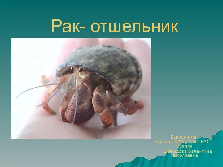 Рак отшельник
 Тема: Биосинтез белка.
 Празитизм как экологическое явление
 Роль цианобактерий в развитии жизни на Земле
 Органы растений: корень, стебель и лист
 Тип молюски
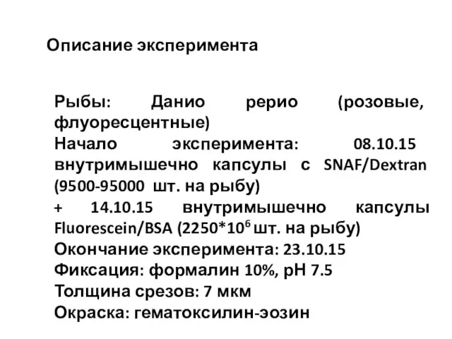 Описание эксперимента. Рыба: Данио рерио
 Значение и охрана пресных водоёмов
 Половое и бесполое размножение Покрытосеменных.
 Роль микроорганизмов в природе
 Каракал
 Бурый медведь
 Дикие животные
 Систематика растений