- Tick borne enciphitalis

Содержание
- 2. General characteristics The disease typically follows a biphasic pattern in 72–87% of patients and the median
- 3. TBE is caused by tick-borne encephalitis virus, a member of the genus Flavivirus in the family
- 4. TRANSMISSION Sheep ticks (Ixodes ricinus), such as this engorged female, transmit the disease It is transmitted
- 6. Transmission
- 7. Epidemiology As of 2011, the disease was most common in Central and Eastern Europe, and Northern
- 8. Treatment The disease is incurable once manifested, so there is no specific drug therapy for TBE.
- 10. Clinical Signs Canine TBE The incubation period for canine TBE in most cases is between 7
- 11. Human Tick Paralysis In humans, tick paralysis is most likely to be seen in children. The
- 12. A recent CDC report showed that vector-borne diseases -- those transmitted by ticks, mosquitoes, and fleas
- 13. Encephalitis is an inflammation of the brain, usually caused by a viral infection. Although rare, it
- 14. Some forms of encephalitis are more severe than others. For example, herpes simplex encephalitis (HSE) has
- 15. Lyme encephalopathy is a rare neuropsychiatric disorder, predominantly affecting memory and concentration [3–7]. Other associated symptoms
- 16. The prognosis for a person with encephalitis varies according to the age of the patient –
- 17. The contagious period and incubation period for encephalitis depends on the underlying cause of encephalitis. For
- 18. LINKS AND REFRENCES https://youtu.be/5bEimiIZ9dA https://youtu.be/Bv3hQZVtE6Y https://youtu.be/ENmVXtwsKE8
- 19. Further information Edlow JA, McGillicuddy DC: Tick paralysis. Inf Dis Clin North Am. 2008, 22, 397-414
- 21. Скачать презентацию

General characteristics
The disease typically follows a biphasic pattern in 72–87% of
General characteristics
The disease typically follows a biphasic pattern in 72–87% of
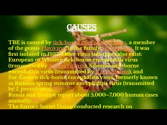
TBE is caused by tick-borne encephalitis virus, a member of the genus Flavivirus in
TBE is caused by tick-borne encephalitis virus, a member of the genus Flavivirus in
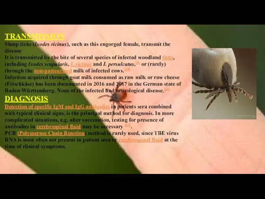
TRANSMISSION
Sheep ticks (Ixodes ricinus), such as this engorged female, transmit the
TRANSMISSION
Sheep ticks (Ixodes ricinus), such as this engorged female, transmit the


Transmission
Transmission
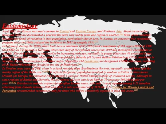
Epidemiology
As of 2011, the disease was most common in Central and Eastern Europe, and
Epidemiology
As of 2011, the disease was most common in Central and Eastern Europe, and

Treatment
The disease is incurable once manifested, so there is no specific drug
Treatment
The disease is incurable once manifested, so there is no specific drug


Clinical Signs
Canine TBE
The incubation period for canine TBE in most cases
Clinical Signs
Canine TBE
The incubation period for canine TBE in most cases
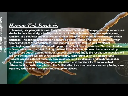
Human Tick Paralysis
In humans, tick paralysis is most likely to be
Human Tick Paralysis
In humans, tick paralysis is most likely to be
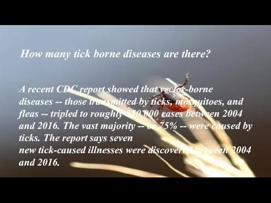
A recent CDC report showed that vector-borne diseases -- those transmitted by
A recent CDC report showed that vector-borne diseases -- those transmitted by
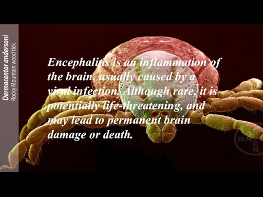
Encephalitis is an inflammation of the brain, usually caused by a viral infection. Although rare, it
Encephalitis is an inflammation of the brain, usually caused by a viral infection. Although rare, it
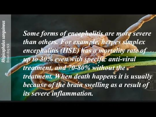
Some forms of encephalitis are more severe than others. For example,
Some forms of encephalitis are more severe than others. For example,
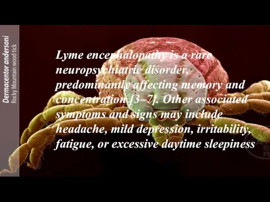
Lyme encephalopathy is a rare neuropsychiatric disorder, predominantly affecting memory and concentration
Lyme encephalopathy is a rare neuropsychiatric disorder, predominantly affecting memory and concentration
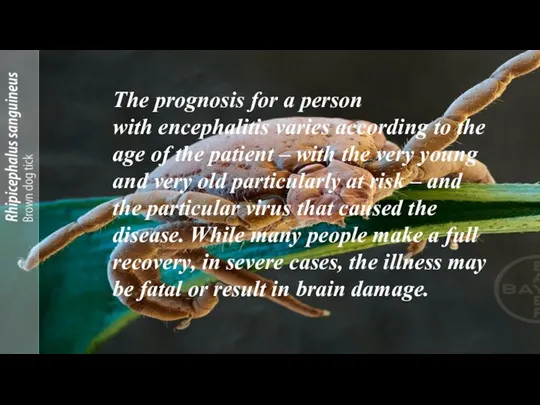
The prognosis for a person with encephalitis varies according to the age of the patient
The prognosis for a person with encephalitis varies according to the age of the patient

The contagious period and incubation period for encephalitis depends on the underlying cause of encephalitis. For
The contagious period and incubation period for encephalitis depends on the underlying cause of encephalitis. For
LINKS AND REFRENCES
https://youtu.be/5bEimiIZ9dA
https://youtu.be/Bv3hQZVtE6Y
https://youtu.be/ENmVXtwsKE8
LINKS AND REFRENCES
https://youtu.be/5bEimiIZ9dA
https://youtu.be/Bv3hQZVtE6Y
https://youtu.be/ENmVXtwsKE8

Further information
Edlow JA, McGillicuddy DC: Tick paralysis. Inf Dis Clin North
Further information
Edlow JA, McGillicuddy DC: Tick paralysis. Inf Dis Clin North
 Презентация на тему "Забота о потомстве у Рыб" - скачать бесплатно презентации по Биологии
 Открытый урок по биологии Размножение и развитие лягушки. Учитель: Шиляева Вера Анатольевна
 Тип Саркодовые
 Историческое развитие растительного мира на Земле
 Генетика человека
 ЁЖ обыкновенный Виноградова Инга 5«а» класс
  Зрительный аппарат хищных птиц
 Презентация Богомол
 Значение и многообразие растений. Мифы и легенды о растениях
 64432-48
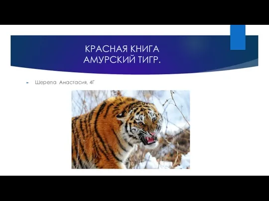 Красная книга. Амурский тигр
 Презентация по биологии Рабдовирусы. (Rhabdoviridae) Возбудители бешенства и вазикулярного стоматита.
 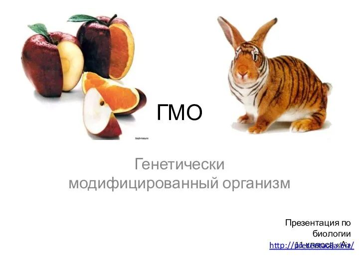 ГМО Генетически модифицированный организм
  Тема урока: Высшие споровые. Плауны, хвощи, папоротники Цель урока: - изучить представителей современных папоротникообразных; -
  Продолговатый мозг. Лекция 5
 Презентация на тему Системы органов человека
  Самая курлыкающая птица. Серый журавль
 Закономерности наследования признаков, установленные Г.Менделем. Первый и второй законы Менделя
 Значение опорнодвигательной системы. Строение костей
 Презентация ученика 5 «б» КЛАССА Хоменко руслана На тему : Тигры
  Хамелеон. Дивовижні факти
 Человек, личность, индивид, индивидуальность
 Островки патогенности. Универсальность факторов патогенности
 Движущие силы эволюции: борьба за существование, наследственная изменчивость, естественный отбор
 Морфология человека. Мягкие части лица. Тотальные размеры тела
 Экологические группы птиц
 Влияние норм минеральных удобрений на урожайность и качество озимой пшеницы в условиях Орловской области
 Осложнения при укусах домашних и диких животных