- Pathological Anatomy Department

Содержание
- 2. Introduction Pathological anatomy and anatomic pathologist History Tasks of the pathological anatomy Biopsy, operational material, autopsy
- 3. Pathological anatomy is the science that studies the structural bases of the disease at different levels
- 4. In 1761 Italian author G. Morgagni wrote the first work on pathological anatomy "About the location
- 5. The founder of modern pathological anatomy is R. Virchow (1821—1902) - German researcher who created the
- 6. Theoretic tasks of the pathological anatomy: 1) Study of the etiology, pathogenesis, morphology and morphogenesis of
- 7. Practical tasks of the pathological anatomy: 1) Control of accuracy and timeliness of clinical diagnosis; 2)
- 8. Methods of the pathological anatomy Macroscopic Microscopic (light microscope) Electron microscope Cytochemistry Histochemistry Immunohistochemistry (IHC) Approaches
- 9. General pathology studies typical pathological processes specific to a particular disease. Systemic pathology studies causes of
- 10. Adaptation limits (reversible/irreversible) depend on tissue type and its functional activity, strength and duration of exposure
- 11. MORPHOLOGY OF THE CELLULAR DAMAGE DEGENERATIONS APOPTOSIS NECROSIS METABOLIC DISORDERS, LEADING TO CHANGES IN THE STRUCTURE
- 12. DEGENERATION Gr.: dys - violation; trophe - nutrition Transformation (ability of some substances turn into the
- 13. I. By localization 1. Intracellular (parenchymal); 2. Extracellular (stromal vascular, mesenchymal); 3. Mixed II. By extent
- 14. INTRACELLULAR PROTEIN DEGENERATIONS Granular degeneration Hyaline-drop degeneration Hydropic degeneration Keratinization degeneration
- 15. INTRACELLULAR FAT DEGENERATIONS CAUSES: Hypoxia (heart diseases, lungs and blood disorders) Infections Chronic intoxications
- 16. INTRACELLULAR FAT DEGENERATIONS CAUSES: Hypoxia (heart diseases, lungs and blood disorders) Infections Chronic intoxications "Tiger's heart"
- 17. INTRACELLULAR CARBOHYDRATE DEGENERATIONS Glycogen metabolic disorders Glycoproteins metabolic disorders Can be revealed using PAS-reaction Glycogen is
- 18. Cystic fibrosis of the pancreas
- 19. EXTRACELLULAR PROTEIN DEGENERATIONS Mucoid swelling Fibrinoid swelling Hyalinosis Amyloidosis
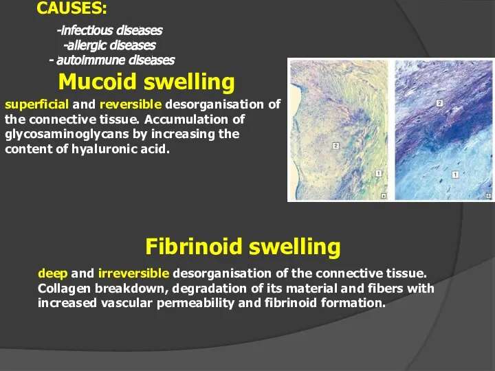
- 20. Mucoid swelling superficial and reversible desorganisation of the connective tissue. Accumulation of glycosaminoglycans by increasing the
- 21. Fibrinoid changes
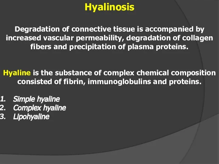
- 22. Hyalinosis Degradation of connective tissue is accompanied by increased vascular permeability, degradation of collagen fibers and
- 23. Hyalinosis Hyalinosis of the spleen vessels in hypertension Hyalinosis of the splenic capsule ("Glased spleen")
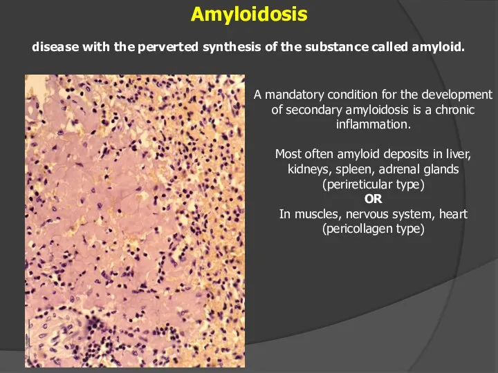
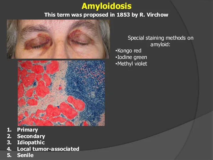
- 24. Amyloidosis disease with the perverted synthesis of the substance called amyloid. A mandatory condition for the
- 25. Amyloidosis Primary Secondary Idiopathic Local tumor-associated Senile This term was proposed in 1853 by R. Virchow
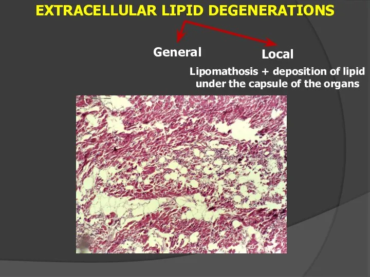
- 26. EXTRACELLULAR LIPID DEGENERATIONS Local Lipomathosis + deposition of lipid under the capsule of the organs General
- 27. I degree of obesity - overweight up to 30%; II degree of obesity - overweight up
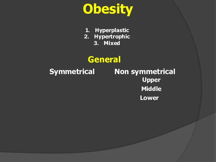
- 28. Obesity Hyperplastic Hypertrophic Mixed
- 29. chronic disease characterized by abnormalities in lipid and protein metabolism, which is manifested by the deposition
- 30. EXTRACELLULAR CARBOHYDRATE DEGENERATIONS
- 31. Pigments metabolism disorders Chromoproteins - endogenous pigments Hemoglobin - derivated Protein - derivated (tyrosine) Lipid -
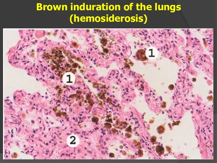
- 32. Pigments metabolism disorders Hemoglobin - derivated Ferritin Hemosiderin is a polymer of ferritin. Hemosiderosis Hematoidin —
- 33. Brown induration of the lungs (hemosiderosis)
- 34. Jaundice is an increase of bilirubin levels in blood, yellowing of the mucous membranes, sclera and
- 35. MELANIN Adrenochrome Others Protein – derivated pigments (tyrosine)
- 37. Скачать презентацию
Introduction
Pathological anatomy and anatomic pathologist
History
Tasks of the pathological anatomy
Biopsy, operational material,
Introduction
Pathological anatomy and anatomic pathologist
History
Tasks of the pathological anatomy
Biopsy, operational material,
Pathological anatomy is the science that studies the structural bases of
Pathological anatomy is the science that studies the structural bases of

In 1761 Italian author G. Morgagni wrote the first work on pathological
In 1761 Italian author G. Morgagni wrote the first work on pathological
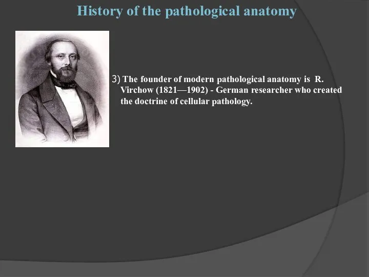
The founder of modern pathological anatomy is R. Virchow (1821—1902) - German
Theoretic tasks of the pathological anatomy:
1) Study of the etiology, pathogenesis, morphology
Theoretic tasks of the pathological anatomy:
1) Study of the etiology, pathogenesis, morphology
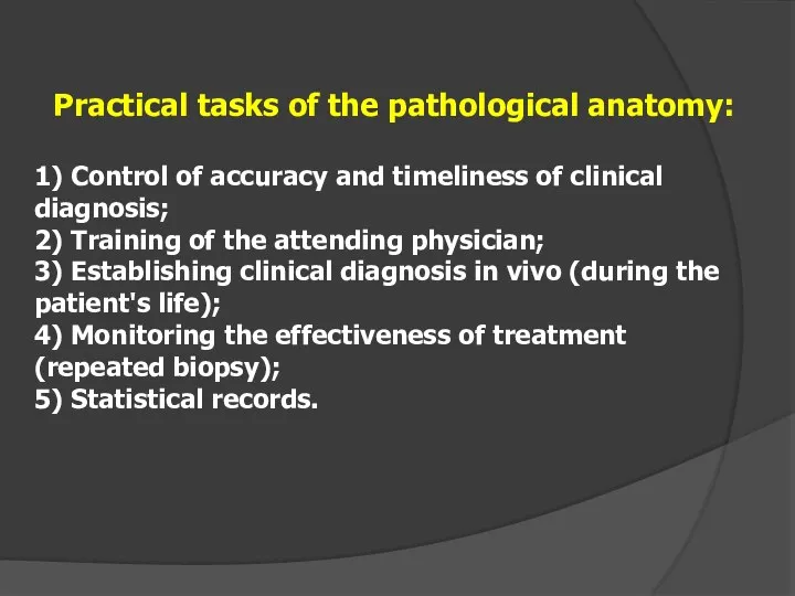
Practical tasks of the pathological anatomy:
1) Control of accuracy and timeliness of
Practical tasks of the pathological anatomy:
1) Control of accuracy and timeliness of
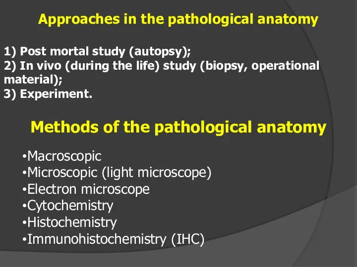
Methods of the pathological anatomy
Macroscopic
Microscopic (light microscope)
Electron microscope
Cytochemistry
Histochemistry
Immunohistochemistry (IHC)
Approaches in the
Methods of the pathological anatomy
Macroscopic
Microscopic (light microscope)
Electron microscope
Cytochemistry
Histochemistry
Immunohistochemistry (IHC)
Approaches in the
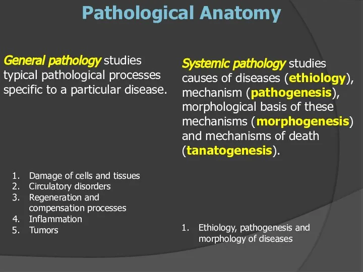
General pathology studies typical pathological processes specific to a particular disease.
General pathology studies typical pathological processes specific to a particular disease.
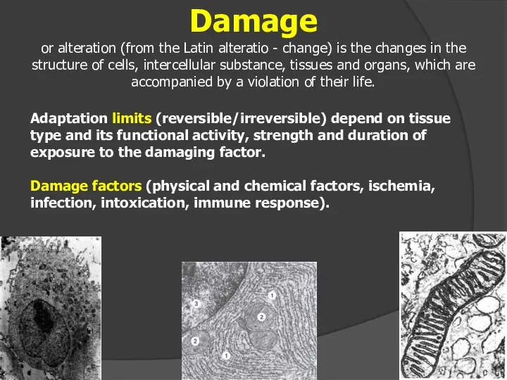
Adaptation limits (reversible/irreversible) depend on tissue type and its functional activity,
Adaptation limits (reversible/irreversible) depend on tissue type and its functional activity,

MORPHOLOGY OF THE CELLULAR DAMAGE
DEGENERATIONS
APOPTOSIS
NECROSIS
METABOLIC DISORDERS, LEADING TO CHANGES IN
DEGENERATIONS
APOPTOSIS
NECROSIS
METABOLIC DISORDERS, LEADING TO CHANGES IN
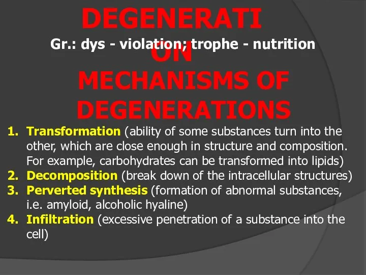
DEGENERATION
Gr.: dys - violation; trophe - nutrition
Transformation (ability of some substances
DEGENERATION
Gr.: dys - violation; trophe - nutrition
Transformation (ability of some substances

I. By localization
1. Intracellular (parenchymal);
2. Extracellular (stromal vascular, mesenchymal);
I. By localization
1. Intracellular (parenchymal);
2. Extracellular (stromal vascular, mesenchymal);
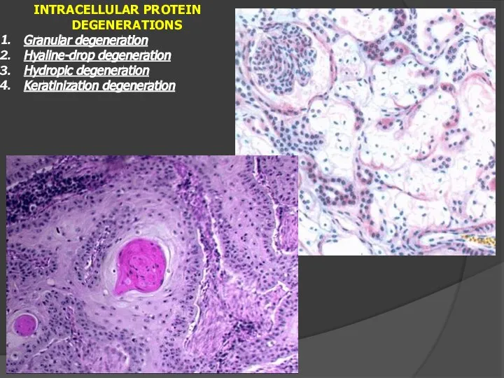
INTRACELLULAR PROTEIN DEGENERATIONS
Granular degeneration
Hyaline-drop degeneration
Hydropic degeneration
Keratinization degeneration
INTRACELLULAR PROTEIN DEGENERATIONS
Granular degeneration
Hyaline-drop degeneration
Hydropic degeneration
Keratinization degeneration
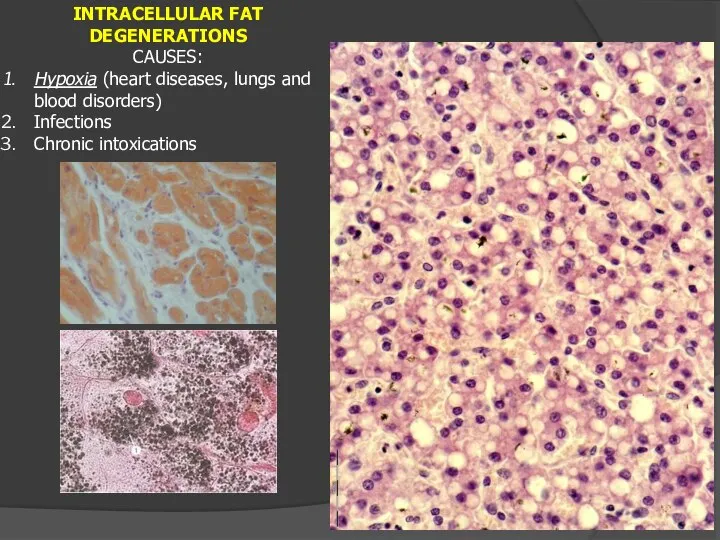
INTRACELLULAR FAT DEGENERATIONS
CAUSES:
Hypoxia (heart diseases, lungs and blood disorders)
Infections
Chronic
INTRACELLULAR FAT DEGENERATIONS
CAUSES:
Hypoxia (heart diseases, lungs and blood disorders)
Infections
Chronic
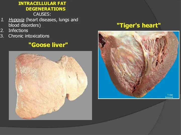
INTRACELLULAR FAT DEGENERATIONS
CAUSES:
Hypoxia (heart diseases, lungs and blood disorders)
Infections
Chronic
INTRACELLULAR FAT DEGENERATIONS
CAUSES:
Hypoxia (heart diseases, lungs and blood disorders)
Infections
Chronic
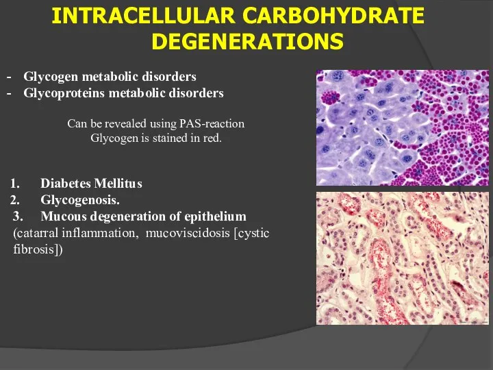
INTRACELLULAR CARBOHYDRATE DEGENERATIONS
Glycogen metabolic disorders
Glycoproteins metabolic disorders
Can be revealed using PAS-reaction
Glycogen
INTRACELLULAR CARBOHYDRATE DEGENERATIONS
Glycogen metabolic disorders
Glycoproteins metabolic disorders
Can be revealed using PAS-reaction
Glycogen
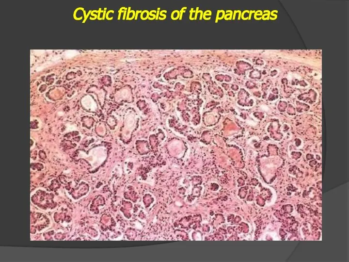
Cystic fibrosis of the pancreas
Cystic fibrosis of the pancreas
EXTRACELLULAR PROTEIN DEGENERATIONS
Mucoid swelling
Fibrinoid swelling
Hyalinosis
Amyloidosis
EXTRACELLULAR PROTEIN DEGENERATIONS
Mucoid swelling
Fibrinoid swelling
Hyalinosis
Amyloidosis
Mucoid swelling
superficial and reversible desorganisation of the connective tissue. Accumulation of
Mucoid swelling
superficial and reversible desorganisation of the connective tissue. Accumulation of
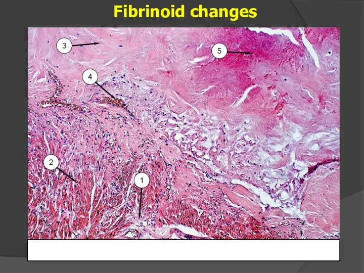
Fibrinoid changes
Fibrinoid changes
Hyalinosis
Degradation of connective tissue is accompanied by increased vascular permeability, degradation
Hyalinosis
Degradation of connective tissue is accompanied by increased vascular permeability, degradation
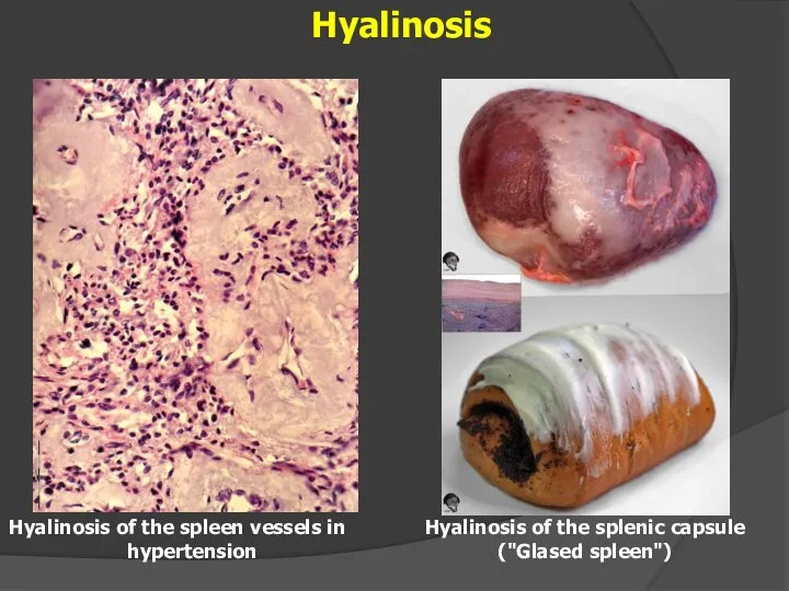
Hyalinosis
Hyalinosis of the spleen vessels in hypertension
Hyalinosis of the splenic capsule
("Glased
Hyalinosis
Hyalinosis of the spleen vessels in hypertension
Hyalinosis of the splenic capsule
("Glased
Amyloidosis
disease with the perverted synthesis of the substance called amyloid.
A mandatory
Amyloidosis
disease with the perverted synthesis of the substance called amyloid.
A mandatory
Amyloidosis
Primary
Secondary
Idiopathic
Local tumor-associated
Senile
This term was proposed in 1853 by R. Virchow
Special staining
Amyloidosis
Primary
Secondary
Idiopathic
Local tumor-associated
Senile
This term was proposed in 1853 by R. Virchow
Special staining
EXTRACELLULAR LIPID DEGENERATIONS
Local
Lipomathosis + deposition of lipid under the capsule of
EXTRACELLULAR LIPID DEGENERATIONS
Local
Lipomathosis + deposition of lipid under the capsule of
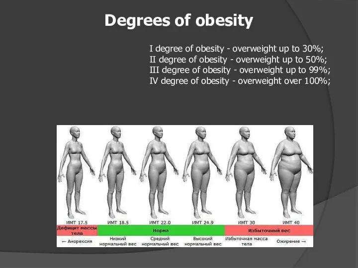
I degree of obesity - overweight up to 30%;
II degree
I degree of obesity - overweight up to 30%; II degree
Obesity
Hyperplastic
Hypertrophic
Mixed
Obesity
Hyperplastic
Hypertrophic
Mixed
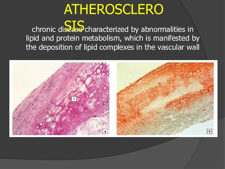
chronic disease characterized by abnormalities in lipid and protein metabolism, which
chronic disease characterized by abnormalities in lipid and protein metabolism, which
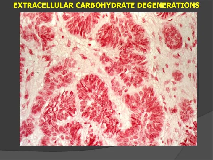
EXTRACELLULAR CARBOHYDRATE DEGENERATIONS
EXTRACELLULAR CARBOHYDRATE DEGENERATIONS
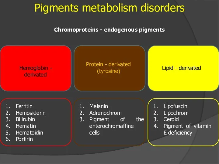
Pigments metabolism disorders
Chromoproteins - endogenous pigments
Hemoglobin - derivated
Protein - derivated
(tyrosine)
Lipid -
Pigments metabolism disorders
Chromoproteins - endogenous pigments
Hemoglobin - derivated
Protein - derivated
(tyrosine)
Lipid -
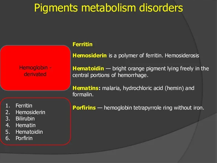
Pigments metabolism disorders
Hemoglobin - derivated
Ferritin
Hemosiderin is a polymer of ferritin. Hemosiderosis
Hematoidin
Pigments metabolism disorders
Hemoglobin - derivated
Ferritin
Hemosiderin is a polymer of ferritin. Hemosiderosis
Hematoidin
Brown induration of the lungs (hemosiderosis)
Brown induration of the lungs (hemosiderosis)
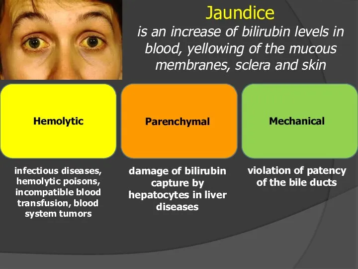
Jaundice
is an increase of bilirubin levels in blood, yellowing of the
Jaundice
is an increase of bilirubin levels in blood, yellowing of the
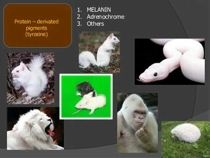
MELANIN
Adrenochrome
Others
Protein – derivated pigments
(tyrosine)
MELANIN
Adrenochrome
Others
Protein – derivated pigments
(tyrosine)
 Цивилизации Древнего мира: Западные цивилизации
 « Культура и искусство первой половины XX века»
  Духовное развитие общества в начале ХХ века. Литература.
 Путешествие швейцарской Милки по России
 Российское государство и общество во второй половине XIX в. Промышленный переворот. Отмена крепостного права Преобразования Алек
 Создание США
 Храмы Владимиро-Суздальской земли и Новгорода
 Київ 60 – 80 рр
  Проект Ломоносовская усадьба: от замысла к реализации
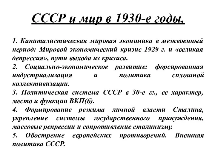 СССР и мир в 1930-е годы
 Античная культура
 Образование СССР
 Штурм Кёнигсберга
 Монгольское нашествие на Русь. Натиск с Запада
 Презентация на тему "Хрущев Никита Сергеевич" - презентации по Истории скачать
  Знаменитые дипломаты России
 Соціально – економічне становище України на початку 20-х років. Неп
  Франция в середине XIX века
 Первая Мировая война
 Культура Древней Руси (X - XIII вв.)
 Генеральный план уездного города София
 Презентация на тему "Через культуру родного края к истокам патриотизма" - презентации по Истории скачать
  Школа № 26. Стерлитамакский городской отдел образования
 Подвиг Александра Матросова
 Древнеримская скульптура
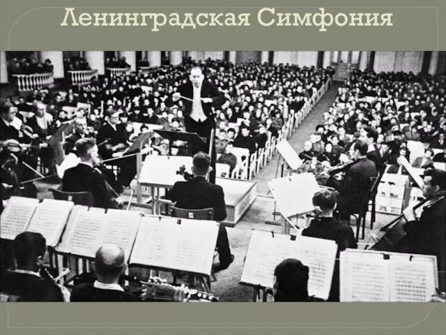 Ленинградская симфония
 9 мая день воинской славы России
 Немецкая оккупационная политика на захваченных территориях ссср в целом, в частности в крыму