- Cephalopelvic disproportion

Содержание
- 2. Disproportion, in relation to the pelvis, is a state where the normal proportion between the size
- 3. Contracted outlet is suspected when the interischial tuberous diameter is 8 cm or less. A contracted
- 4. As the head is the largest part of the fetus, it is more important to know
- 5. DIAGNOSIS OF CEPHALOPELVIC DISPROPORTION (CPD) AT THE BRIM The presence and degree of cephalopelvic disproportion at
- 6. Inferences: The head can be pushed down in the pelvis without overlapping of the parietal
- 7. Abdominovaginal method (Muller-Munro Kerr): This bimanual method is superior to the abdominal method as the pelvic
- 8. Inferences: (1) The head can be pushed down up to the level of ischial spines and
- 9. X-ray pelvimetry: Lateral X-ray view with the patient in standing position is helpful in assessing cephalopelvic
- 10. Degree of disproportion and contracted pelvis: Based on the clinical and supplemented by imaging pelvimetry, the
- 11. Management: Unlike inlet disproportion, clinical diagnosis of midpelvic and outlet disproportion can only be made after
- 13. Скачать презентацию
Disproportion, in relation to the pelvis, is a state where the
Disproportion, in relation to the pelvis, is a state where the
Contracted outlet is suspected when the interischial tuberous diameter is 8
Contracted outlet is suspected when the interischial tuberous diameter is 8
As the head is the largest part of the fetus, it
As the head is the largest part of the fetus, it
DIAGNOSIS OF CEPHALOPELVIC DISPROPORTION (CPD) AT THE BRIM The presence and
DIAGNOSIS OF CEPHALOPELVIC DISPROPORTION (CPD) AT THE BRIM The presence and
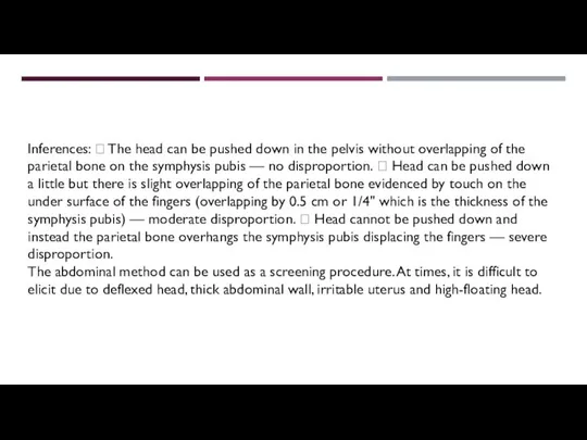
Inferences: The head can be pushed down in the pelvis
Inferences: The head can be pushed down in the pelvis
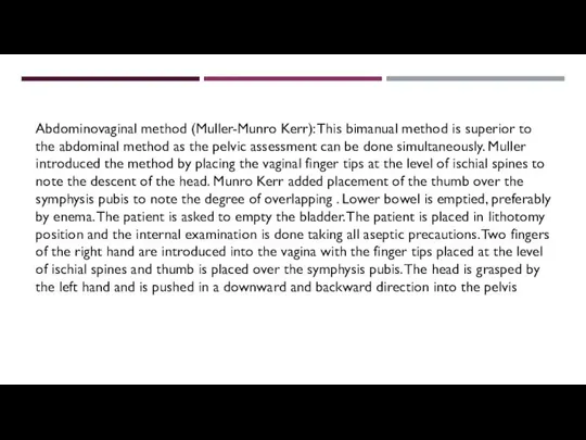
Abdominovaginal method (Muller-Munro Kerr): This bimanual method is superior to the
Abdominovaginal method (Muller-Munro Kerr): This bimanual method is superior to the
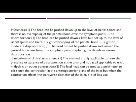
Inferences: (1) The head can be pushed down up to the
Inferences: (1) The head can be pushed down up to the
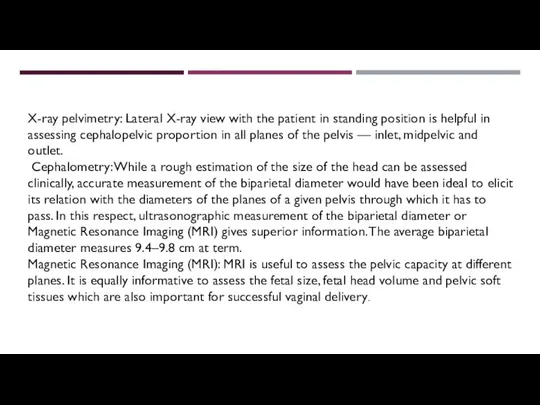
X-ray pelvimetry: Lateral X-ray view with the patient in standing position
X-ray pelvimetry: Lateral X-ray view with the patient in standing position
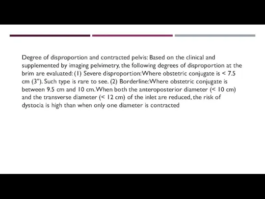
Degree of disproportion and contracted pelvis: Based on the clinical and
Degree of disproportion and contracted pelvis: Based on the clinical and
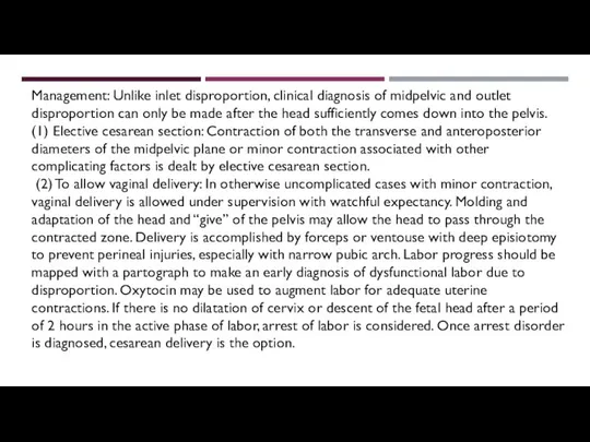
Management: Unlike inlet disproportion, clinical diagnosis of midpelvic and outlet disproportion
Management: Unlike inlet disproportion, clinical diagnosis of midpelvic and outlet disproportion
 Юные экологи - краеведы
 Austria, Kirillova Alina 342 group
 Sredstvo-upravleniya
 Электроэнергетика России
 Использование новых видов деревьев в озеленении петербурга в условиях потепления климата
 Расчет прочности наклонных сечений изгибаемых элементов
 Арт.Родина_Карта_25.07 — копия
 35e4f50ff12c8d471433d164663eb0fd
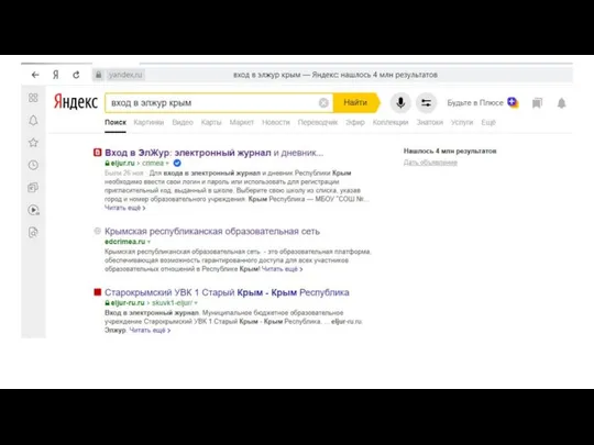 Регистрация электронный журнал (2)
 Обзор возможностей календаря Google
 Коллаж фото от встречи до свадьбы
 Revoland Presentation
 Преза для сайта
 20120310_predl_k_ot.ppt_5
 Изучение требований профессиональных стандартов к работникам сферы электроэнергетики и электротехники
 Путь поэта
 Времена года
 Сюрприз для Виталика
 Что такое дым и почему копчености такие вкусные
 Загадки для мальчиков. С днём защитника отечества
 Народный календарь на апрель месяц
 Prezentatsia1
 Основы религиозных культур и светской этики
 Мой класс и моя школа. (1 класс)
 Производственная практика
 Шашлык по-кавказки
 20170822_opasnye_internet_soobshchestva
 20160205_mumu_moya_prezentatsiya