- Chapter 12. The Cell Cycle

Содержание
- 2. Overview: The Key Roles of Cell Division The ability of organisms to reproduce best distinguishes living
- 3. Fig. 12-1
- 4. In unicellular organisms, division of one cell reproduces the entire organism Multicellular organisms depend on cell
- 5. Fig. 12-2 100 µm 200 µm 20 µm (a) Reproduction (b) Growth and development (c) Tissue
- 6. Fig. 12-2a 100 µm (a) Reproduction
- 7. Fig. 12-2b 200 µm (b) Growth and development
- 8. Fig. 12-2c 20 µm (c) Tissue renewal
- 9. Concept 12.1: Cell division results in genetically identical daughter cells Most cell division results in daughter
- 10. Cellular Organization of the Genetic Material All the DNA in a cell constitutes the cell’s genome
- 11. Fig. 12-3 20 µm
- 12. Every eukaryotic species has a characteristic number of chromosomes in each cell nucleus Somatic cells (nonreproductive
- 13. Distribution of Chromosomes During Eukaryotic Cell Division In preparation for cell division, DNA is replicated and
- 14. Fig. 12-4 0.5 µm Chromosomes Chromosome duplication (including DNA synthesis) Chromo- some arm Centromere Sister chromatids
- 15. Eukaryotic cell division consists of: Mitosis, the division of the nucleus Cytokinesis, the division of the
- 16. Concept 12.2: The mitotic phase alternates with interphase in the cell cycle In 1882, the German
- 17. Phases of the Cell Cycle The cell cycle consists of Mitotic (M) phase (mitosis and cytokinesis)
- 18. Interphase (about 90% of the cell cycle) can be divided into subphases: G1 phase (“first gap”)
- 19. Fig. 12-5 S (DNA synthesis) MITOTIC (M) PHASE Mitosis Cytokinesis G1 G2 INTERPHASE
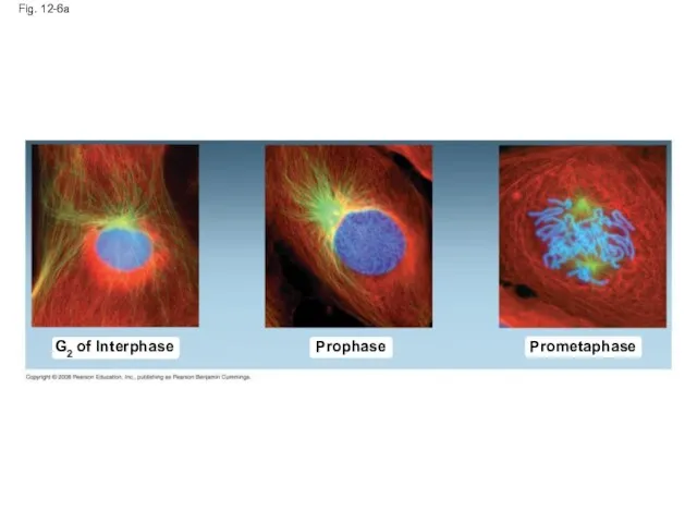
- 20. Mitosis is conventionally divided into five phases: Prophase Prometaphase Metaphase Anaphase Telophase Cytokinesis is well underway
- 21. Fig. 12-6 G2 of Interphase Centrosomes (with centriole pairs) Chromatin (duplicated) Nucleolus Nuclear envelope Plasma membrane
- 22. Prophase Fig. 12-6a Prometaphase G2 of Interphase
- 23. Fig. 12-6b Prometaphase Prophase G2 of Interphase Nonkinetochore microtubules Fragments of nuclear envelope Aster Centromere Early
- 24. Fig. 12-6c Metaphase Anaphase Telophase and Cytokinesis
- 25. Fig. 12-6d Metaphase Anaphase Telophase and Cytokinesis Cleavage furrow Nucleolus forming Metaphase plate Centrosome at one
- 26. The Mitotic Spindle: A Closer Look The mitotic spindle is an apparatus of microtubules that controls
- 27. An aster (a radial array of short microtubules) extends from each centrosome The spindle includes the
- 28. During prometaphase, some spindle microtubules attach to the kinetochores of chromosomes and begin to move the
- 29. Fig. 12-7 Microtubules Chromosomes Sister chromatids Aster Metaphase plate Centrosome Kineto- chores Kinetochore microtubules Overlapping nonkinetochore
- 30. In anaphase, sister chromatids separate and move along the kinetochore microtubules toward opposite ends of the
- 31. Fig. 12-8 EXPERIMENT Kinetochore RESULTS CONCLUSION Spindle pole Mark Chromosome movement Kinetochore Microtubule Motor protein Chromosome
- 32. Fig. 12-8a Kinetochore Spindle pole Mark EXPERIMENT RESULTS
- 33. Fig. 12-8b Kinetochore Microtubule Tubulin Subunits Chromosome Chromosome movement Motor protein CONCLUSION
- 34. Nonkinetochore microtubules from opposite poles overlap and push against each other, elongating the cell In telophase,
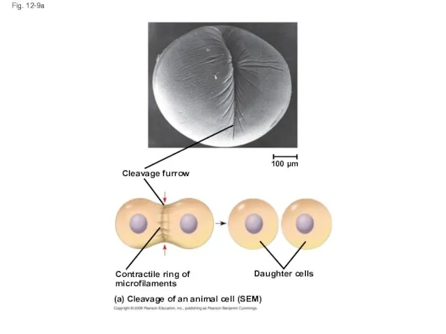
- 35. Cytokinesis: A Closer Look In animal cells, cytokinesis occurs by a process known as cleavage, forming
- 36. Video: Sea Urchin (Time Lapse) Video: Animal Mitosis Copyright © 2008 Pearson Education, Inc., publishing as
- 37. Fig. 12-9 Cleavage furrow 100 µm Contractile ring of microfilaments Daughter cells (a) Cleavage of an
- 38. Cleavage furrow Fig. 12-9a 100 µm Daughter cells (a) Cleavage of an animal cell (SEM) Contractile
- 39. Fig. 12-9b Daughter cells (b) Cell plate formation in a plant cell (TEM) Vesicles forming cell
- 40. Fig. 12-10 Chromatin condensing Metaphase Anaphase Telophase Prometaphase Nucleus Prophase 1 2 3 5 4 Nucleolus
- 41. Fig. 12-10a Nucleus Prophase 1 Nucleolus Chromatin condensing
- 42. Fig. 12-10b Prometaphase 2 Chromosomes
- 43. Fig. 12-10c Metaphase 3
- 44. Fig. 12-10d Anaphase 4
- 45. Fig. 12-10e Telophase 5 Cell plate 10 µm
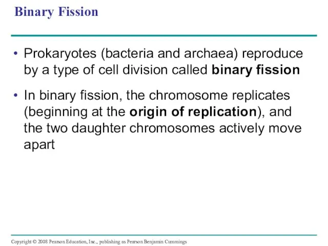
- 46. Binary Fission Prokaryotes (bacteria and archaea) reproduce by a type of cell division called binary fission
- 47. Fig. 12-11-1 Origin of replication Two copies of origin E. coli cell Bacterial chromosome Plasma membrane
- 48. Fig. 12-11-2 Origin of replication Two copies of origin E. coli cell Bacterial chromosome Plasma membrane
- 49. Fig. 12-11-3 Origin of replication Two copies of origin E. coli cell Bacterial chromosome Plasma membrane
- 50. Fig. 12-11-4 Origin of replication Two copies of origin E. coli cell Bacterial chromosome Plasma membrane
- 51. The Evolution of Mitosis Since prokaryotes evolved before eukaryotes, mitosis probably evolved from binary fission Certain
- 52. Fig. 12-12 (a) Bacteria Bacterial chromosome Chromosomes Microtubules Intact nuclear envelope (b) Dinoflagellates Kinetochore microtubule Intact
- 53. Fig. 12-12ab Bacterial chromosome Chromosomes Microtubules (a) Bacteria (b) Dinoflagellates Intact nuclear envelope
- 54. Fig. 12-12cd Kinetochore microtubule (c) Diatoms and yeasts Kinetochore microtubule (d) Most eukaryotes Fragments of nuclear
- 55. Concept 12.3: The eukaryotic cell cycle is regulated by a molecular control system The frequency of
- 56. Evidence for Cytoplasmic Signals The cell cycle appears to be driven by specific chemical signals present
- 57. Fig. 12-13 Experiment 1 Experiment 2 EXPERIMENT RESULTS S G1 M G1 M M S S
- 58. The Cell Cycle Control System The sequential events of the cell cycle are directed by a
- 59. Fig. 12-14 S G1 M checkpoint G2 M Control system G1 checkpoint G2 checkpoint
- 60. For many cells, the G1 checkpoint seems to be the most important one If a cell
- 61. Fig. 12-15 G1 G0 G1 checkpoint Cell receives a go-ahead signal G1 (b) Cell does not
- 62. The Cell Cycle Clock: Cyclins and Cyclin-Dependent Kinases Two types of regulatory proteins are involved in
- 63. Fig. 12-16 Protein kinase activity (– ) % of dividing cells (– ) Time (min) 300
- 64. Fig. 12-17 M G1 S G2 M G1 S G2 M G1 MPF activity Cyclin concentration
- 65. Fig. 12-17a Time (a) Fluctuation of MPF activity and cyclin concentration during the cell cycle Cyclin
- 66. Fig. 12-17b Cyclin is degraded Cdk MPF Cdk M S G1 G2 checkpoint Degraded cyclin Cyclin
- 67. Stop and Go Signs: Internal and External Signals at the Checkpoints An example of an internal
- 68. Fig. 12-18 Petri plate Scalpels Cultured fibroblasts Without PDGF cells fail to divide With PDGF cells
- 69. Another example of external signals is density-dependent inhibition, in which crowded cells stop dividing Most animal
- 70. Fig. 12-19 Anchorage dependence Density-dependent inhibition Density-dependent inhibition (a) Normal mammalian cells (b) Cancer cells 25
- 71. Cancer cells exhibit neither density-dependent inhibition nor anchorage dependence Copyright © 2008 Pearson Education, Inc., publishing
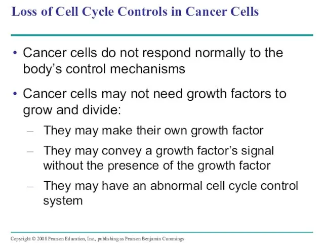
- 72. Loss of Cell Cycle Controls in Cancer Cells Cancer cells do not respond normally to the
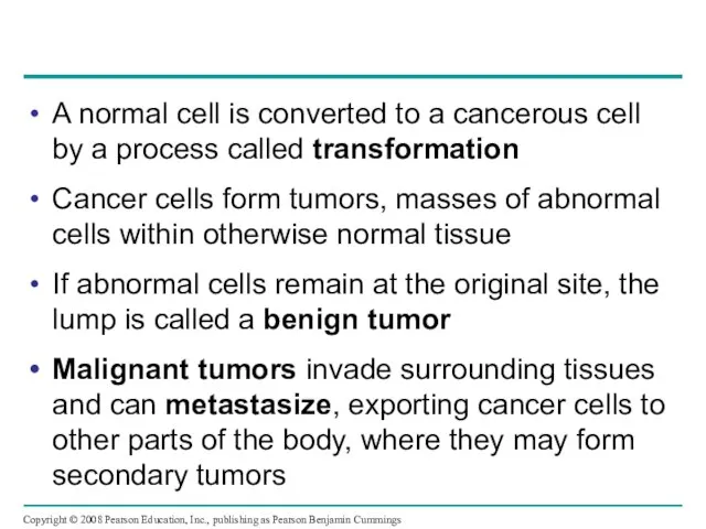
- 73. A normal cell is converted to a cancerous cell by a process called transformation Cancer cells
- 74. Fig. 12-20 Tumor A tumor grows from a single cancer cell. Glandular tissue Lymph vessel Blood
- 75. Fig. 12-UN1 Telophase and Cytokinesis Anaphase Metaphase Prometaphase Prophase MITOTIC (M) PHASE Cytokinesis Mitosis S G1
- 76. Fig. 12-UN2
- 77. Fig. 12-UN3
- 78. Fig. 12-UN4
- 79. Fig. 12-UN5
- 80. Fig. 12-UN6
- 81. You should now be able to: Describe the structural organization of the prokaryotic genome and the
- 83. Скачать презентацию
Overview: The Key Roles of Cell Division
The ability of organisms to
Overview: The Key Roles of Cell Division
The ability of organisms to
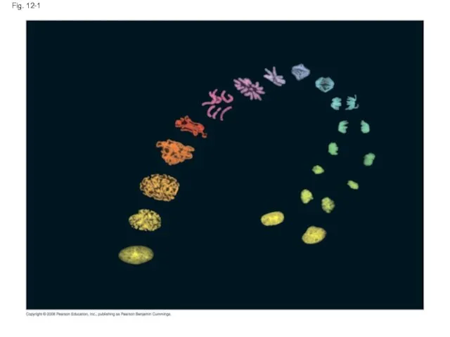
Fig. 12-1
Fig. 12-1
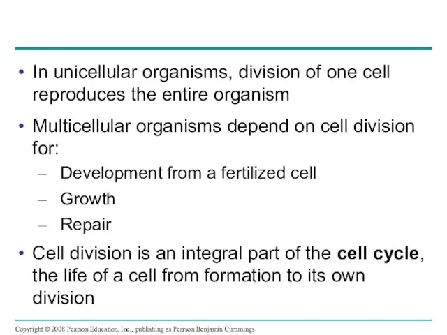
In unicellular organisms, division of one cell reproduces the entire organism
Multicellular
In unicellular organisms, division of one cell reproduces the entire organism
Multicellular
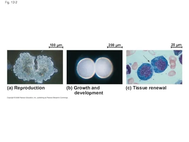
Fig. 12-2
100 µm
200 µm
20 µm
(a) Reproduction
(b) Growth and
development
(c) Tissue renewal
Fig. 12-2
100 µm
200 µm
20 µm
(a) Reproduction
(b) Growth and
development
(c) Tissue renewal
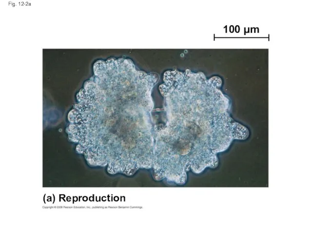
Fig. 12-2a
100 µm
(a) Reproduction
Fig. 12-2a
100 µm
(a) Reproduction
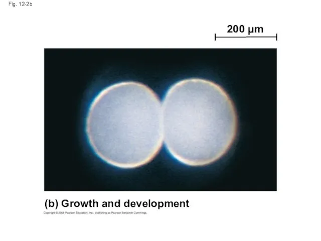
Fig. 12-2b
200 µm
(b) Growth and development
Fig. 12-2b
200 µm
(b) Growth and development
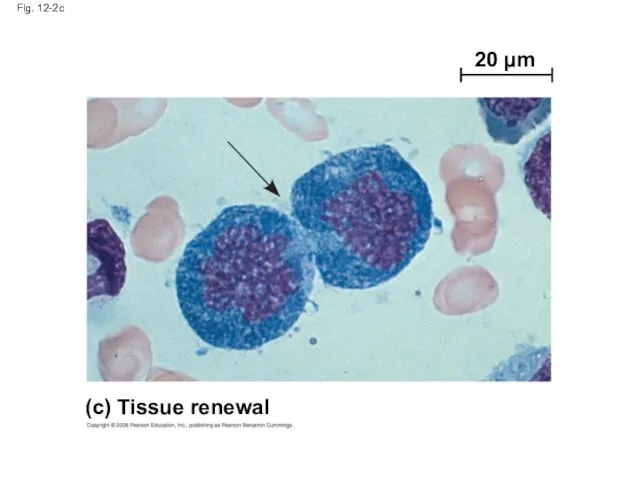
Fig. 12-2c
20 µm
(c) Tissue renewal
Fig. 12-2c
20 µm
(c) Tissue renewal
Concept 12.1: Cell division results in genetically
identical daughter cells
Most cell
Concept 12.1: Cell division results in genetically
identical daughter cells
Most cell
Cellular Organization of the Genetic Material
All the DNA in a cell
Cellular Organization of the Genetic Material
All the DNA in a cell
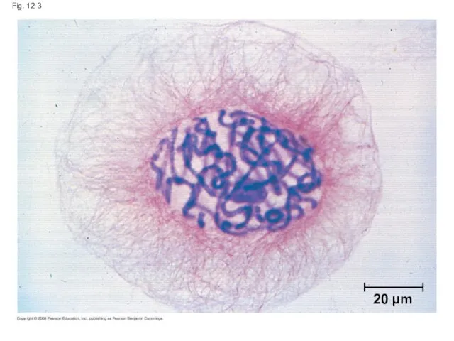
Fig. 12-3
20 µm
Fig. 12-3
20 µm
Every eukaryotic species has a characteristic number of chromosomes in each
Every eukaryotic species has a characteristic number of chromosomes in each
Distribution of Chromosomes During Eukaryotic Cell Division
In preparation for cell division,
Distribution of Chromosomes During Eukaryotic Cell Division
In preparation for cell division,
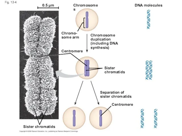
Fig. 12-4
0.5 µm
Chromosomes
Chromosome
duplication
(including DNA
synthesis)
Chromo-
some arm
Centromere
Sister
chromatids
DNA molecules
Separation of
sister chromatids
Centromere
Sister chromatids
Fig. 12-4
0.5 µm
Chromosomes
Chromosome
duplication
(including DNA
synthesis)
Chromo-
some arm
Centromere
Sister
chromatids
DNA molecules
Separation of
sister chromatids
Centromere
Sister chromatids
Eukaryotic cell division consists of:
Mitosis, the division of the nucleus
Cytokinesis, the
Eukaryotic cell division consists of:
Mitosis, the division of the nucleus
Cytokinesis, the
Concept 12.2: The mitotic phase alternates with
interphase in the cell cycle
In
Concept 12.2: The mitotic phase alternates with
interphase in the cell cycle
In
Phases of the Cell Cycle
The cell cycle consists of
Mitotic (M) phase
Phases of the Cell Cycle
The cell cycle consists of
Mitotic (M) phase
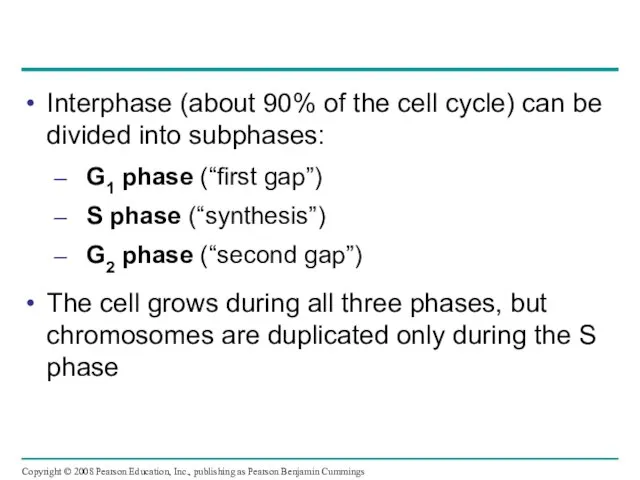
Interphase (about 90% of the cell cycle) can be divided into
Interphase (about 90% of the cell cycle) can be divided into
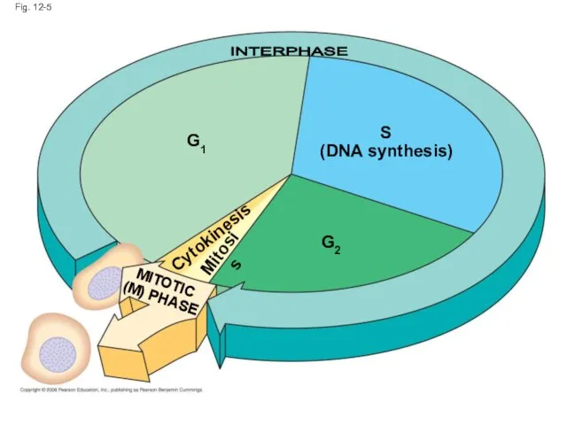
Fig. 12-5
S
(DNA synthesis)
MITOTIC
(M) PHASE
Mitosis
Cytokinesis
G1
G2
INTERPHASE
Fig. 12-5
S
(DNA synthesis)
MITOTIC
(M) PHASE
Mitosis
Cytokinesis
G1
G2
INTERPHASE
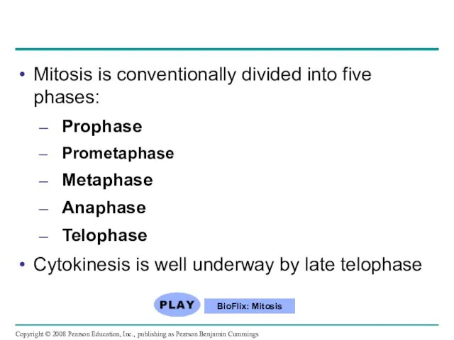
Mitosis is conventionally divided into five phases:
Prophase
Prometaphase
Metaphase
Anaphase
Telophase
Cytokinesis is well underway by
Mitosis is conventionally divided into five phases:
Prophase
Prometaphase
Metaphase
Anaphase
Telophase
Cytokinesis is well underway by
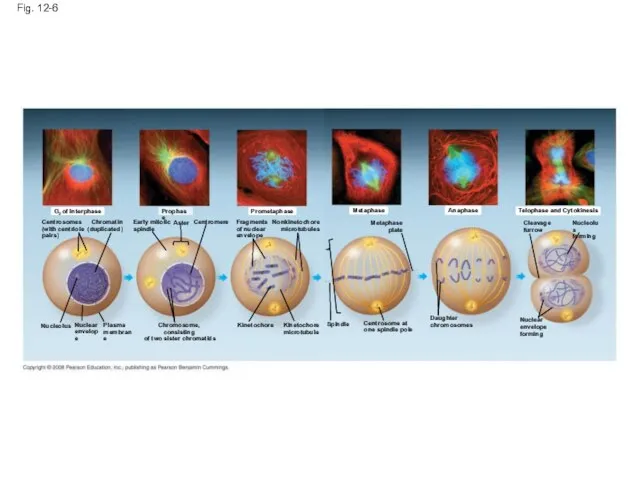
Fig. 12-6
G2 of Interphase
Centrosomes
(with centriole
pairs)
Chromatin
(duplicated)
Nucleolus
Nuclear
envelope
Plasma
membrane
Early mitotic
spindle
Aster
Centromere
Chromosome, consisting
of two sister chromatids
Prophase
Prometaphase
Fragments
of
Fig. 12-6
G2 of Interphase
Centrosomes
(with centriole
pairs)
Chromatin
(duplicated)
Nucleolus
Nuclear
envelope
Plasma
membrane
Early mitotic
spindle
Aster
Centromere
Chromosome, consisting
of two sister chromatids
Prophase
Prometaphase
Fragments
of
Prophase
Fig. 12-6a
Prometaphase
G2 of Interphase
Prophase
Fig. 12-6a
Prometaphase
G2 of Interphase
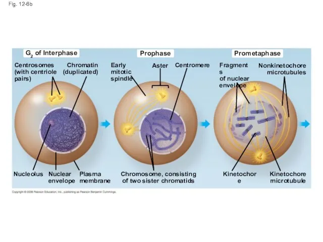
Fig. 12-6b
Prometaphase
Prophase
G2 of Interphase
Nonkinetochore
microtubules
Fragments
of nuclear
envelope
Aster
Centromere
Early mitotic
spindle
Chromatin
(duplicated)
Centrosomes
(with centriole
pairs)
Nucleolus
Nuclear
envelope
Plasma
membrane
Chromosome, consisting
of two sister chromatids
Kinetochore
Kinetochore
microtubule
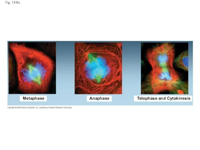
Fig. 12-6c
Metaphase
Anaphase
Telophase and Cytokinesis
Fig. 12-6c
Metaphase
Anaphase
Telophase and Cytokinesis
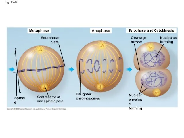
Fig. 12-6d
Metaphase
Anaphase
Telophase and Cytokinesis
Cleavage
furrow
Nucleolus
forming
Metaphase
plate
Centrosome at
one spindle pole
Spindle
Daughter
chromosomes
Nuclear
envelope
forming
Fig. 12-6d
Metaphase
Anaphase
Telophase and Cytokinesis
Cleavage
furrow
Nucleolus
forming
Metaphase
plate
Centrosome at
one spindle pole
Spindle
Daughter
chromosomes
Nuclear
envelope
forming
The Mitotic Spindle: A Closer Look
The mitotic spindle is an apparatus
The Mitotic Spindle: A Closer Look
The mitotic spindle is an apparatus
An aster (a radial array of short microtubules) extends from each
An aster (a radial array of short microtubules) extends from each
During prometaphase, some spindle microtubules attach to the kinetochores of chromosomes
During prometaphase, some spindle microtubules attach to the kinetochores of chromosomes
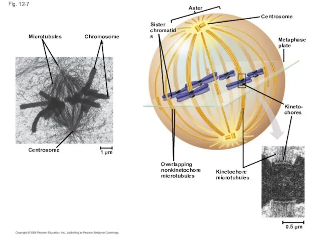
Fig. 12-7
Microtubules
Chromosomes
Sister
chromatids
Aster
Metaphase
plate
Centrosome
Kineto-
chores
Kinetochore
microtubules
Overlapping
nonkinetochore
microtubules
Centrosome
1 µm
0.5 µm
Fig. 12-7
Microtubules
Chromosomes
Sister
chromatids
Aster
Metaphase
plate
Centrosome
Kineto-
chores
Kinetochore
microtubules
Overlapping
nonkinetochore
microtubules
Centrosome
1 µm
0.5 µm
In anaphase, sister chromatids separate and move along the kinetochore microtubules
In anaphase, sister chromatids separate and move along the kinetochore microtubules
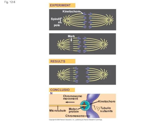
Fig. 12-8
EXPERIMENT
Kinetochore
RESULTS
CONCLUSION
Spindle
pole
Mark
Chromosome
movement
Kinetochore
Microtubule
Motor
protein
Chromosome
Tubulin
subunits
Fig. 12-8
EXPERIMENT
Kinetochore
RESULTS
CONCLUSION
Spindle
pole
Mark
Chromosome
movement
Kinetochore
Microtubule
Motor
protein
Chromosome
Tubulin
subunits
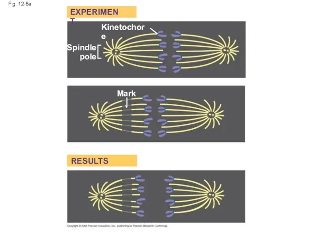
Fig. 12-8a
Kinetochore
Spindle
pole
Mark
EXPERIMENT
RESULTS
Fig. 12-8a
Kinetochore
Spindle
pole
Mark
EXPERIMENT
RESULTS
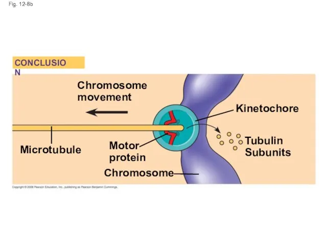
Fig. 12-8b
Kinetochore
Microtubule
Tubulin
Subunits
Chromosome
Chromosome
movement
Motor
protein
CONCLUSION
Fig. 12-8b
Kinetochore
Microtubule
Tubulin
Subunits
Chromosome
Chromosome
movement
Motor
protein
CONCLUSION
Nonkinetochore microtubules from opposite poles overlap and push against each other,
Nonkinetochore microtubules from opposite poles overlap and push against each other,
Cytokinesis: A Closer Look
In animal cells, cytokinesis occurs by a process
Cytokinesis: A Closer Look
In animal cells, cytokinesis occurs by a process
Video: Sea Urchin (Time Lapse)
Video: Animal Mitosis
Copyright © 2008 Pearson Education,
Video: Sea Urchin (Time Lapse)
Video: Animal Mitosis
Copyright © 2008 Pearson Education,
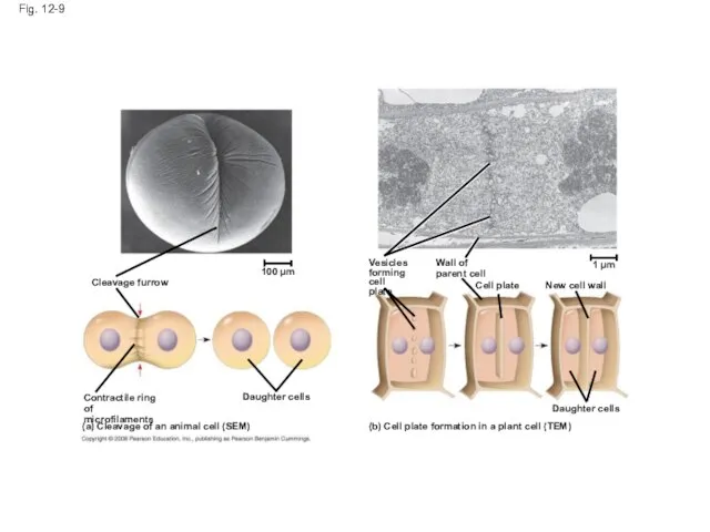
Fig. 12-9
Cleavage furrow
100 µm
Contractile ring of
microfilaments
Daughter cells
(a) Cleavage of an animal
Fig. 12-9
Cleavage furrow
100 µm
Contractile ring of
microfilaments
Daughter cells
(a) Cleavage of an animal
Cleavage furrow
Fig. 12-9a
100 µm
Daughter cells
(a) Cleavage of an animal cell (SEM)
Contractile
Cleavage furrow
Fig. 12-9a
100 µm
Daughter cells
(a) Cleavage of an animal cell (SEM)
Contractile
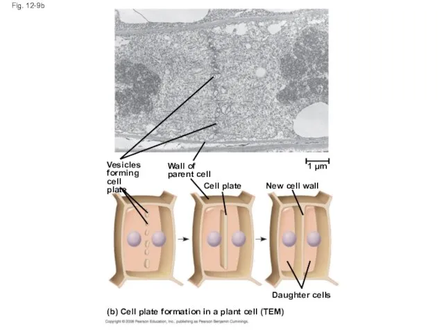
Fig. 12-9b
Daughter cells
(b) Cell plate formation in a plant cell (TEM)
Vesicles
forming
cell
Fig. 12-9b
Daughter cells
(b) Cell plate formation in a plant cell (TEM)
Vesicles
forming
cell
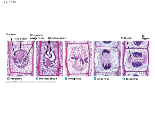
Fig. 12-10
Chromatin
condensing
Metaphase
Anaphase
Telophase
Prometaphase
Nucleus
Prophase
1
2
3
5
4
Nucleolus
Chromosomes
Cell plate
10 µm
Fig. 12-10
Chromatin
condensing
Metaphase
Anaphase
Telophase
Prometaphase
Nucleus
Prophase
1
2
3
5
4
Nucleolus
Chromosomes
Cell plate
10 µm
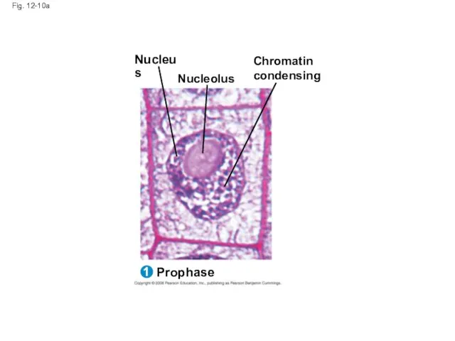
Fig. 12-10a
Nucleus
Prophase
1
Nucleolus
Chromatin
condensing
Fig. 12-10a
Nucleus
Prophase
1
Nucleolus
Chromatin
condensing
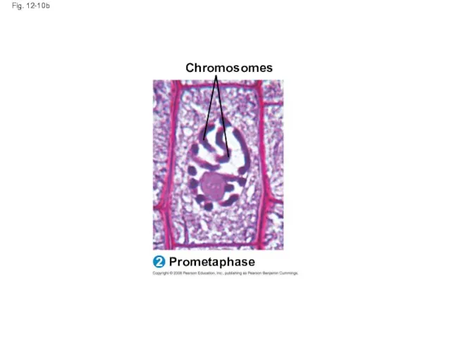
Fig. 12-10b
Prometaphase
2
Chromosomes
Fig. 12-10b
Prometaphase
2
Chromosomes
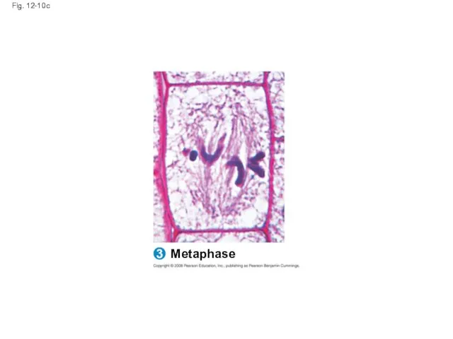
Fig. 12-10c
Metaphase
3
Fig. 12-10c
Metaphase
3
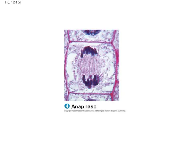
Fig. 12-10d
Anaphase
4
Fig. 12-10d
Anaphase
4
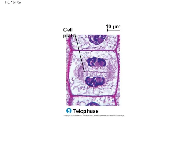
Fig. 12-10e
Telophase
5
Cell plate
10 µm
Fig. 12-10e
Telophase
5
Cell plate
10 µm
Binary Fission
Prokaryotes (bacteria and archaea) reproduce by a type of cell
Binary Fission
Prokaryotes (bacteria and archaea) reproduce by a type of cell
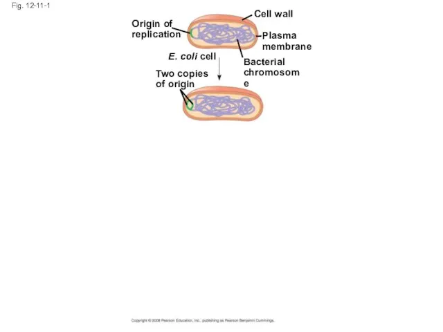
Fig. 12-11-1
Origin of
replication
Two copies
of origin
E. coli cell
Bacterial
chromosome
Plasma
membrane
Cell wall
Fig. 12-11-1
Origin of
replication
Two copies
of origin
E. coli cell
Bacterial
chromosome
Plasma
membrane
Cell wall
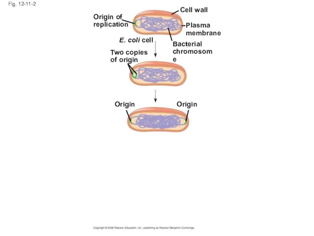
Fig. 12-11-2
Origin of
replication
Two copies
of origin
E. coli cell
Bacterial
chromosome
Plasma
membrane
Cell wall
Origin
Origin
Fig. 12-11-2
Origin of
replication
Two copies
of origin
E. coli cell
Bacterial
chromosome
Plasma
membrane
Cell wall
Origin
Origin
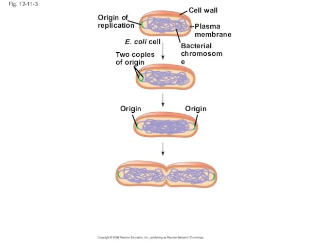
Fig. 12-11-3
Origin of
replication
Two copies
of origin
E. coli cell
Bacterial
chromosome
Plasma
membrane
Cell wall
Origin
Origin
Fig. 12-11-3
Origin of
replication
Two copies
of origin
E. coli cell
Bacterial
chromosome
Plasma
membrane
Cell wall
Origin
Origin
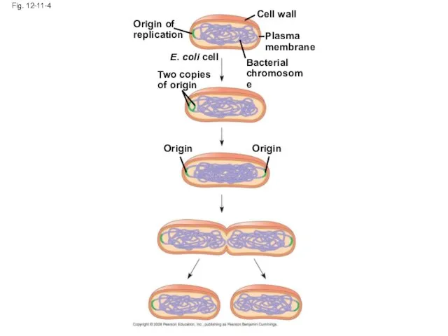
Fig. 12-11-4
Origin of
replication
Two copies
of origin
E. coli cell
Bacterial
chromosome
Plasma
membrane
Cell wall
Origin
Origin
Fig. 12-11-4
Origin of
replication
Two copies
of origin
E. coli cell
Bacterial
chromosome
Plasma
membrane
Cell wall
Origin
Origin
The Evolution of Mitosis
Since prokaryotes evolved before eukaryotes, mitosis probably evolved
The Evolution of Mitosis
Since prokaryotes evolved before eukaryotes, mitosis probably evolved
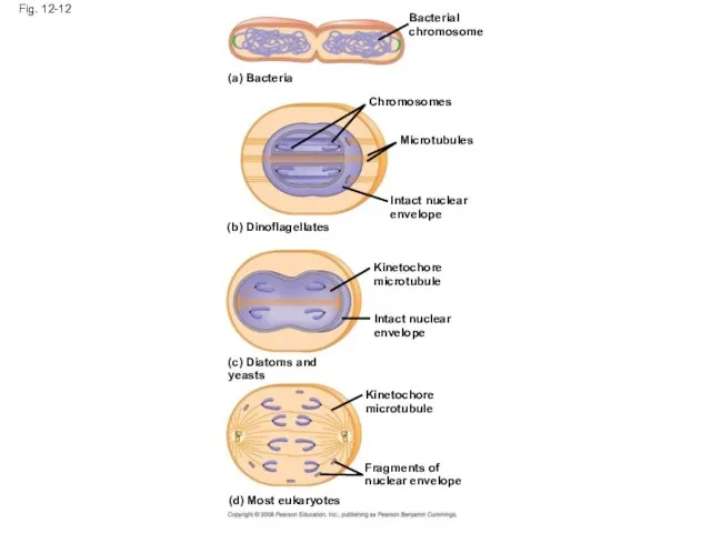
Fig. 12-12
(a) Bacteria
Bacterial
chromosome
Chromosomes
Microtubules
Intact nuclear
envelope
(b) Dinoflagellates
Kinetochore
microtubule
Intact nuclear
envelope
(c) Diatoms and yeasts
Kinetochore
microtubule
Fragments of
nuclear envelope
(d)
Fig. 12-12
(a) Bacteria
Bacterial
chromosome
Chromosomes
Microtubules
Intact nuclear
envelope
(b) Dinoflagellates
Kinetochore
microtubule
Intact nuclear
envelope
(c) Diatoms and yeasts
Kinetochore
microtubule
Fragments of
nuclear envelope
(d)
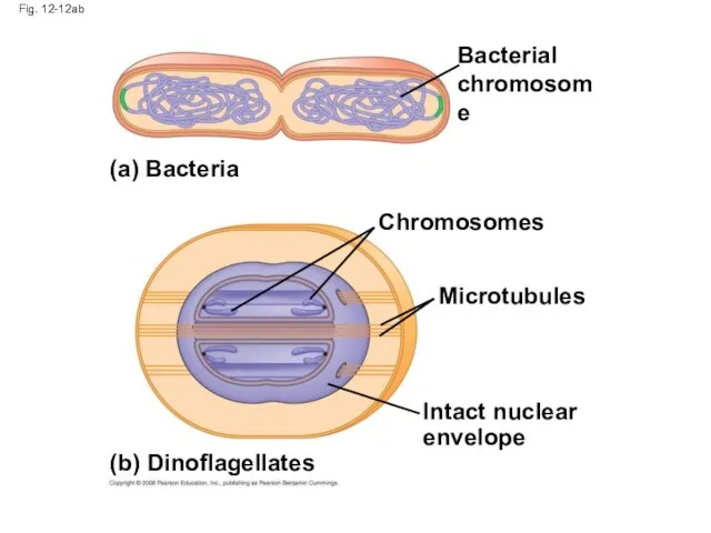
Fig. 12-12ab
Bacterial
chromosome
Chromosomes
Microtubules
(a) Bacteria
(b) Dinoflagellates
Intact nuclear
envelope
Fig. 12-12ab
Bacterial
chromosome
Chromosomes
Microtubules
(a) Bacteria
(b) Dinoflagellates
Intact nuclear
envelope
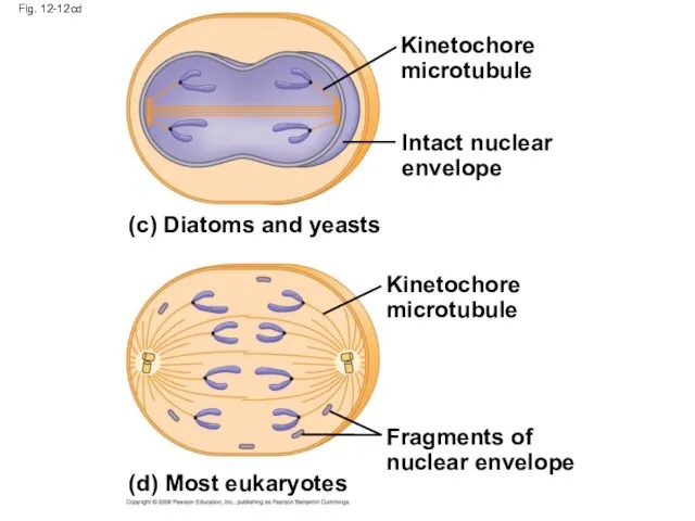
Fig. 12-12cd
Kinetochore
microtubule
(c) Diatoms and yeasts
Kinetochore
microtubule
(d) Most eukaryotes
Fragments of
nuclear envelope
Intact nuclear
envelope
Fig. 12-12cd
Kinetochore
microtubule
(c) Diatoms and yeasts
Kinetochore
microtubule
(d) Most eukaryotes
Fragments of
nuclear envelope
Intact nuclear
envelope
Concept 12.3: The eukaryotic cell cycle is regulated by a molecular
Concept 12.3: The eukaryotic cell cycle is regulated by a molecular
Evidence for Cytoplasmic Signals
The cell cycle appears to be driven by
Evidence for Cytoplasmic Signals
The cell cycle appears to be driven by
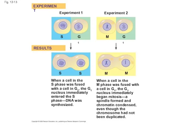
Fig. 12-13
Experiment 1
Experiment 2
EXPERIMENT
RESULTS
S
G1
M
G1
M
M
S
S
When a cell in the
S phase was fused
Fig. 12-13
Experiment 1
Experiment 2
EXPERIMENT
RESULTS
S
G1
M
G1
M
M
S
S
When a cell in the
S phase was fused
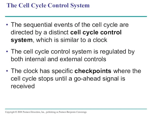
The Cell Cycle Control System
The sequential events of the cell cycle
The Cell Cycle Control System
The sequential events of the cell cycle
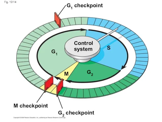
Fig. 12-14
S
G1
M checkpoint
G2
M
Control
system
G1 checkpoint
G2 checkpoint
Fig. 12-14
S
G1
M checkpoint
G2
M
Control
system
G1 checkpoint
G2 checkpoint
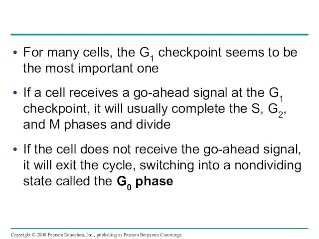
For many cells, the G1 checkpoint seems to be the most
For many cells, the G1 checkpoint seems to be the most
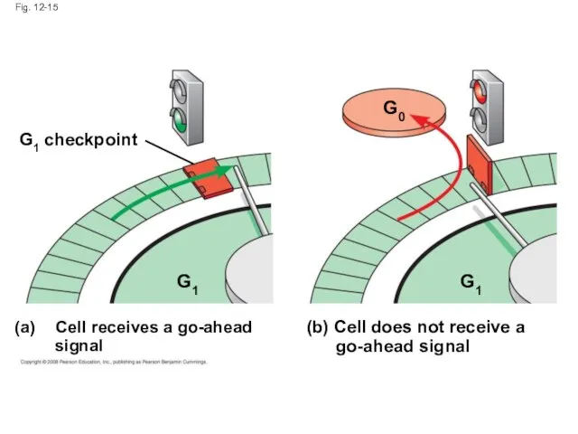
Fig. 12-15
G1
G0
G1 checkpoint
Cell receives a go-ahead
signal
G1
(b) Cell does not receive
Fig. 12-15
G1
G0
G1 checkpoint
Cell receives a go-ahead
signal
G1
(b) Cell does not receive
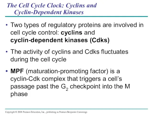
The Cell Cycle Clock: Cyclins and
Cyclin-Dependent Kinases
Two types of regulatory
The Cell Cycle Clock: Cyclins and
Cyclin-Dependent Kinases
Two types of regulatory
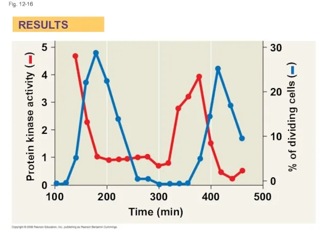
Fig. 12-16
Protein kinase activity (– )
% of dividing cells (– )
Time
Fig. 12-16
Protein kinase activity (– )
% of dividing cells (– )
Time
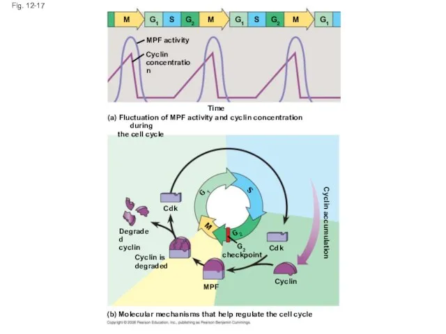
Fig. 12-17
M
G1
S
G2
M
G1
S
G2
M
G1
MPF activity
Cyclin
concentration
Time
(a) Fluctuation of MPF activity and cyclin concentration during
Fig. 12-17
M
G1
S
G2
M
G1
S
G2
M
G1
MPF activity
Cyclin
concentration
Time
(a) Fluctuation of MPF activity and cyclin concentration during
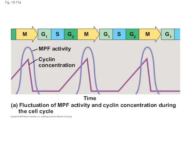
Fig. 12-17a
Time
(a) Fluctuation of MPF activity and cyclin concentration during
the
Fig. 12-17a
Time
(a) Fluctuation of MPF activity and cyclin concentration during
the
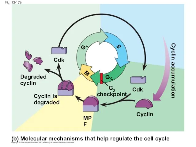
Fig. 12-17b
Cyclin is
degraded
Cdk
MPF
Cdk
M
S
G1
G2
checkpoint
Degraded
cyclin
Cyclin
(b) Molecular mechanisms that help regulate the cell cycle
G2
Cyclin
Fig. 12-17b
Cyclin is
degraded
Cdk
MPF
Cdk
M
S
G1
G2
checkpoint
Degraded
cyclin
Cyclin
(b) Molecular mechanisms that help regulate the cell cycle
G2
Cyclin
Stop and Go Signs: Internal and External Signals at the Checkpoints
An
Stop and Go Signs: Internal and External Signals at the Checkpoints
An
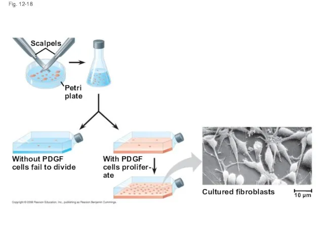
Fig. 12-18
Petri
plate
Scalpels
Cultured fibroblasts
Without PDGF
cells fail to divide
With PDGF
cells prolifer-
ate
10 µm
Fig. 12-18
Petri
plate
Scalpels
Cultured fibroblasts
Without PDGF
cells fail to divide
With PDGF
cells prolifer-
ate
10 µm
Another example of external signals is density-dependent inhibition, in which crowded
Another example of external signals is density-dependent inhibition, in which crowded
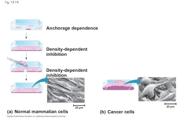
Fig. 12-19
Anchorage dependence
Density-dependent inhibition
Density-dependent inhibition
(a) Normal mammalian cells
(b) Cancer cells
25 µm
25
Fig. 12-19
Anchorage dependence
Density-dependent inhibition
Density-dependent inhibition
(a) Normal mammalian cells
(b) Cancer cells
25 µm
25
Cancer cells exhibit neither density-dependent inhibition nor anchorage dependence
Copyright © 2008
Cancer cells exhibit neither density-dependent inhibition nor anchorage dependence
Copyright © 2008
Loss of Cell Cycle Controls in Cancer Cells
Cancer cells do not
Loss of Cell Cycle Controls in Cancer Cells
Cancer cells do not
A normal cell is converted to a cancerous cell by a
A normal cell is converted to a cancerous cell by a

Fig. 12-20
Tumor
A tumor grows
from a single
cancer cell.
Glandular
tissue
Lymph
vessel
Blood
vessel
Metastatic
tumor
Cancer
cell
Cancer cells
invade neigh-
boring tissue.
Cancer cells
Fig. 12-20
Tumor
A tumor grows
from a single
cancer cell.
Glandular
tissue
Lymph
vessel
Blood
vessel
Metastatic
tumor
Cancer
cell
Cancer cells
invade neigh-
boring tissue.
Cancer cells
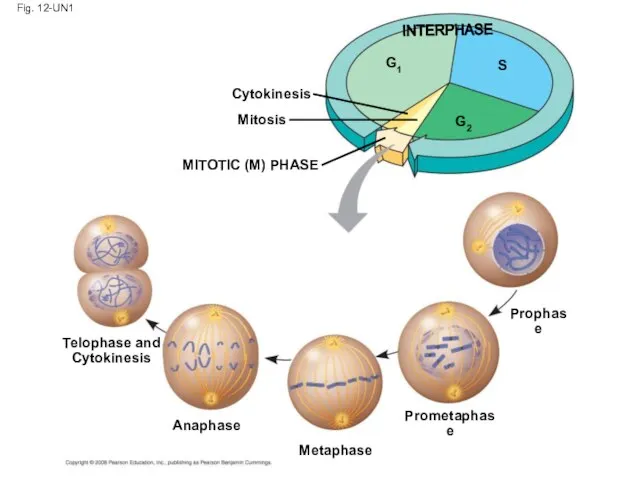
Fig. 12-UN1
Telophase and
Cytokinesis
Anaphase
Metaphase
Prometaphase
Prophase
MITOTIC (M) PHASE
Cytokinesis
Mitosis
S
G1
G2
INTERPHASE
Fig. 12-UN1
Telophase and
Cytokinesis
Anaphase
Metaphase
Prometaphase
Prophase
MITOTIC (M) PHASE
Cytokinesis
Mitosis
S
G1
G2
INTERPHASE
Fig. 12-UN2
Fig. 12-UN2
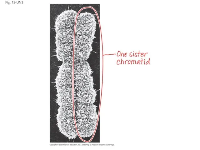
Fig. 12-UN3
Fig. 12-UN3
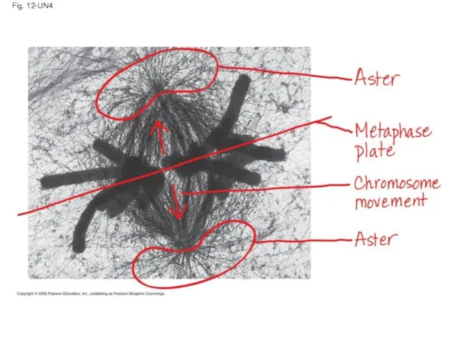
Fig. 12-UN4
Fig. 12-UN4
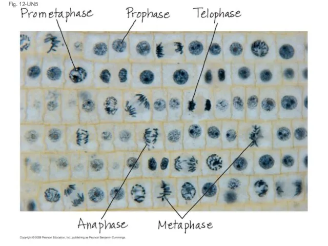
Fig. 12-UN5
Fig. 12-UN5
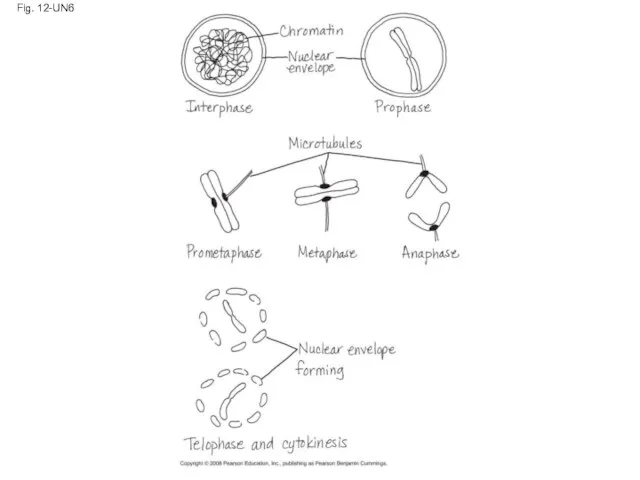
Fig. 12-UN6
Fig. 12-UN6
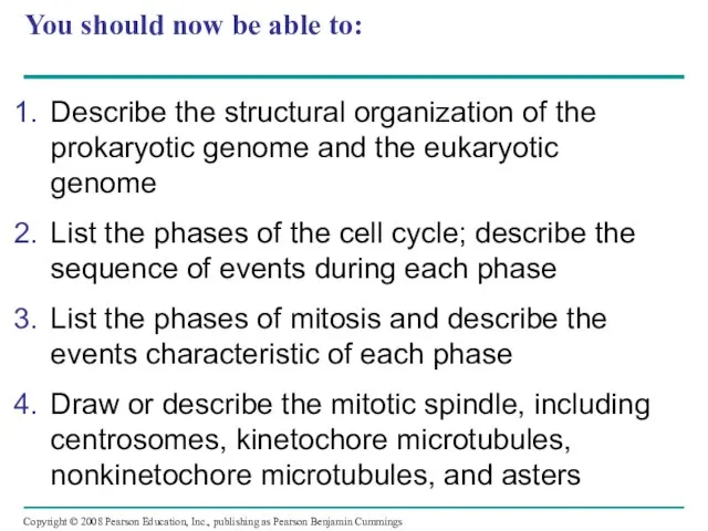
You should now be able to:
Describe the structural organization of the
You should now be able to:
Describe the structural organization of the
 Функции тонкого и толстого кишечника. Всасывание. Барьерная роль печени. Аппендицит.
 Презентация на тему "Типы взаимоотношений в биогеоценозе" - скачать презентации по Биологии
 Презентация на тему "Назови детёнышей зверей" - презентации по Биологии
 Общая характеристика проводящих путей ЦНС
 Посвящается Чарльзу Дарвину к 200-летию со Дня его Рождения
  Физиология дыхания. Зачем мы дышим?
 Шоколад
 Метод генной и клеточной инженерии Выполнила ученица 11 класса Деева Нелли Учитель Надежда Борисовна Лобова
 Обмен веществ. Фотосинтез. Урок биологии в 10 классе
 Каракал
 Физиология питания.
  Знакомство с профессией Пчеловод (Пасечник)
 Луг и человек. К уроку по окружающему миру в 3 классе
 Medical Academy named after S.I. Georgievsky of Vernadsky CFU
 Презентация на тему Значение растений и их охрана Биология, 7 класс
 100 тысяч видов
 Особенности скелета человека связанные с прямохождением и трудовой деятельностью
 Организм человека как единая биологическая система
 Презентация на тему "Взгляд на эволюцию" - скачать презентации по Биологии
 Гризуни
 Николай Иванович Вавилов. (13(25)ноября – 26 января 1943г) Подготовила Карпова Вероника 11б
  Психофизиология мотиваций и эмоций
 Ядро.Будова ядра та його функції
  Мохнатая азбука. Б.Заходер
 Отряд Рукокрылые (Chiroptera)
 Микробные объекты в биотехнологии - история развития, предмет, задачи, методы
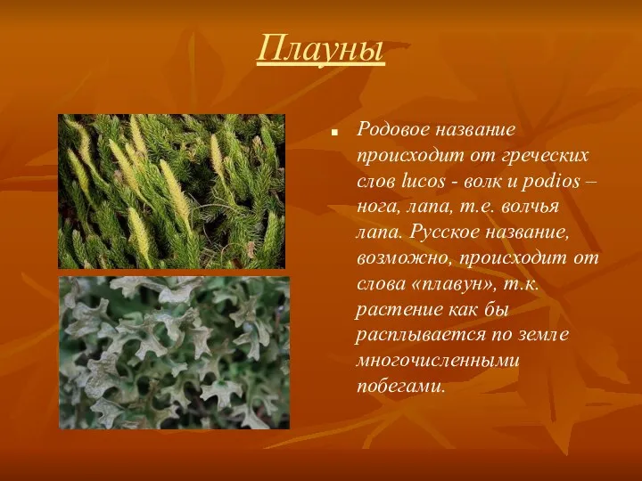 Плауны
 Презентация на тему "Миграции животных" - скачать презентации по Биологии