- Meiosis and Sexual Life Cycles

Содержание
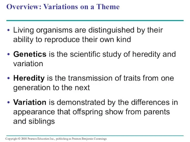
- 2. Overview: Variations on a Theme Living organisms are distinguished by their ability to reproduce their own
- 3. Fig. 13-1
- 4. Concept 13.1: Offspring acquire genes from parents by inheriting chromosomes In a literal sense, children do
- 5. Inheritance of Genes Genes are the units of heredity, and are made up of segments of
- 6. Comparison of Asexual and Sexual Reproduction In asexual reproduction, one parent produces genetically identical offspring by
- 7. Fig. 13-2 (a) Hydra (b) Redwoods Parent Bud 0.5 mm
- 8. Fig. 13-2a (a) Hydra 0.5 mm Bud Parent
- 9. Fig. 13-2b (b) Redwoods
- 10. Concept 13.2: Fertilization and meiosis alternate in sexual life cycles A life cycle is the generation-to-generation
- 11. Sets of Chromosomes in Human Cells Human somatic cells (any cell other than a gamete) have
- 12. Fig. 13-3 APPLICATION TECHNIQUE Pair of homologous replicated chromosomes 5 µm Centromere Sister chromatids Metaphase chromosome
- 13. Fig. 13-3a APPLICATION
- 14. Fig. 13-3b TECHNIQUE Pair of homologous replicated chromosomes Centromere Sister chromatids Metaphase chromosome 5 µm
- 15. The sex chromosomes are called X and Y Human females have a homologous pair of X
- 16. Each pair of homologous chromosomes includes one chromosome from each parent The 46 chromosomes in a
- 17. In a cell in which DNA synthesis has occurred, each chromosome is replicated Each replicated chromosome
- 18. Fig. 13-4 Key Maternal set of chromosomes (n = 3) Paternal set of chromosomes (n =
- 19. A gamete (sperm or egg) contains a single set of chromosomes, and is haploid (n) For
- 20. Fertilization is the union of gametes (the sperm and the egg) The fertilized egg is called
- 21. At sexual maturity, the ovaries and testes produce haploid gametes Gametes are the only types of
- 22. Fig. 13-5 Key Haploid (n) Diploid (2n) Haploid gametes (n = 23) Egg (n) Sperm (n)
- 23. The Variety of Sexual Life Cycles The alternation of meiosis and fertilization is common to all
- 24. In animals, meiosis produces gametes, which undergo no further cell division before fertilization Gametes are the
- 25. Fig. 13-6 Key Haploid (n) Diploid (2n) n n Gametes n n n Mitosis MEIOSIS FERTILIZATION
- 26. Fig. 13-6a Key Haploid (n) Diploid (2n) Gametes n n n 2n 2n Zygote MEIOSIS FERTILIZATION
- 27. Plants and some algae exhibit an alternation of generations This life cycle includes both a diploid
- 28. Each spore grows by mitosis into a haploid organism called a gametophyte A gametophyte makes haploid
- 29. Fig. 13-6b Key Haploid (n) Diploid (2n) n n n n n 2n 2n Mitosis Mitosis
- 30. In most fungi and some protists, the only diploid stage is the single-celled zygote; there is
- 31. Fig. 13-6c Key Haploid (n) Diploid (2n) Mitosis Mitosis Gametes Zygote Haploid unicellular or multicellular organism
- 32. Depending on the type of life cycle, either haploid or diploid cells can divide by mitosis
- 33. Concept 13.3: Meiosis reduces the number of chromosome sets from diploid to haploid Like mitosis, meiosis
- 34. The Stages of Meiosis In the first cell division (meiosis I), homologous chromosomes separate Meiosis I
- 35. Fig. 13-7-1 Interphase Homologous pair of chromosomes in diploid parent cell Chromosomes replicate Homologous pair of
- 36. Fig. 13-7-2 Interphase Homologous pair of chromosomes in diploid parent cell Chromosomes replicate Homologous pair of
- 37. Fig. 13-7-3 Interphase Homologous pair of chromosomes in diploid parent cell Chromosomes replicate Homologous pair of
- 38. Meiosis I is preceded by interphase, in which chromosomes are replicated to form sister chromatids The
- 39. Fig. 13-8 Prophase I Metaphase I Anaphase I Telophase I and Cytokinesis Prophase II Metaphase II
- 40. Division in meiosis I occurs in four phases: – Prophase I – Metaphase I – Anaphase
- 41. Metaphase I Fig. 13-8a Prophase I Anaphase I Telophase I and Cytokinesis Centrosome (with centriole pair)
- 42. Prophase I Prophase I typically occupies more than 90% of the time required for meiosis Chromosomes
- 43. In crossing over, nonsister chromatids exchange DNA segments Each pair of chromosomes forms a tetrad, a
- 44. Metaphase I In metaphase I, tetrads line up at the metaphase plate, with one chromosome facing
- 45. Fig. 13-8b Prophase I Metaphase I Centrosome (with centriole pair) Sister chromatids Chiasmata Spindle Centromere (with
- 46. Anaphase I In anaphase I, pairs of homologous chromosomes separate One chromosome moves toward each pole,
- 47. Telophase I and Cytokinesis In the beginning of telophase I, each half of the cell has
- 48. In animal cells, a cleavage furrow forms; in plant cells, a cell plate forms No chromosome
- 49. Fig. 13-8c Anaphase I Telophase I and Cytokinesis Sister chromatids remain attached Homologous chromosomes separate Cleavage
- 50. Division in meiosis II also occurs in four phases: – Prophase II – Metaphase II –
- 51. Fig. 13-8d Prophase II Metaphase II Anaphase II Telophase II and Cytokinesis Sister chromatids separate Haploid
- 52. Prophase II In prophase II, a spindle apparatus forms In late prophase II, chromosomes (each still
- 53. Metaphase II In metaphase II, the sister chromatids are arranged at the metaphase plate Because of
- 54. Fig. 13-8e Prophase II Metaphase II
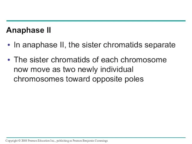
- 55. Anaphase II In anaphase II, the sister chromatids separate The sister chromatids of each chromosome now
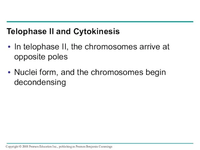
- 56. Telophase II and Cytokinesis In telophase II, the chromosomes arrive at opposite poles Nuclei form, and
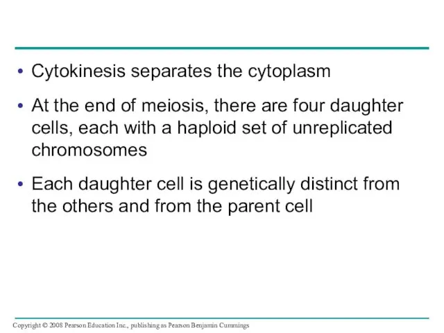
- 57. Cytokinesis separates the cytoplasm At the end of meiosis, there are four daughter cells, each with
- 58. Fig. 13-8f Anaphase II Telephase II and Cytokinesis Sister chromatids separate Haploid daughter cells forming
- 59. A Comparison of Mitosis and Meiosis Mitosis conserves the number of chromosome sets, producing cells that
- 60. Fig. 13-9 MITOSIS MEIOSIS MEIOSIS I Prophase I Chiasma Homologous chromosome pair Chromosome replication Parent cell
- 61. Fig. 13-9a MITOSIS MEIOSIS MEIOSIS I Prophase I Chiasma Chromosome replication Homologous chromosome pair Chromosome replication
- 62. Fig. 13-9b SUMMARY Meiosis Mitosis Property DNA replication Number of divisions Occurs during interphase before mitosis
- 63. Three events are unique to meiosis, and all three occur in meiosis l: – Synapsis and
- 64. Sister chromatid cohesion allows sister chromatids of a single chromosome to stay together through meiosis I
- 65. Fig. 13-10 EXPERIMENT RESULTS Shugoshin+ (normal)+ Spore case Fluorescent label Metaphase I Shugoshin– Anaphase I Metaphase
- 66. Fig. 13-10a EXPERIMENT Shugoshin+ (normal) Spore case Fluorescent label Metaphase I Anaphase I Metaphase II Anaphase
- 67. Fig. 13-10b RESULTS Shugoshin+ Shugoshin– Spore cases (%) 100 80 60 40 20 0
- 68. Concept 13.4: Genetic variation produced in sexual life cycles contributes to evolution Mutations (changes in an
- 69. Origins of Genetic Variation Among Offspring The behavior of chromosomes during meiosis and fertilization is responsible
- 70. Independent Assortment of Chromosomes Homologous pairs of chromosomes orient randomly at metaphase I of meiosis In
- 71. The number of combinations possible when chromosomes assort independently into gametes is 2n, where n is
- 72. Fig. 13-11-1 Possibility 1 Possibility 2 Two equally probable arrangements of chromosomes at metaphase I
- 73. Fig. 13-11-2 Possibility 1 Possibility 2 Two equally probable arrangements of chromosomes at metaphase I Metaphase
- 74. Fig. 13-11-3 Possibility 1 Possibility 2 Two equally probable arrangements of chromosomes at metaphase I Metaphase
- 75. Crossing Over Crossing over produces recombinant chromosomes, which combine genes inherited from each parent Crossing over
- 76. In crossing over, homologous portions of two nonsister chromatids trade places Crossing over contributes to genetic
- 77. Fig. 13-12-1 Prophase I of meiosis Pair of homologs Nonsister chromatids held together during synapsis
- 78. Fig. 13-12-2 Prophase I of meiosis Pair of homologs Nonsister chromatids held together during synapsis Chiasma
- 79. Fig. 13-12-3 Prophase I of meiosis Pair of homologs Nonsister chromatids held together during synapsis Chiasma
- 80. Fig. 13-12-4 Prophase I of meiosis Pair of homologs Nonsister chromatids held together during synapsis Chiasma
- 81. Fig. 13-12-5 Prophase I of meiosis Pair of homologs Nonsister chromatids held together during synapsis Chiasma
- 82. Random Fertilization Random fertilization adds to genetic variation because any sperm can fuse with any ovum
- 83. Crossing over adds even more variation Each zygote has a unique genetic identity Animation: Genetic Variation
- 84. The Evolutionary Significance of Genetic Variation Within Populations Natural selection results in the accumulation of genetic
- 85. Fig. 13-UN1 Prophase I: Each homologous pair undergoes synapsis and crossing over between nonsister chromatids. Metaphase
- 86. Fig. 13-UN2 F H
- 87. Fig. 13-UN3
- 88. Fig. 13-UN4
- 90. Скачать презентацию
Overview: Variations on a Theme
Living organisms are distinguished by their ability
Overview: Variations on a Theme
Living organisms are distinguished by their ability
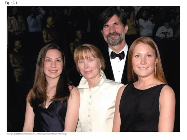
Fig. 13-1
Fig. 13-1
Concept 13.1: Offspring acquire genes from parents by inheriting chromosomes
In a
Concept 13.1: Offspring acquire genes from parents by inheriting chromosomes
In a
Inheritance of Genes
Genes are the units of heredity, and are made
Inheritance of Genes
Genes are the units of heredity, and are made
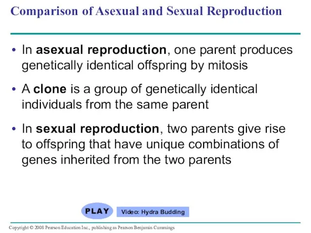
Comparison of Asexual and Sexual Reproduction
In asexual reproduction, one parent
Comparison of Asexual and Sexual Reproduction
In asexual reproduction, one parent
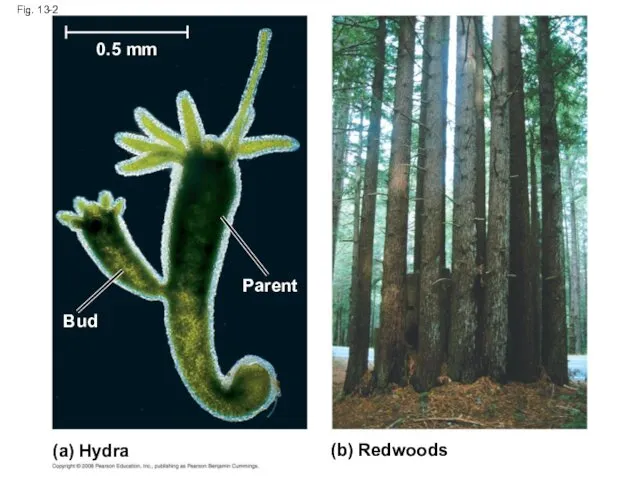
Fig. 13-2
(a) Hydra
(b) Redwoods
Parent
Bud
0.5 mm
Fig. 13-2
(a) Hydra
(b) Redwoods
Parent
Bud
0.5 mm
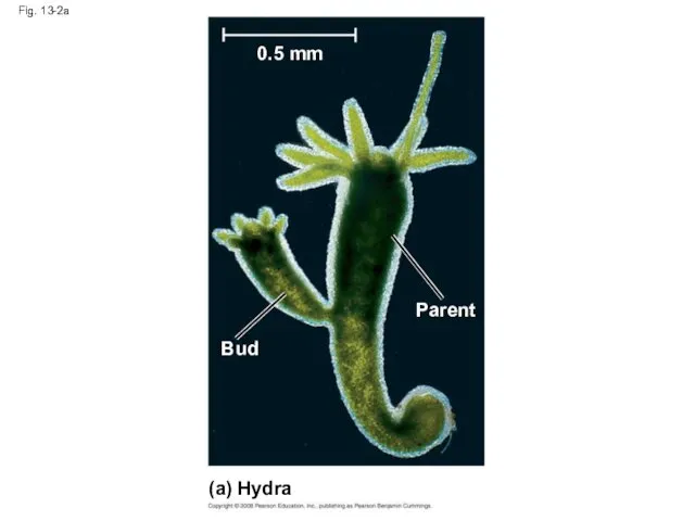
Fig. 13-2a
(a) Hydra
0.5 mm
Bud
Parent
Fig. 13-2a
(a) Hydra
0.5 mm
Bud
Parent
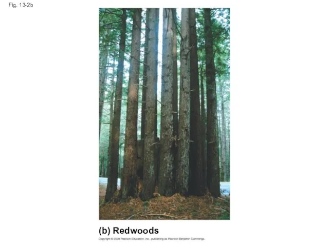
Fig. 13-2b
(b) Redwoods
Fig. 13-2b
(b) Redwoods
Concept 13.2: Fertilization and meiosis alternate in sexual life cycles
A life
Concept 13.2: Fertilization and meiosis alternate in sexual life cycles
A life
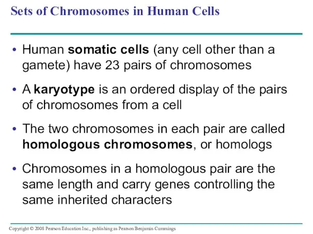
Sets of Chromosomes in Human Cells
Human somatic cells (any cell other
Sets of Chromosomes in Human Cells
Human somatic cells (any cell other
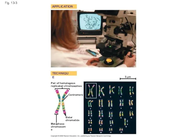
Fig. 13-3
APPLICATION
TECHNIQUE
Pair of homologous
replicated chromosomes
5 µm
Centromere
Sister
chromatids
Metaphase
chromosome
Fig. 13-3
APPLICATION
TECHNIQUE
Pair of homologous
replicated chromosomes
5 µm
Centromere
Sister
chromatids
Metaphase
chromosome
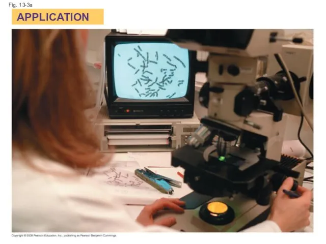
Fig. 13-3a
APPLICATION
Fig. 13-3a
APPLICATION
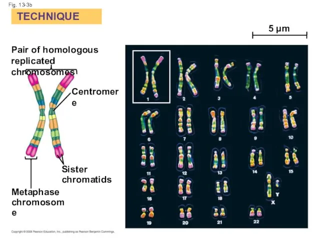
Fig. 13-3b
TECHNIQUE
Pair of homologous
replicated chromosomes
Centromere
Sister
chromatids
Metaphase
chromosome
5 µm
Fig. 13-3b
TECHNIQUE
Pair of homologous
replicated chromosomes
Centromere
Sister
chromatids
Metaphase
chromosome
5 µm
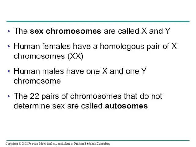
The sex chromosomes are called X and Y
Human females have a
The sex chromosomes are called X and Y
Human females have a
Each pair of homologous chromosomes includes one chromosome from each parent
The
Each pair of homologous chromosomes includes one chromosome from each parent
The
In a cell in which DNA synthesis has occurred, each chromosome
In a cell in which DNA synthesis has occurred, each chromosome
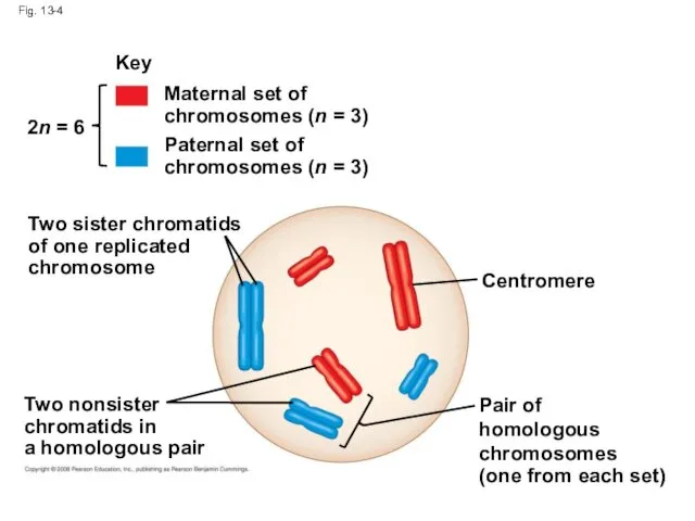
Fig. 13-4
Key
Maternal set of
chromosomes (n = 3)
Paternal set of
chromosomes (n =
Fig. 13-4
Key
Maternal set of
chromosomes (n = 3)
Paternal set of
chromosomes (n =
A gamete (sperm or egg) contains a single set of chromosomes,
A gamete (sperm or egg) contains a single set of chromosomes,
Fertilization is the union of gametes (the sperm and the egg)
The
Fertilization is the union of gametes (the sperm and the egg)
The
At sexual maturity, the ovaries and testes produce haploid gametes
Gametes are
At sexual maturity, the ovaries and testes produce haploid gametes
Gametes are
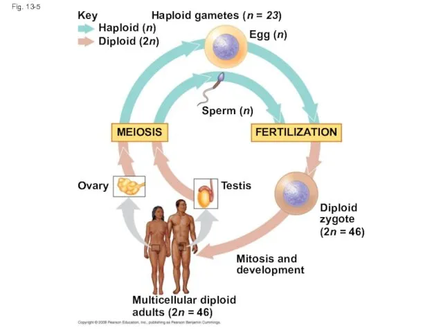
Fig. 13-5
Key
Haploid (n)
Diploid (2n)
Haploid gametes (n = 23)
Egg (n)
Sperm (n)
MEIOSIS
FERTILIZATION
Ovary
Testis
Diploid
zygote
(2n =
Fig. 13-5
Key
Haploid (n)
Diploid (2n)
Haploid gametes (n = 23)
Egg (n)
Sperm (n)
MEIOSIS
FERTILIZATION
Ovary
Testis
Diploid
zygote
(2n =
The Variety of Sexual Life Cycles
The alternation of meiosis and fertilization
The Variety of Sexual Life Cycles
The alternation of meiosis and fertilization
In animals, meiosis produces gametes, which undergo no further cell division
In animals, meiosis produces gametes, which undergo no further cell division
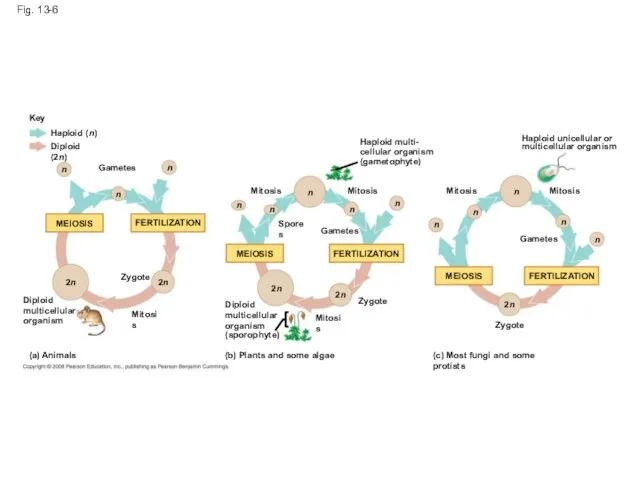
Fig. 13-6
Key
Haploid (n)
Diploid (2n)
n
n
Gametes
n
n
n
Mitosis
MEIOSIS
FERTILIZATION
MEIOSIS
2n
2n
Zygote
2n
Mitosis
Diploid
multicellular
organism
(a) Animals
Spores
Diploid
multicellular
organism
(sporophyte)
(b) Plants and some algae
2n
Mitosis
Gametes
Mitosis
n
n
n
Zygote
FERTILIZATION
n
n
n
Mitosis
Zygote
(c) Most fungi
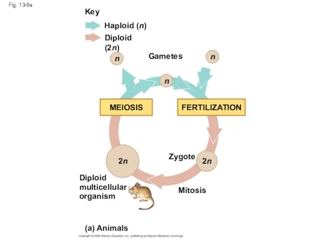
Fig. 13-6a
Key
Haploid (n)
Diploid (2n)
Gametes
n
n
n
2n
2n
Zygote
MEIOSIS
FERTILIZATION
Mitosis
Diploid
multicellular
organism
(a) Animals
Fig. 13-6a
Key
Haploid (n)
Diploid (2n)
Gametes
n
n
n
2n
2n
Zygote
MEIOSIS
FERTILIZATION
Mitosis
Diploid
multicellular
organism
(a) Animals
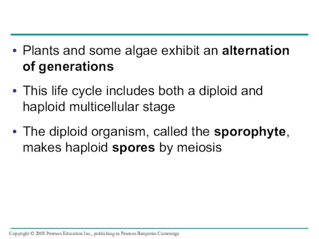
Plants and some algae exhibit an alternation of generations
This life cycle
Plants and some algae exhibit an alternation of generations
This life cycle
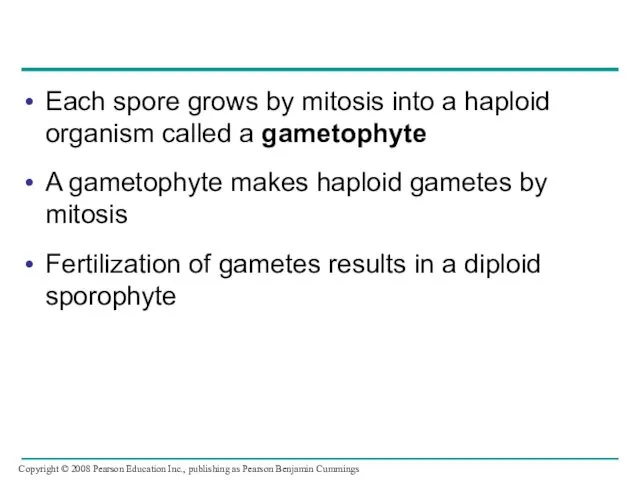
Each spore grows by mitosis into a haploid organism called a
Each spore grows by mitosis into a haploid organism called a
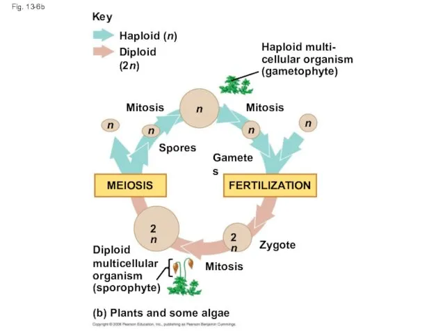
Fig. 13-6b
Key
Haploid (n)
Diploid (2n)
n
n
n
n
n
2n
2n
Mitosis
Mitosis
Mitosis
Zygote
Spores
Gametes
MEIOSIS
FERTILIZATION
Diploid
multicellular
organism
(sporophyte)
Haploid multi-
cellular organism
(gametophyte)
(b) Plants and some algae
Fig. 13-6b
Key
Haploid (n)
Diploid (2n)
n
n
n
n
n
2n
2n
Mitosis
Mitosis
Mitosis
Zygote
Spores
Gametes
MEIOSIS
FERTILIZATION
Diploid
multicellular
organism
(sporophyte)
Haploid multi-
cellular organism
(gametophyte)
(b) Plants and some algae
In most fungi and some protists, the only diploid stage is
In most fungi and some protists, the only diploid stage is
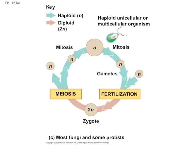
Fig. 13-6c
Key
Haploid (n)
Diploid (2n)
Mitosis
Mitosis
Gametes
Zygote
Haploid unicellular or
multicellular organism
MEIOSIS
FERTILIZATION
n
n
n
n
n
2n
(c) Most fungi and some
Fig. 13-6c
Key
Haploid (n)
Diploid (2n)
Mitosis
Mitosis
Gametes
Zygote
Haploid unicellular or
multicellular organism
MEIOSIS
FERTILIZATION
n
n
n
n
n
2n
(c) Most fungi and some
Depending on the type of life cycle, either haploid or diploid
Depending on the type of life cycle, either haploid or diploid
Concept 13.3: Meiosis reduces the number of chromosome sets from diploid
Concept 13.3: Meiosis reduces the number of chromosome sets from diploid
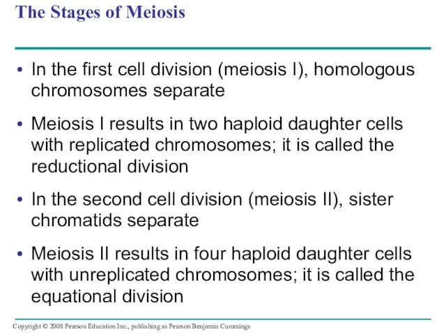
The Stages of Meiosis
In the first cell division (meiosis I), homologous
The Stages of Meiosis
In the first cell division (meiosis I), homologous
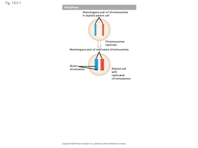
Fig. 13-7-1
Interphase
Homologous pair of chromosomes
in diploid parent cell
Chromosomes
replicate
Homologous pair of replicated
Fig. 13-7-1
Interphase
Homologous pair of chromosomes
in diploid parent cell
Chromosomes
replicate
Homologous pair of replicated
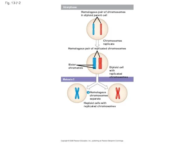
Fig. 13-7-2
Interphase
Homologous pair of chromosomes
in diploid parent cell
Chromosomes
replicate
Homologous pair of replicated
Fig. 13-7-2
Interphase
Homologous pair of chromosomes
in diploid parent cell
Chromosomes
replicate
Homologous pair of replicated
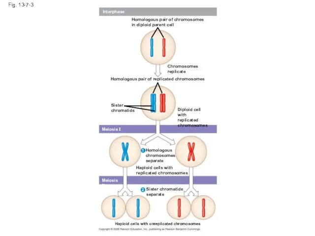
Fig. 13-7-3
Interphase
Homologous pair of chromosomes
in diploid parent cell
Chromosomes
replicate
Homologous pair of replicated
Fig. 13-7-3
Interphase
Homologous pair of chromosomes
in diploid parent cell
Chromosomes
replicate
Homologous pair of replicated
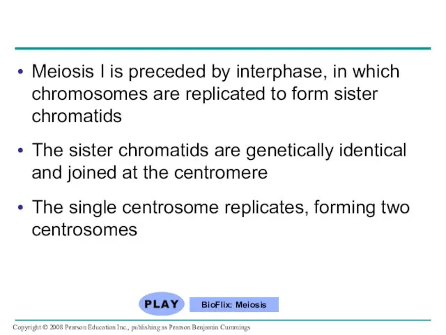
Meiosis I is preceded by interphase, in which chromosomes are replicated
Meiosis I is preceded by interphase, in which chromosomes are replicated
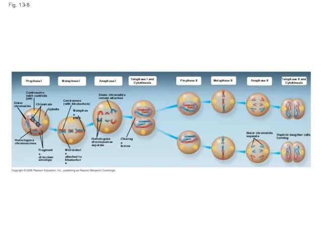
Fig. 13-8
Prophase I
Metaphase I
Anaphase I
Telophase I and
Cytokinesis
Prophase II
Metaphase II
Anaphase II
Telophase II
Fig. 13-8
Prophase I
Metaphase I
Anaphase I
Telophase I and
Cytokinesis
Prophase II
Metaphase II
Anaphase II
Telophase II
Division in meiosis I occurs in four phases:
– Prophase I
– Metaphase I
– Anaphase I
– Telophase
Division in meiosis I occurs in four phases:
– Prophase I
– Metaphase I
– Anaphase I
– Telophase
Metaphase I
Fig. 13-8a
Prophase I
Anaphase I
Telophase I and
Cytokinesis
Centrosome
(with centriole pair)
Sister
chromatids
Chiasmata
Spindle
Homologous
chromosomes
Fragments
of nuclear
envelope
Centromere
(with kinetochore)
Metaphase
plate
Microtubule
attached
Metaphase I
Fig. 13-8a
Prophase I
Anaphase I
Telophase I and
Cytokinesis
Centrosome
(with centriole pair)
Sister
chromatids
Chiasmata
Spindle
Homologous
chromosomes
Fragments
of nuclear
envelope
Centromere
(with kinetochore)
Metaphase
plate
Microtubule
attached
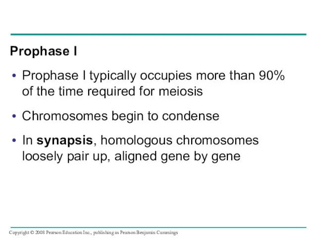
Prophase I
Prophase I typically occupies more than 90% of the time
Prophase I
Prophase I typically occupies more than 90% of the time
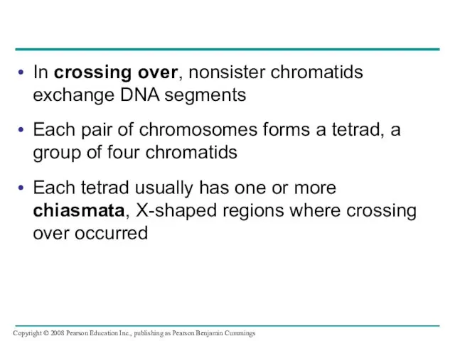
In crossing over, nonsister chromatids exchange DNA segments
Each pair of chromosomes
In crossing over, nonsister chromatids exchange DNA segments
Each pair of chromosomes
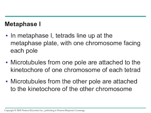
Metaphase I
In metaphase I, tetrads line up at the metaphase plate,
Metaphase I
In metaphase I, tetrads line up at the metaphase plate,
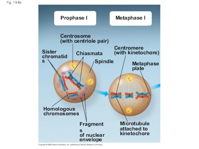
Fig. 13-8b
Prophase I
Metaphase I
Centrosome
(with centriole pair)
Sister
chromatids
Chiasmata
Spindle
Centromere
(with kinetochore)
Metaphase
plate
Homologous
chromosomes
Fragments
of nuclear
envelope
Microtubule
attached to
kinetochore
Fig. 13-8b
Prophase I
Metaphase I
Centrosome
(with centriole pair)
Sister
chromatids
Chiasmata
Spindle
Centromere
(with kinetochore)
Metaphase
plate
Homologous
chromosomes
Fragments
of nuclear
envelope
Microtubule
attached to
kinetochore
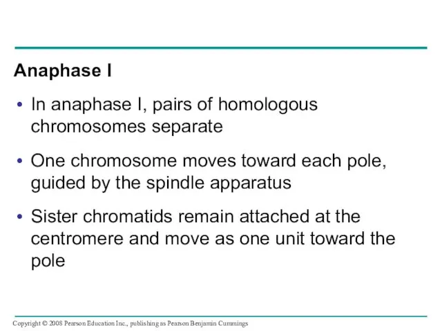
Anaphase I
In anaphase I, pairs of homologous chromosomes separate
One chromosome moves
Anaphase I
In anaphase I, pairs of homologous chromosomes separate
One chromosome moves
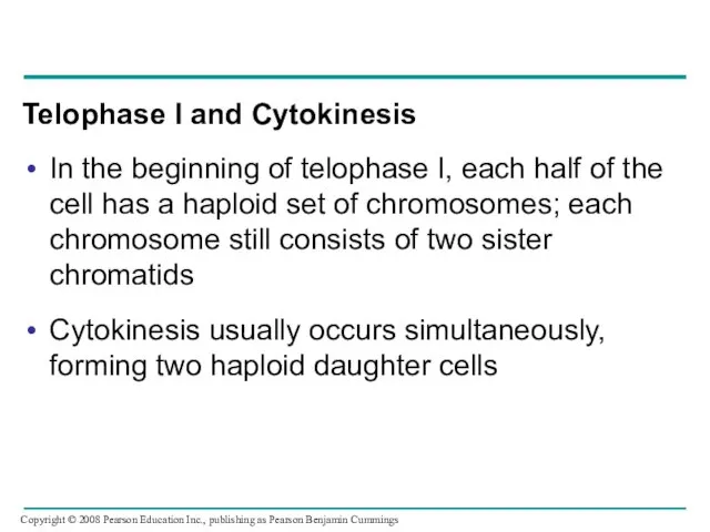
Telophase I and Cytokinesis
In the beginning of telophase I, each half
Telophase I and Cytokinesis
In the beginning of telophase I, each half
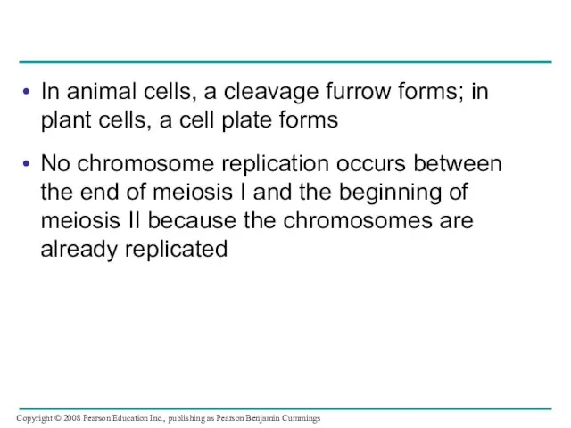
In animal cells, a cleavage furrow forms; in plant cells, a
In animal cells, a cleavage furrow forms; in plant cells, a
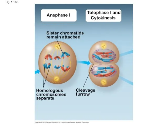
Fig. 13-8c
Anaphase I
Telophase I and
Cytokinesis
Sister chromatids
remain attached
Homologous
chromosomes
separate
Cleavage
furrow
Fig. 13-8c
Anaphase I
Telophase I and
Cytokinesis
Sister chromatids
remain attached
Homologous
chromosomes
separate
Cleavage
furrow
Division in meiosis II also occurs in four phases:
– Prophase II
– Metaphase II
– Anaphase
Division in meiosis II also occurs in four phases:
– Prophase II
– Metaphase II
– Anaphase
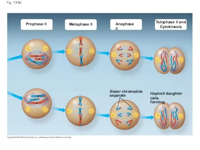
Fig. 13-8d
Prophase II
Metaphase II
Anaphase II
Telophase II and
Cytokinesis
Sister chromatids
separate
Haploid daughter cells
forming
Fig. 13-8d
Prophase II
Metaphase II
Anaphase II
Telophase II and
Cytokinesis
Sister chromatids
separate
Haploid daughter cells
forming
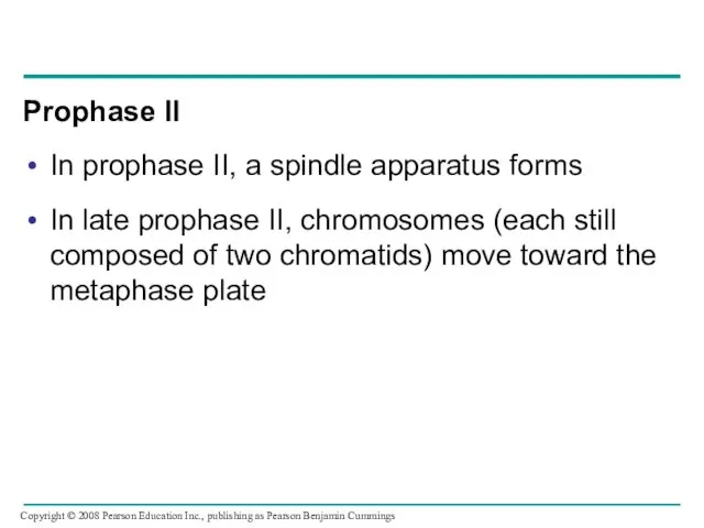
Prophase II
In prophase II, a spindle apparatus forms
In late prophase II,
Prophase II
In prophase II, a spindle apparatus forms
In late prophase II,
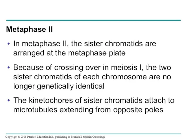
Metaphase II
In metaphase II, the sister chromatids are arranged at the
Metaphase II
In metaphase II, the sister chromatids are arranged at the
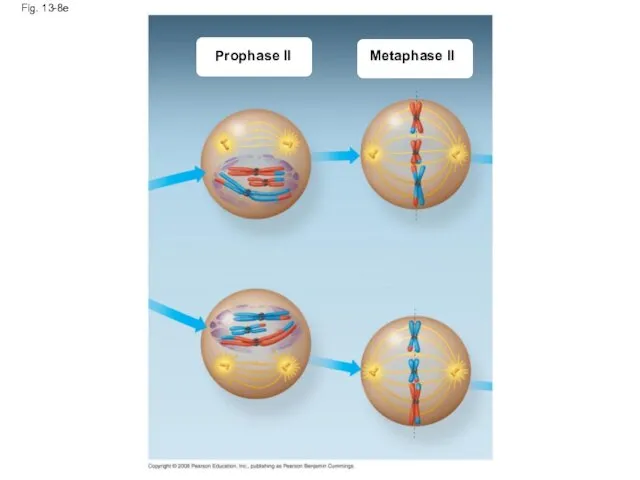
Fig. 13-8e
Prophase II
Metaphase II
Fig. 13-8e
Prophase II
Metaphase II
Anaphase II
In anaphase II, the sister chromatids separate
The sister chromatids of
Anaphase II
In anaphase II, the sister chromatids separate
The sister chromatids of
Telophase II and Cytokinesis
In telophase II, the chromosomes arrive at opposite
Telophase II and Cytokinesis
In telophase II, the chromosomes arrive at opposite
Cytokinesis separates the cytoplasm
At the end of meiosis, there are four
Cytokinesis separates the cytoplasm
At the end of meiosis, there are four
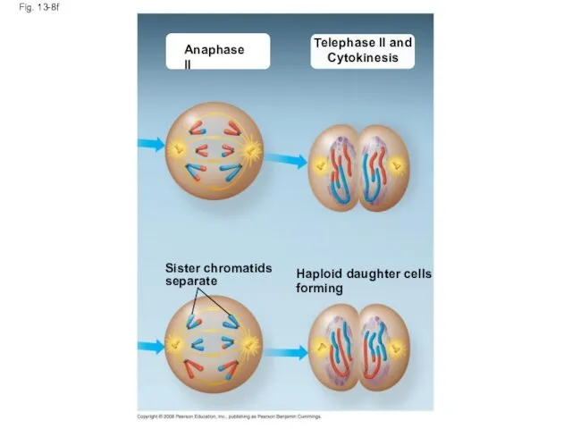
Fig. 13-8f
Anaphase II
Telephase II and
Cytokinesis
Sister chromatids
separate
Haploid daughter cells
forming
Fig. 13-8f
Anaphase II
Telephase II and
Cytokinesis
Sister chromatids
separate
Haploid daughter cells
forming
A Comparison of Mitosis and Meiosis
Mitosis conserves the number of chromosome
A Comparison of Mitosis and Meiosis
Mitosis conserves the number of chromosome
Fig. 13-9
MITOSIS
MEIOSIS
MEIOSIS I
Prophase I
Chiasma
Homologous
chromosome
pair
Chromosome
replication
Parent cell
2n = 6
Chromosome
replication
Replicated chromosome
Prophase
Metaphase
Metaphase I
Anaphase I
Telophase I
Haploid
n
Fig. 13-9
MITOSIS
MEIOSIS
MEIOSIS I
Prophase I
Chiasma
Homologous
chromosome
pair
Chromosome
replication
Parent cell
2n = 6
Chromosome
replication
Replicated chromosome
Prophase
Metaphase
Metaphase I
Anaphase I
Telophase I
Haploid
n
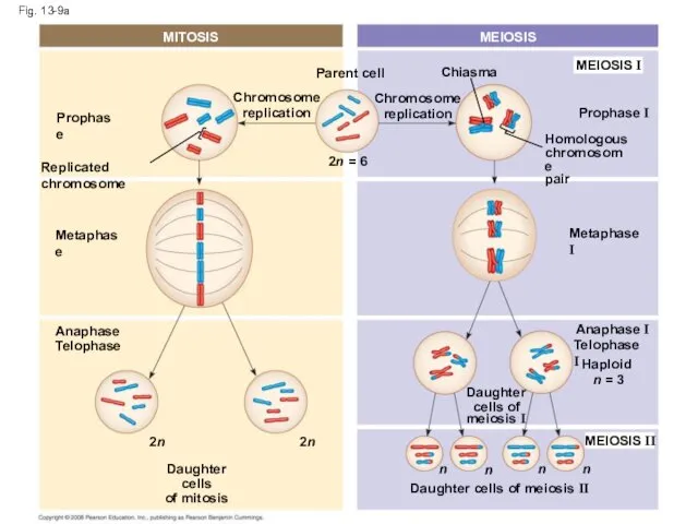
Fig. 13-9a
MITOSIS
MEIOSIS
MEIOSIS I
Prophase I
Chiasma
Chromosome
replication
Homologous
chromosome
pair
Chromosome
replication
2n = 6
Parent cell
Prophase
Replicated chromosome
Metaphase
Metaphase I
Anaphase I
Telophase I
Haploid
Fig. 13-9a
MITOSIS
MEIOSIS
MEIOSIS I
Prophase I
Chiasma
Chromosome
replication
Homologous
chromosome
pair
Chromosome
replication
2n = 6
Parent cell
Prophase
Replicated chromosome
Metaphase
Metaphase I
Anaphase I
Telophase I
Haploid
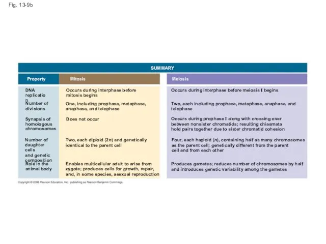
Fig. 13-9b
SUMMARY
Meiosis
Mitosis
Property
DNA
replication
Number of
divisions
Occurs during interphase before
mitosis begins
One, including prophase, metaphase,
anaphase, and
Fig. 13-9b
SUMMARY
Meiosis
Mitosis
Property
DNA
replication
Number of
divisions
Occurs during interphase before
mitosis begins
One, including prophase, metaphase,
anaphase, and
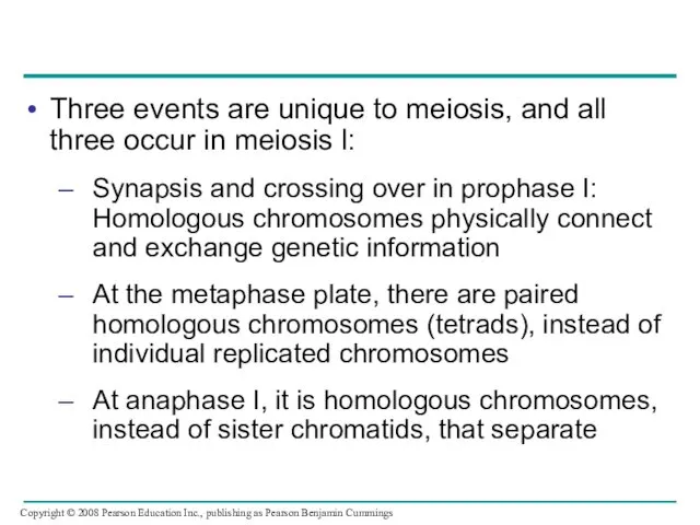
Three events are unique to meiosis, and all three occur in
Three events are unique to meiosis, and all three occur in
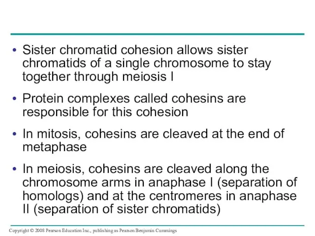
Sister chromatid cohesion allows sister chromatids of a single chromosome to
Sister chromatid cohesion allows sister chromatids of a single chromosome to
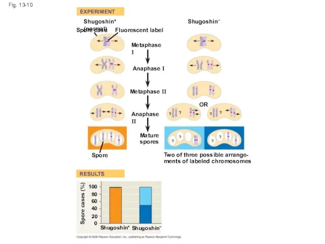
Fig. 13-10
EXPERIMENT
RESULTS
Shugoshin+ (normal)+
Spore case
Fluorescent label
Metaphase I
Shugoshin–
Anaphase I
Metaphase II
Anaphase II
Mature
spores
OR
Spore
Two of three
Fig. 13-10
EXPERIMENT
RESULTS
Shugoshin+ (normal)+
Spore case
Fluorescent label
Metaphase I
Shugoshin–
Anaphase I
Metaphase II
Anaphase II
Mature
spores
OR
Spore
Two of three
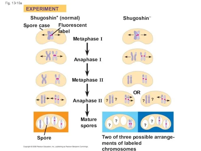
Fig. 13-10a
EXPERIMENT
Shugoshin+ (normal)
Spore case
Fluorescent label
Metaphase I
Anaphase I
Metaphase II
Anaphase II
Mature
spores
Spore
OR
Two of three
Fig. 13-10a
EXPERIMENT
Shugoshin+ (normal)
Spore case
Fluorescent label
Metaphase I
Anaphase I
Metaphase II
Anaphase II
Mature
spores
Spore
OR
Two of three
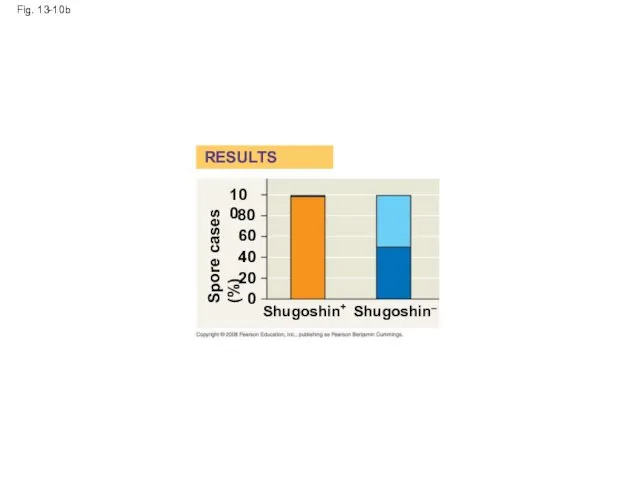
Fig. 13-10b
RESULTS
Shugoshin+
Shugoshin–
Spore cases (%)
100
80
60
40
20
0
Fig. 13-10b
RESULTS
Shugoshin+
Shugoshin–
Spore cases (%)
100
80
60
40
20
0
Concept 13.4: Genetic variation produced in sexual life cycles contributes to
Concept 13.4: Genetic variation produced in sexual life cycles contributes to
Origins of Genetic Variation Among Offspring
The behavior of chromosomes during meiosis
Origins of Genetic Variation Among Offspring
The behavior of chromosomes during meiosis
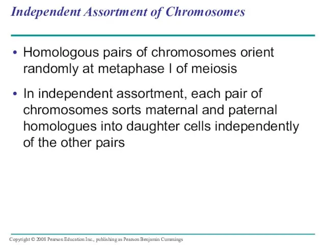
Independent Assortment of Chromosomes
Homologous pairs of chromosomes orient randomly at metaphase
Independent Assortment of Chromosomes
Homologous pairs of chromosomes orient randomly at metaphase
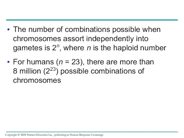
The number of combinations possible when chromosomes assort independently into gametes
The number of combinations possible when chromosomes assort independently into gametes
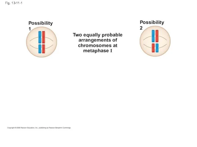
Fig. 13-11-1
Possibility 1
Possibility 2
Two equally probable
arrangements of
chromosomes at
metaphase I
Fig. 13-11-1
Possibility 1
Possibility 2
Two equally probable
arrangements of
chromosomes at
metaphase I
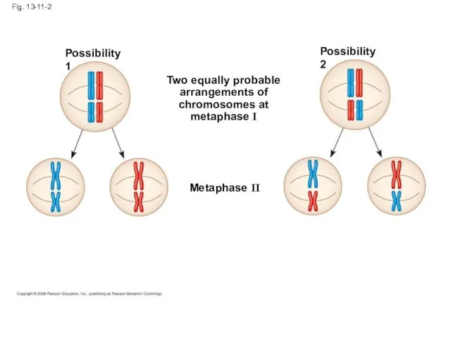
Fig. 13-11-2
Possibility 1
Possibility 2
Two equally probable
arrangements of
chromosomes at
metaphase I
Metaphase II
Fig. 13-11-2
Possibility 1
Possibility 2
Two equally probable
arrangements of
chromosomes at
metaphase I
Metaphase II
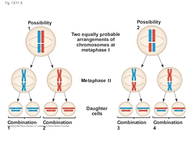
Fig. 13-11-3
Possibility 1
Possibility 2
Two equally probable
arrangements of
chromosomes at
metaphase I
Metaphase II
Daughter
cells
Combination 1
Combination
Fig. 13-11-3
Possibility 1
Possibility 2
Two equally probable
arrangements of
chromosomes at
metaphase I
Metaphase II
Daughter
cells
Combination 1
Combination
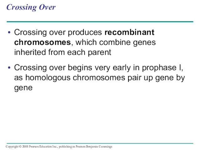
Crossing Over
Crossing over produces recombinant chromosomes, which combine genes inherited from
Crossing Over
Crossing over produces recombinant chromosomes, which combine genes inherited from
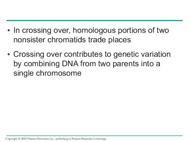
In crossing over, homologous portions of two nonsister chromatids trade places
Crossing
In crossing over, homologous portions of two nonsister chromatids trade places
Crossing
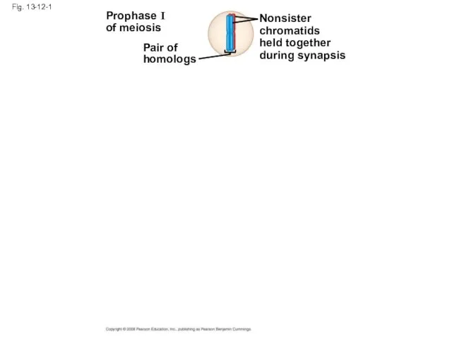
Fig. 13-12-1
Prophase I
of meiosis
Pair of
homologs
Nonsister
chromatids
held together
during synapsis
Fig. 13-12-1
Prophase I
of meiosis
Pair of
homologs
Nonsister
chromatids
held together
during synapsis
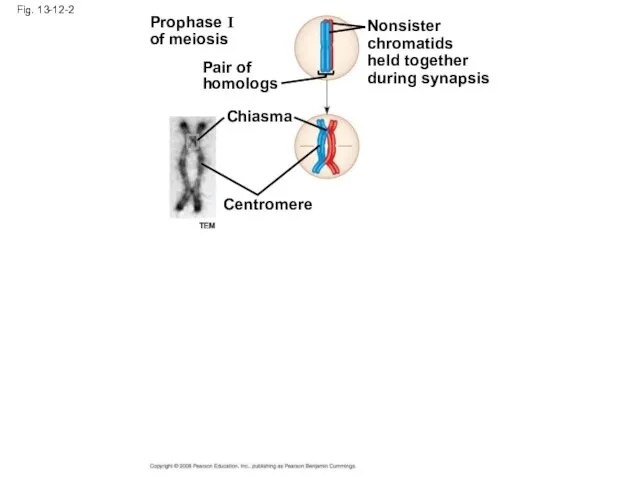
Fig. 13-12-2
Prophase I
of meiosis
Pair of
homologs
Nonsister
chromatids
held together
during synapsis
Chiasma
Centromere
TEM
Fig. 13-12-2
Prophase I
of meiosis
Pair of
homologs
Nonsister
chromatids
held together
during synapsis
Chiasma
Centromere
TEM
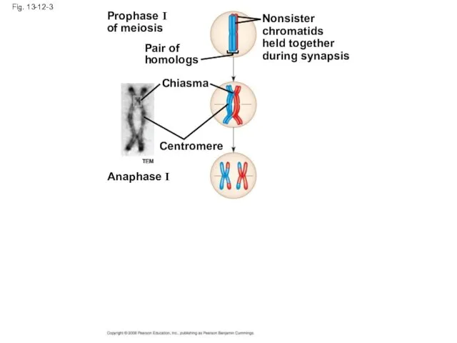
Fig. 13-12-3
Prophase I
of meiosis
Pair of
homologs
Nonsister
chromatids
held together
during synapsis
Chiasma
Centromere
Anaphase I
TEM
Fig. 13-12-3
Prophase I
of meiosis
Pair of
homologs
Nonsister
chromatids
held together
during synapsis
Chiasma
Centromere
Anaphase I
TEM
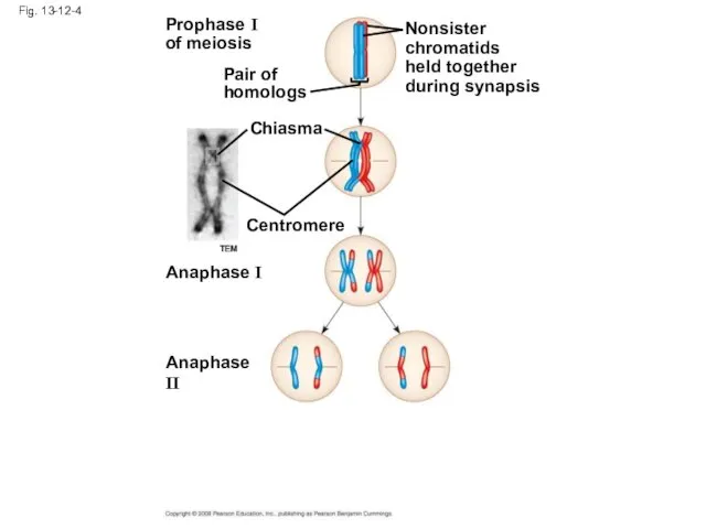
Fig. 13-12-4
Prophase I
of meiosis
Pair of
homologs
Nonsister
chromatids
held together
during synapsis
Chiasma
Centromere
Anaphase I
Anaphase II
TEM
Fig. 13-12-4
Prophase I
of meiosis
Pair of
homologs
Nonsister
chromatids
held together
during synapsis
Chiasma
Centromere
Anaphase I
Anaphase II
TEM
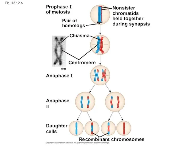
Fig. 13-12-5
Prophase I
of meiosis
Pair of
homologs
Nonsister
chromatids
held together
during synapsis
Chiasma
Centromere
Anaphase I
Anaphase II
Daughter
cells
Recombinant chromosomes
TEM
Fig. 13-12-5
Prophase I
of meiosis
Pair of
homologs
Nonsister
chromatids
held together
during synapsis
Chiasma
Centromere
Anaphase I
Anaphase II
Daughter
cells
Recombinant chromosomes
TEM
Random Fertilization
Random fertilization adds to genetic variation because any sperm can
Random Fertilization
Random fertilization adds to genetic variation because any sperm can
Crossing over adds even more variation
Each zygote has a unique genetic
Crossing over adds even more variation
Each zygote has a unique genetic
The Evolutionary Significance of Genetic Variation Within Populations
Natural selection results in
The Evolutionary Significance of Genetic Variation Within Populations
Natural selection results in
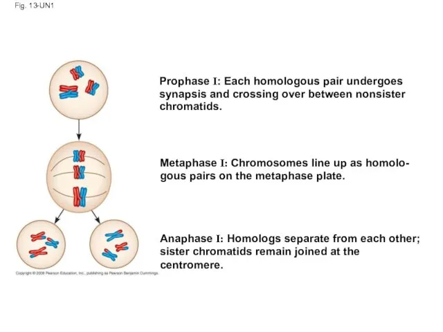
Fig. 13-UN1
Prophase I: Each homologous pair undergoes
synapsis and crossing over between
Fig. 13-UN1
Prophase I: Each homologous pair undergoes
synapsis and crossing over between
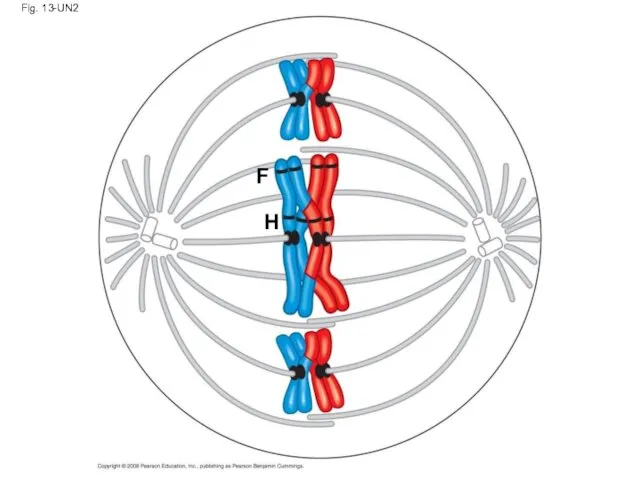
Fig. 13-UN2
F
H
Fig. 13-UN2
F
H
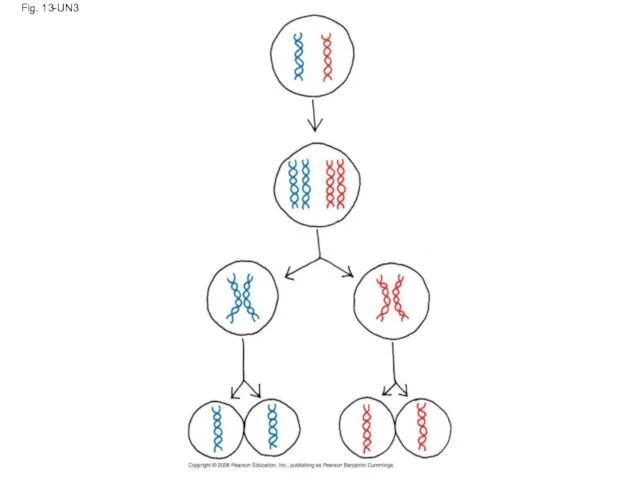
Fig. 13-UN3
Fig. 13-UN3
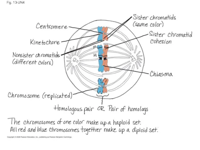
Fig. 13-UN4
Fig. 13-UN4
 Мой внутренний мир. Мир звуков
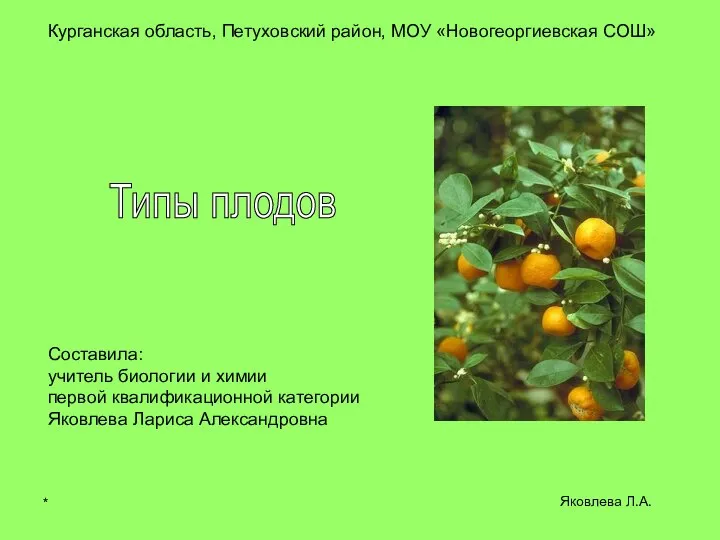 Презентация на тему "Типы плодов" - скачать бесплатно презентации по Биологии
 Общие сведения о животном мире Вводная презентация для учащихся 7 класса составитель Проценко Л.В._
 Декоративные кролики
 Значение воды в жизнедеятельности растений
 Презентация Бобры Воспитатель: Глотова Ирина Сергеевна I квалификационная категория
 ОРГАНИЗАЦИЯ НАСЛЕДСТВЕННОГО МАТЕРИАЛА Лекция 4
 Признаки растений
 Белки. Структура. Уровни организации
 Презентация на тему "Водорості" - скачать бесплатно презентации по Биологии
 Многообразие млекопитающих. (Часть 2)
 Сорные растения
 Обмен белков. Общие пути обмена аминокислот
 Основные этапы промышленного получения антибиотиков
 Строение клетки
 Что? Где? Когда? Экологическая викторина для старшеклассников
 Составитель: Серебрянская Н.А., учитель биологии МОУ «Образцовская СОШ» Фроловского муниципального района Вольгоградской области
 Знатоки природы. Турнир
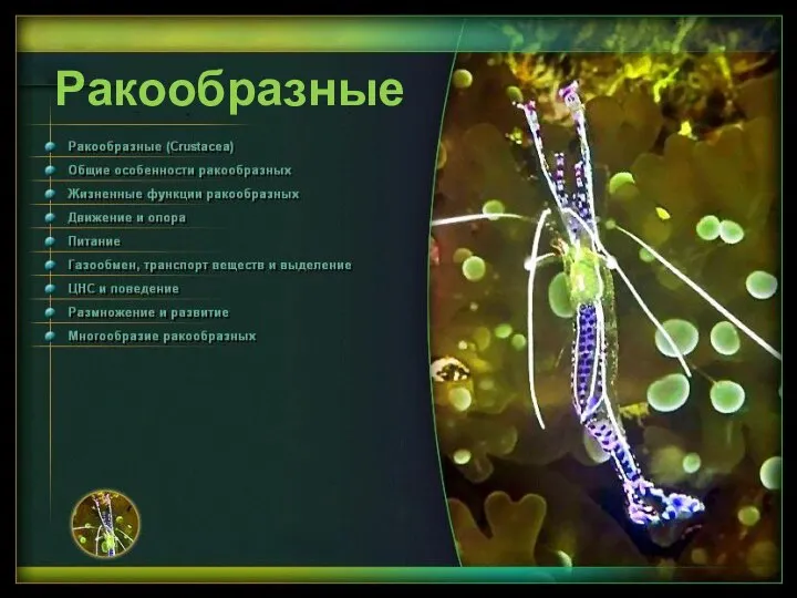 Презентация на тему Ракообразные
  АТМОСФЕРНОЕ ДАВЛЕНИЕ Как атмосферное давление влияет на организм человека?
 Ткани
Ткани Живое прошлое Земли
 Класс Млекопитающие .
 «В конце концов, жизнь - это отношения между организмами и внешней средой». Клод Бернар
 Триасовый период
 Қозғыштық. Қозу үрдісі
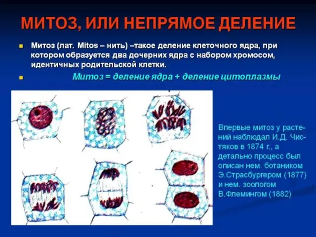 Презентация на тему "митоз" - скачать презентации по Биологии_
 Строение и возрастные особенности сердца. Сердечный цикл и его фазы