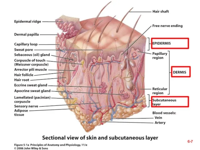
- The integumentary system

Содержание
- 2. 6- Ch. 6 Study Guide Critically read Chapter 6– pp. 187-194 before “Skin Color” section Skip
- 3. 6- § Quotable Quotes (Skin) Some guys say beauty is only skin deep. But when you
- 4. 6- I. Introduction 6-
- 5. 6- § Overview (1) Dermatology– scientific study and medical treatment of this system Largest organ (skin)
- 6. 6- 6- § Overview (2) Thickness variable, based on thickness of Epidermis, two categories-- Thick skin–
- 7. 6-
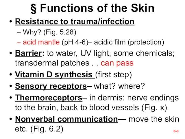
- 8. 6- 6- § Functions of the Skin Resistance to trauma/infection Why? (Fig. 5.28) acid mantle (pH
- 9. 6-
- 10. In hot environment In cold environment 6- vasodilation vasoconstriction Heat loss Less Heat loss Thermoregulation
- 11. 6- Social functions-- Figure 6.2 Skeletal muscles attach to dermal collagen fibers and produce expressions as
- 12. 6- II. Epidermis 6-
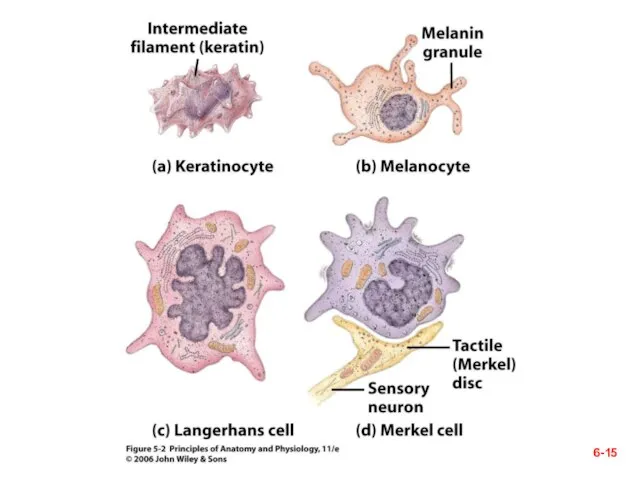
- 13. 6- 6- § Cells of the Epidermis (1) Five types of cells-- Keratinocytes – most of
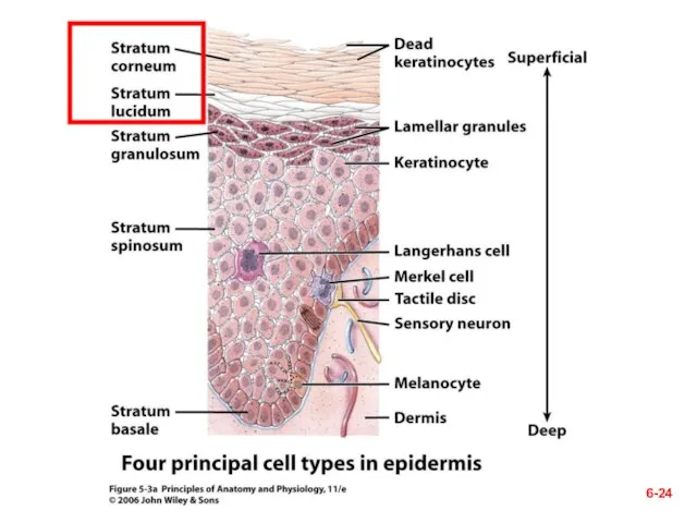
- 14. The Epidermis— Fig. 6.2 6-
- 15. 6-
- 16. 6- 6- § Cells of the Epidermis (2) Location of the following types of cells— stratum
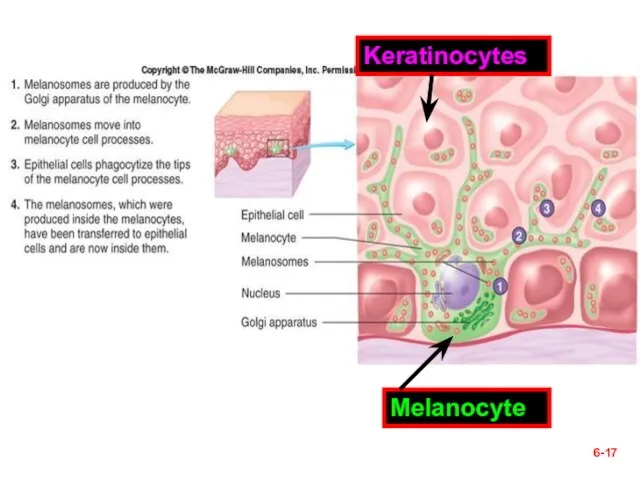
- 17. 6- Melanocyte Keratinocytes
- 18. 6- § Layers of the Epidermis— Next five slides (1-5) from deep to superficial and from
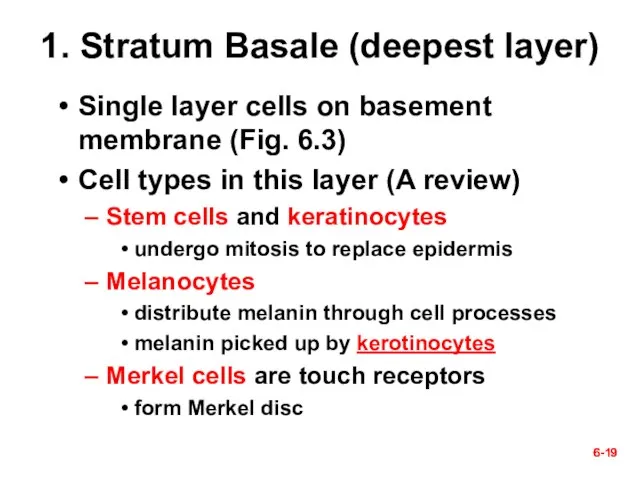
- 19. 6- 6- 1. Stratum Basale (deepest layer) Single layer cells on basement membrane (Fig. 6.3) Cell
- 20. Figure 6.2a 6-
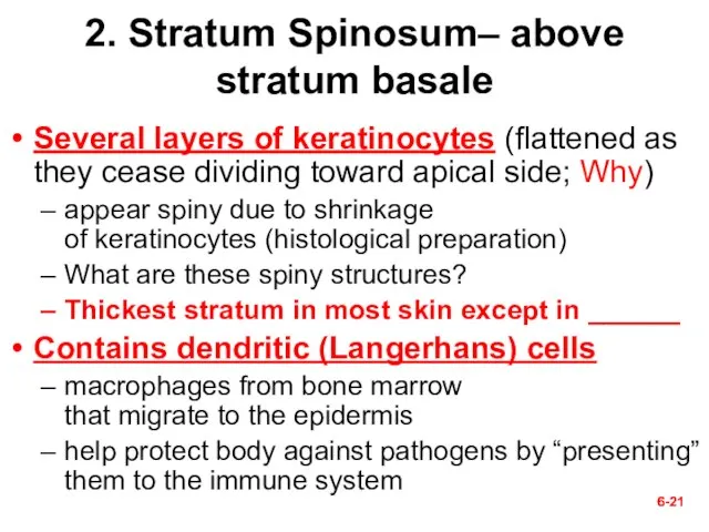
- 21. 6- 6- 2. Stratum Spinosum– above stratum basale Several layers of keratinocytes (flattened as they cease
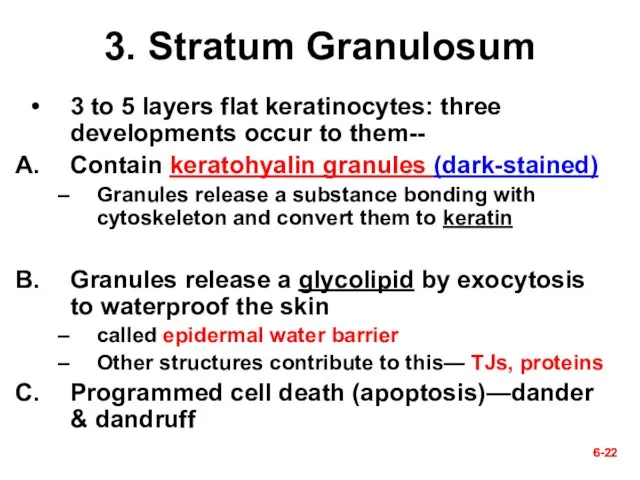
- 22. 6- 6- 3. Stratum Granulosum 3 to 5 layers flat keratinocytes: three developments occur to them--
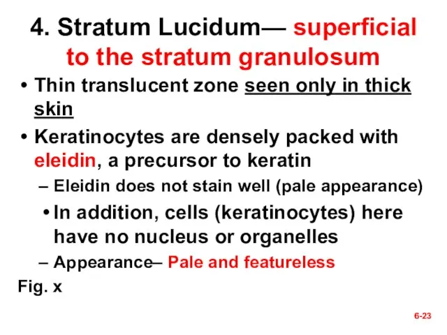
- 23. 6- 6- 4. Stratum Lucidum— superficial to the stratum granulosum Thin translucent zone seen only in
- 24. 6-
- 25. 6- 6- 5. Stratum Corneum Up to 30 layers of dead, scaly, keratinized cells surface cells
- 26. 6- 6- § Life History of Keratinocytes Produced by stem cells in stratum basale New cells
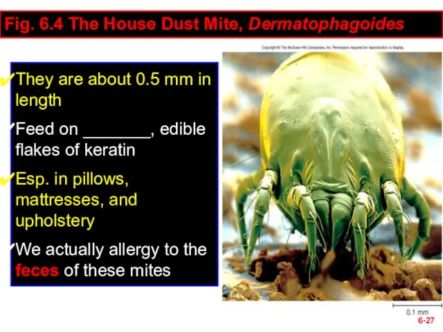
- 27. 6- Fig. 6.4 The House Dust Mite, Dermatophagoides They are about 0.5 mm in length Feed
- 28. 6- Questions (muddiest points)? Next section– III. Dermis & Hypodermis 6-
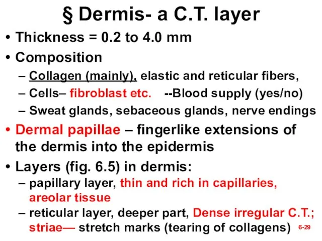
- 29. 6- 6- § Dermis- a C.T. layer Thickness = 0.2 to 4.0 mm Composition Collagen (mainly),
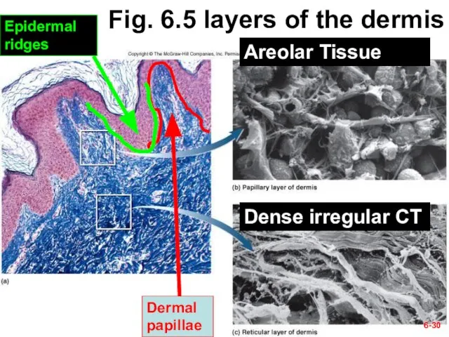
- 30. 6- Fig. 6.5 layers of the dermis Dermal papillae Epidermal ridges Areolar Tissue Dense irregular CT
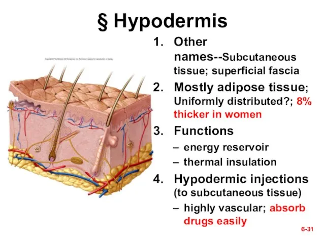
- 31. 6- 6- § Hypodermis Other names--Subcutaneous tissue; superficial fascia Mostly adipose tissue; Uniformly distributed?; 8% thicker
- 32. 6- Questions? Next section— IV. Cutaneous Glands 6-
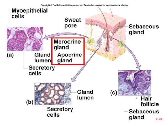
- 33. 6- Table 6.2— summary of cutaneous glands 1. Sweat glands 2. Oil glands 3. Ceruminous glands
- 34. § Cutaneous Glands 6-
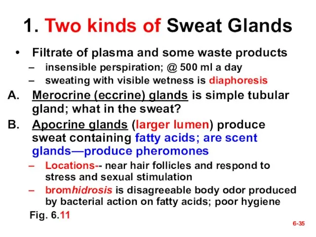
- 35. 6- 6- 1. Two kinds of Sweat Glands Filtrate of plasma and some waste products insensible
- 36. 6-
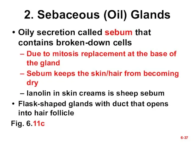
- 37. 6- 6- 2. Sebaceous (Oil) Glands Oily secretion called sebum that contains broken-down cells Due to
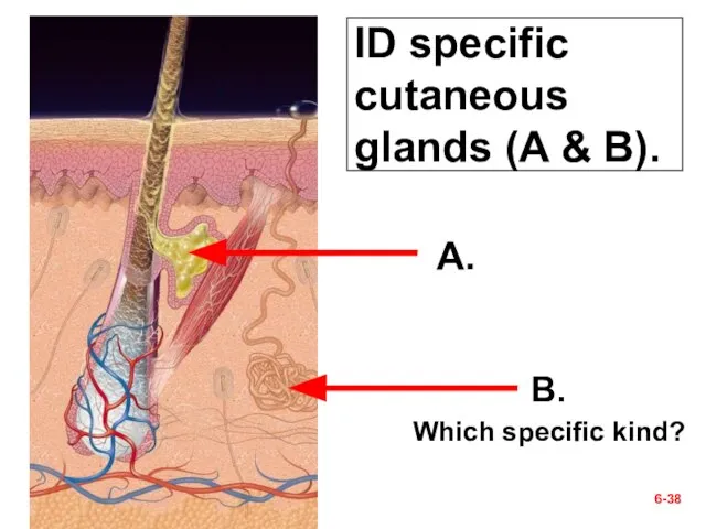
- 38. ID specific cutaneous glands (A & B). 6- A. B. Which specific kind?
- 39. 6- 6- 3. Ceruminous Glands Found only in external ear canal Their secretion combines with sebum
- 40. 6- Ceruminous glands—inappropriate interventions
- 41. 6- ? Cotton-tipped applicator (a no-no)
- 42. 6- ᵡ Ear Candling!?
- 43. 6- 6- 4. Mammary Glands Breasts of both sexes rarely contain mammary glands secondary sexual characteristic
- 44. Mammary Glands 6- Areola Nipple
- 45. Check Point Questions (True/False) The three layers of the skin are the epidermis, dermis, and hypodermis.
- 46. 6- Questions (muddiest points)? Next section— V. Skin Disorders 6-
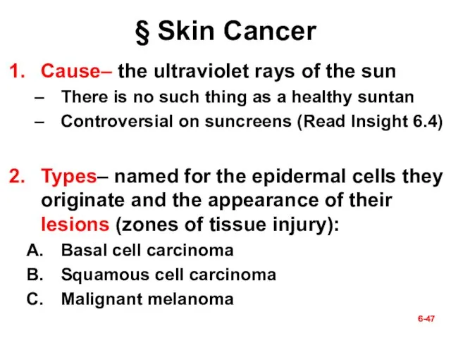
- 47. 6- 6- § Skin Cancer Cause– the ultraviolet rays of the sun There is no such
- 48. 6- 6- A. Basal cell carcinoma Most common type and the least dangerous one Origination- by
- 49. Fig. 6.12a A. Basal cell carcinoma 6-
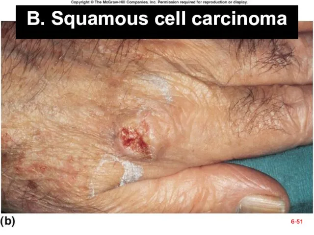
- 50. 6- 6- B. Squamous cell carcinoma Chance of recovery is good with early detection and surgical
- 51. B. Squamous cell carcinoma 6-
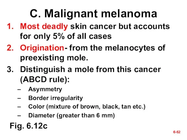
- 52. 6- 6- C. Malignant melanoma Most deadly skin cancer but accounts for only 5% of all
- 53. C. Malignant melanoma; which of the ABCD rules can you identify 6-
- 55. Скачать презентацию
6-
Ch. 6 Study Guide
Critically read Chapter 6–
pp. 187-194 before
6-
Ch. 6 Study Guide
Critically read Chapter 6–
pp. 187-194 before
6-
§ Quotable Quotes (Skin)
Some guys say beauty is only skin deep.
6-
§ Quotable Quotes (Skin)
Some guys say beauty is only skin deep.

6-
I. Introduction
6-
6-
I. Introduction
6-
6-
§ Overview (1)
Dermatology– scientific study and medical treatment of this system
Largest
6-
§ Overview (1)
Dermatology– scientific study and medical treatment of this system
Largest
6-
6-
§ Overview (2)
Thickness variable, based on thickness of Epidermis, two categories--
Thick
6-
6-
§ Overview (2)
Thickness variable, based on thickness of Epidermis, two categories--
Thick
6-
6-
6-
6-
§ Functions of the Skin
Resistance to trauma/infection
Why? (Fig. 5.28)
acid mantle (pH
6-
6-
§ Functions of the Skin
Resistance to trauma/infection
Why? (Fig. 5.28)
acid mantle (pH
6-
6-
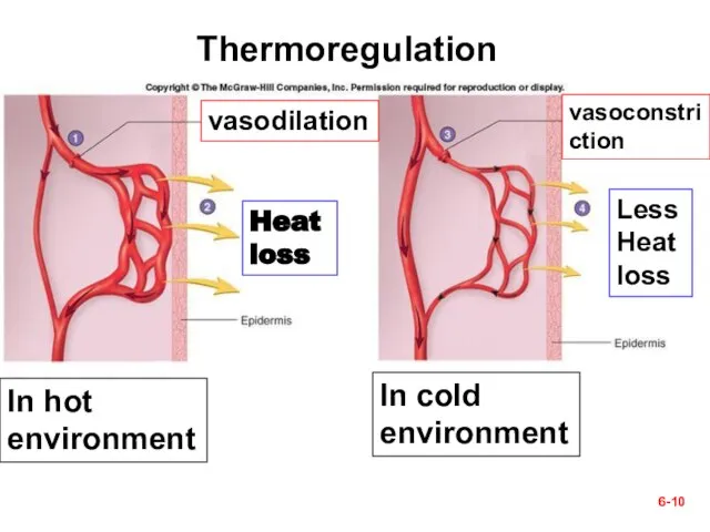
In hot environment
In cold environment
6-
vasodilation
vasoconstriction
Heat loss
Less
Heat loss
Thermoregulation
In hot environment
In cold environment
6-
vasodilation
vasoconstriction
Heat loss
Less
Heat loss
Thermoregulation
6-
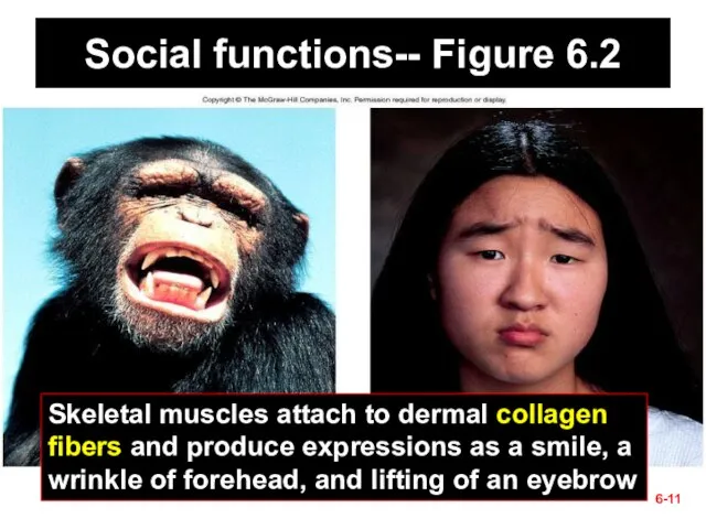
Social functions-- Figure 6.2
Skeletal muscles attach to dermal collagen fibers and
6-
Social functions-- Figure 6.2
Skeletal muscles attach to dermal collagen fibers and
6-
II. Epidermis
6-
6-
II. Epidermis
6-
6-
6-
§ Cells of the Epidermis (1)
Five types of cells--
Keratinocytes – most
6-
6-
§ Cells of the Epidermis (1)
Five types of cells--
Keratinocytes – most
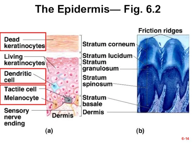
The Epidermis— Fig. 6.2
6-
The Epidermis— Fig. 6.2
6-
6-
6-
6-
6-
§ Cells of the Epidermis (2)
Location of the following types of
6-
6-
§ Cells of the Epidermis (2)
Location of the following types of
6-
Melanocyte
Keratinocytes
6-
Melanocyte
Keratinocytes
6-
§ Layers of the Epidermis—
Next five slides (1-5)
from deep to superficial
6-
§ Layers of the Epidermis— Next five slides (1-5) from deep to superficial
6-
6-
1. Stratum Basale (deepest layer)
Single layer cells on basement membrane (Fig.
6-
6-
1. Stratum Basale (deepest layer)
Single layer cells on basement membrane (Fig.
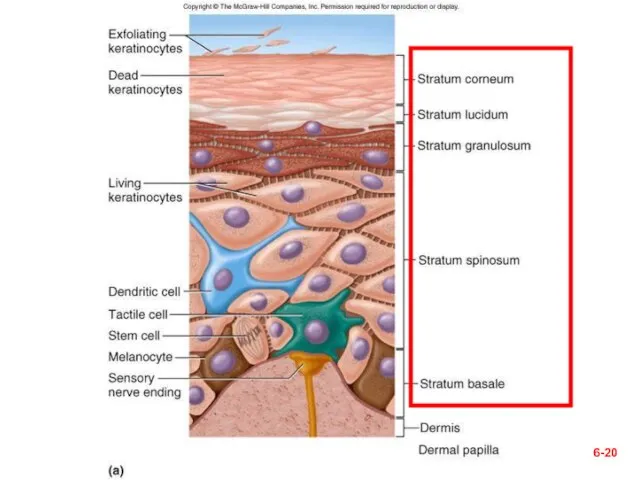
Figure 6.2a
6-
Figure 6.2a
6-
6-
6-
2. Stratum Spinosum– above stratum basale
Several layers of keratinocytes (flattened as
6-
6-
2. Stratum Spinosum– above stratum basale
Several layers of keratinocytes (flattened as
6-
6-
3. Stratum Granulosum
3 to 5 layers flat keratinocytes: three developments occur
6-
6-
3. Stratum Granulosum
3 to 5 layers flat keratinocytes: three developments occur
6-
6-
4. Stratum Lucidum— superficial to the stratum granulosum
Thin translucent zone seen
6-
6-
4. Stratum Lucidum— superficial to the stratum granulosum
Thin translucent zone seen
6-
6-
6-
6-
5. Stratum Corneum
Up to 30 layers of dead, scaly,
keratinized cells
surface cells
6-
6-
5. Stratum Corneum
Up to 30 layers of dead, scaly,
keratinized cells
surface cells
6-
6-
§ Life History of Keratinocytes
Produced by stem cells in stratum basale
New
6-
6-
§ Life History of Keratinocytes
Produced by stem cells in stratum basale
New
6-
Fig. 6.4 The House Dust Mite, Dermatophagoides
They are about 0.5 mm
6-
Fig. 6.4 The House Dust Mite, Dermatophagoides
They are about 0.5 mm
6-
Questions (muddiest points)?
Next section–
III. Dermis & Hypodermis
6-
6-
Questions (muddiest points)?
Next section–
III. Dermis & Hypodermis
6-
6-
6-
§ Dermis- a C.T. layer
Thickness = 0.2 to 4.0 mm
Composition
Collagen (mainly),
6-
6-
§ Dermis- a C.T. layer
Thickness = 0.2 to 4.0 mm
Composition
Collagen (mainly),
6-
Fig. 6.5 layers of the dermis
Dermal papillae
Epidermal ridges
Areolar Tissue
Dense irregular CT
6-
6-
Fig. 6.5 layers of the dermis
Dermal papillae
Epidermal ridges
Areolar Tissue
Dense irregular CT
6-
6-
6-
§ Hypodermis
Other names--Subcutaneous tissue; superficial fascia
Mostly adipose tissue; Uniformly distributed?; 8%
6-
6-
§ Hypodermis
Other names--Subcutaneous tissue; superficial fascia
Mostly adipose tissue; Uniformly distributed?; 8%
6-
Questions?
Next section—
IV. Cutaneous Glands
6-
6-
Questions?
Next section—
IV. Cutaneous Glands
6-
6-
Table 6.2— summary of cutaneous glands
1. Sweat glands
2. Oil glands
3. Ceruminous
6-
Table 6.2— summary of cutaneous glands 1. Sweat glands 2. Oil glands 3. Ceruminous
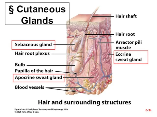
§ Cutaneous Glands
6-
§ Cutaneous Glands
6-
6-
6-
1. Two kinds of Sweat Glands
Filtrate of plasma and some waste
6-
6-
1. Two kinds of Sweat Glands
Filtrate of plasma and some waste
6-
6-
6-
6-
2. Sebaceous (Oil) Glands
Oily secretion called sebum that contains broken-down cells
Due
6-
6-
2. Sebaceous (Oil) Glands
Oily secretion called sebum that contains broken-down cells
Due
ID specific cutaneous glands (A & B).
6-
A.
B.
Which specific kind?
ID specific cutaneous glands (A & B).
6-
A.
B.
Which specific kind?
6-
6-
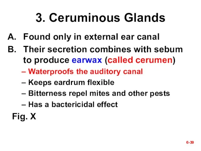
3. Ceruminous Glands
Found only in external ear canal
Their secretion combines with
6-
6-
3. Ceruminous Glands
Found only in external ear canal
Their secretion combines with
6-
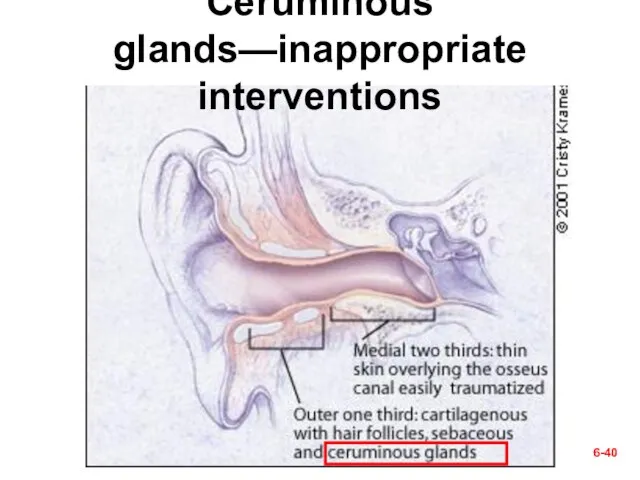
Ceruminous glands—inappropriate interventions
6-
Ceruminous glands—inappropriate interventions
6-
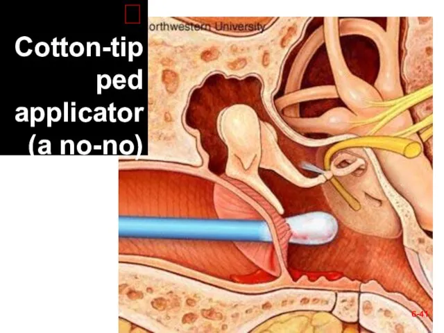
? Cotton-tipped applicator (a no-no)
6-
? Cotton-tipped applicator (a no-no)

6-
ᵡ Ear Candling!?
6-
ᵡ Ear Candling!?
6-
6-
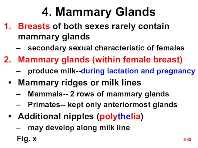
4. Mammary Glands
Breasts of both sexes rarely contain mammary glands
secondary sexual
6-
6-
4. Mammary Glands
Breasts of both sexes rarely contain mammary glands
secondary sexual
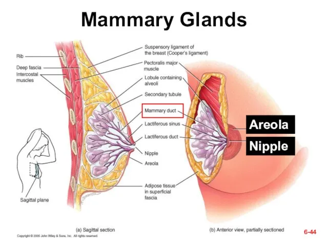
Mammary Glands
6-
Areola
Nipple
Mammary Glands
6-
Areola
Nipple
Check Point Questions
(True/False) The three layers of the skin are the
Check Point Questions
(True/False) The three layers of the skin are the
6-
Questions (muddiest points)?
Next section—
V. Skin Disorders
6-
6-
Questions (muddiest points)?
Next section—
V. Skin Disorders
6-
6-
6-
§ Skin Cancer
Cause– the ultraviolet rays of the sun
There is
6-
6-
§ Skin Cancer
Cause– the ultraviolet rays of the sun
There is
6-
6-
A. Basal cell carcinoma
Most common type and the least dangerous one
Origination-
6-
6-
A. Basal cell carcinoma
Most common type and the least dangerous one
Origination-
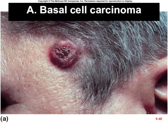
Fig. 6.12a
A. Basal cell carcinoma
6-
Fig. 6.12a
A. Basal cell carcinoma
6-
6-
6-
B. Squamous cell carcinoma
Chance of recovery is good with early detection
6-
6-
B. Squamous cell carcinoma
Chance of recovery is good with early detection
B. Squamous cell carcinoma
6-
B. Squamous cell carcinoma
6-
6-
6-
C. Malignant melanoma
Most deadly skin cancer but accounts for only 5%
6-
6-
C. Malignant melanoma
Most deadly skin cancer but accounts for only 5%
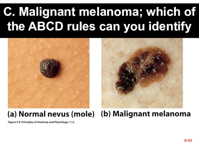
C. Malignant melanoma; which of the ABCD rules can you identify
6-
C. Malignant melanoma; which of the ABCD rules can you identify
6-
 Биология в современном естествознании
 Самые агрессивные породы собак Собаки бывают не только милыми, но и опасными. И отнюдь не только бойцовые породы, как может пока
 Презентация на тему "История микробиологии" - скачать презентации по Биологии
 Память - удивительное свойство человеческого разума
 Презентация на тему Класс двудольные семейство крестоцветные
  Мышцы, фасции и топография шеи
 Поясничное и крестцовое сплетения
 Водоросли в косметике: свойства и особенности применения
 Морфофункциональная характеристика аппарата движения. Исследование двигательных функций методом активных и пассивных движений
 Эмоции. Физиология сна
 Генотип – целостная система Сахаров Н.Н. – учитель биологии МОУ Нехаевская СОШ Волгоградская область
  Биология, как наука
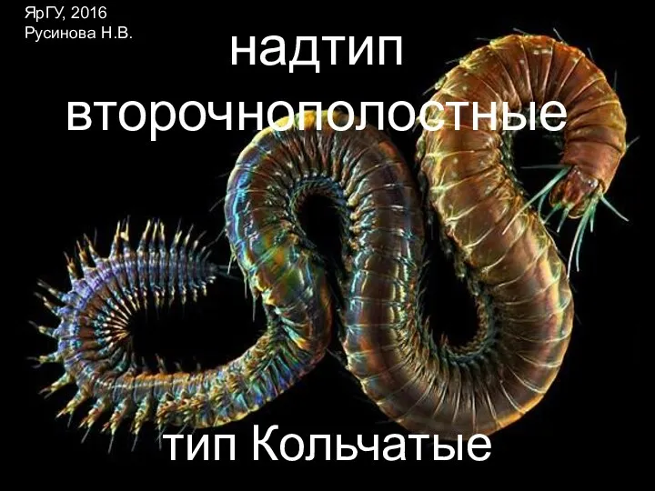 Надтип вторичноротые, тип кольчатые черви
 Презентация на тему "Класс Двудольные, Семейство Крестоцветные (Капустные)" - скачать бесплатно презентации по Биологии
 Антропогенез. Человеческая триада: высокоразвитый головной мозг, умелая рука, прямохождение
 Фрукты (для детей)
 Анализаторы. Сенсорная система
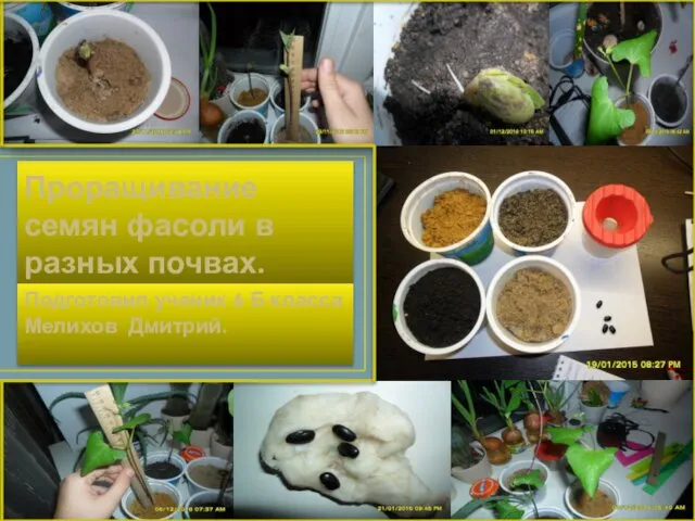 Проращивание семян фасоли в разных почвах
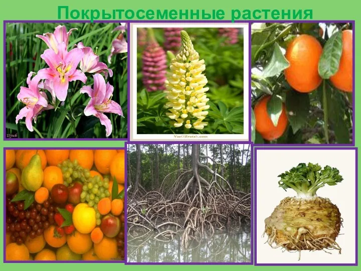 Покрытосеменные растения
 Особенности обмена аминокислот
 Биологические ритмы человека
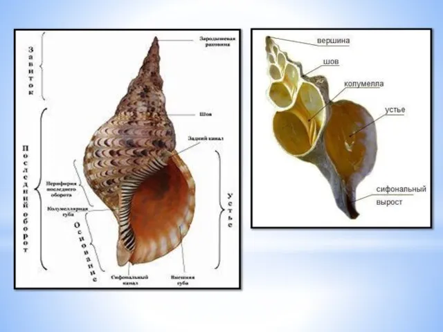 Класс Брюхоногие моллюски
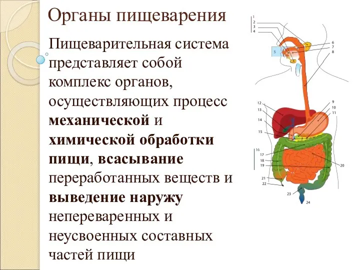 Органы пищеварения
 Механизмы вдоха и выдоха. Регуляция дыхания
 Отруйні та шкідливі рослини для сільськогосподарських тварин
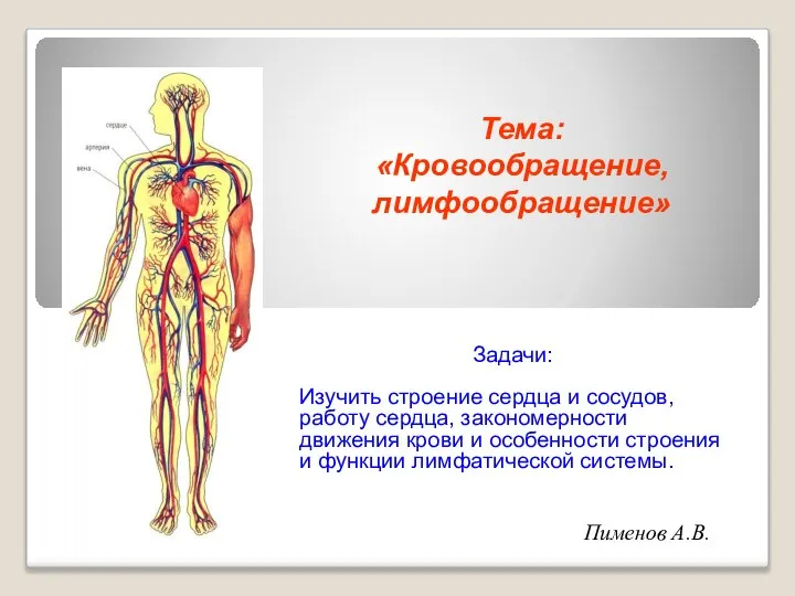 Кровообращение и лимфообращение
 Нервная система человека
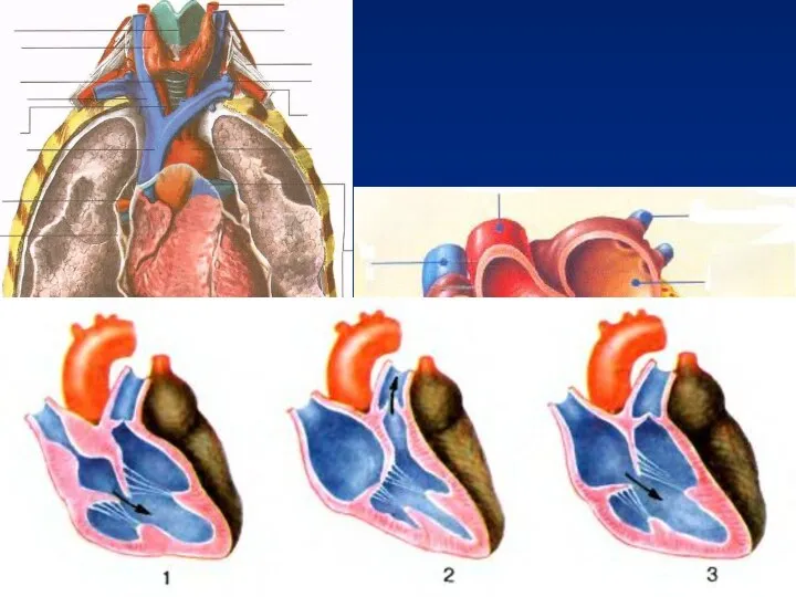 Движение крови по сосудам. Регуляция кровообращения