- Tsetse flies

Содержание
- 3. EXTERNAL ANATOMY OF GLOSSINA The word anatomy means the structure of the body, in this case,
- 4. EXTERNAL APPEARANCE The tsetse flies are nearly always some shade of brown or grey-brown; sometimes there
- 5. Compound eyes On the head is a pair of large compound eyes. Each of these eyes
- 6. The arista is a long thin structure like an eyelash, but has a row of branched
- 7. Mouthparts The mouthparts are very important to the life of the fly. They are long and
- 8. Labium The labium is the thickest of these very thin structures. At the free end it
- 9. THORAX The thorax in the mature fly is covered by stiff cuticle. The three pairs of
- 10. ABDOMEN In the resting fly, the abdomen is covered over by the wings. It has seven
- 11. DIGESTIVE SYSTEM Salivary glands and saliva The tsetse fly has two salivary glands. The main part
- 12. Labellar teeth There are hundreds of very small sharp labellar teeth at the end of the
- 13. NERVOUS SYSTEM The senses and the behaviour of the tsetse fly are coordinated by the nervous
- 14. REPRODUCTIVE SYSTEM Glossina reproduces by the female fly producing eggs which hatch into larvae inside the
- 15. LARVAL STAGES As with other flies, the larva in Glossinapasses through several stages or instars, as
- 16. First instar larva This is the stage that emerges from the egg. It breaks out of
- 18. Female tsetse mate just once. After 7 - 9 days she produces a single egg which
- 20. Скачать презентацию
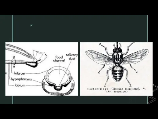
EXTERNAL ANATOMY OF GLOSSINA
The word anatomy means the structure of the
EXTERNAL ANATOMY OF GLOSSINA
The word anatomy means the structure of the
EXTERNAL APPEARANCE
The tsetse flies are nearly always some shade of brown
EXTERNAL APPEARANCE
The tsetse flies are nearly always some shade of brown
Compound eyes On the head is a pair of large compound eyes.
Compound eyes On the head is a pair of large compound eyes.
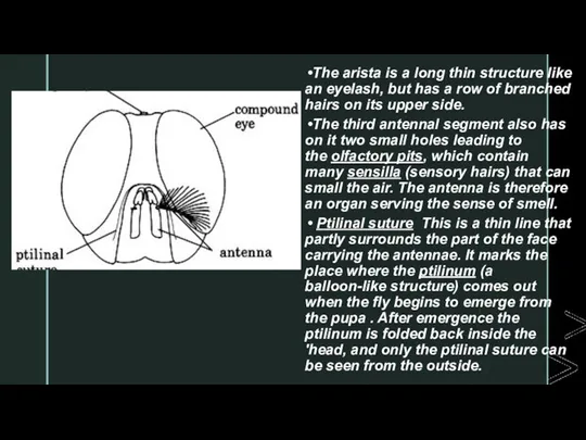
The arista is a long thin structure like an eyelash, but
The arista is a long thin structure like an eyelash, but
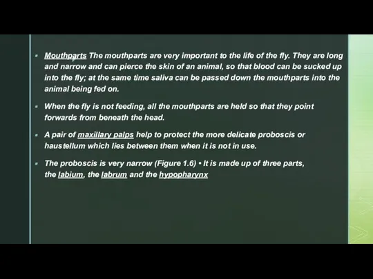
Mouthparts The mouthparts are very important to the life of the fly.
Mouthparts The mouthparts are very important to the life of the fly.
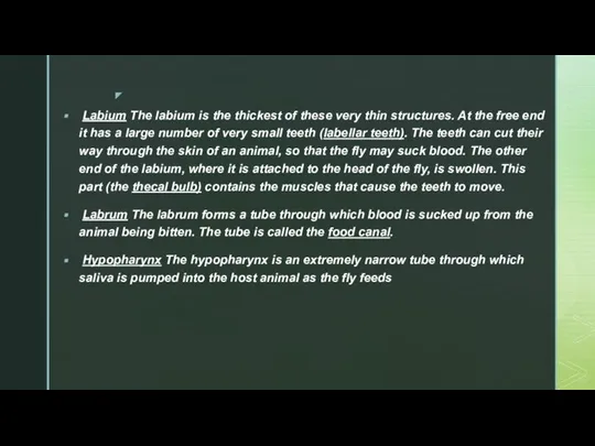
Labium The labium is the thickest of these very thin structures. At
Labium The labium is the thickest of these very thin structures. At
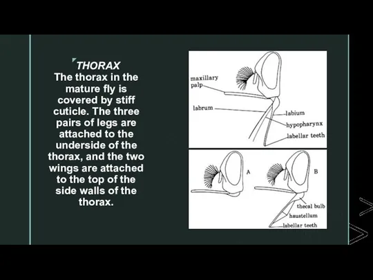
THORAX
The thorax in the mature fly is covered by stiff cuticle.
THORAX
The thorax in the mature fly is covered by stiff cuticle.
ABDOMEN
In the resting fly, the abdomen is covered over by the
ABDOMEN
In the resting fly, the abdomen is covered over by the
DIGESTIVE SYSTEM
Salivary glands and saliva The tsetse fly has two salivary glands. The
DIGESTIVE SYSTEM
Salivary glands and saliva The tsetse fly has two salivary glands. The
Labellar teeth
There are hundreds of very small sharp labellar teeth at the end of the
Labellar teeth
There are hundreds of very small sharp labellar teeth at the end of the
NERVOUS SYSTEM
The senses and the behaviour of the tsetse fly are
NERVOUS SYSTEM
The senses and the behaviour of the tsetse fly are
REPRODUCTIVE SYSTEM
Glossina reproduces by the female fly producing eggs which hatch into
REPRODUCTIVE SYSTEM
Glossina reproduces by the female fly producing eggs which hatch into
LARVAL STAGES
As with other flies, the larva in Glossinapasses through several stages
LARVAL STAGES
As with other flies, the larva in Glossinapasses through several stages
First instar larva This is the stage that emerges from the egg.
First instar larva This is the stage that emerges from the egg.

Female tsetse mate just once. After 7 - 9 days she
Female tsetse mate just once. After 7 - 9 days she
 Совместная жизнь видов в биогеоценозе
 РОЖДЕНИЕ БАБОЧКИ Составитель: Л.В.Ветчинкина, учитель начальных классов МКОУ Лаптево-Логовская СОШ
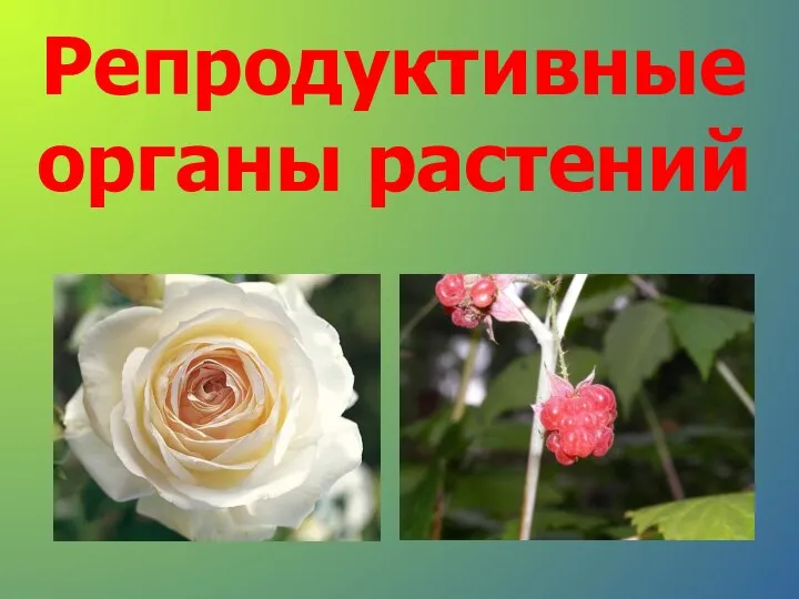 Презентация на тему Репродуктивные органы растений
  Анализаторы (сенсорные системы)
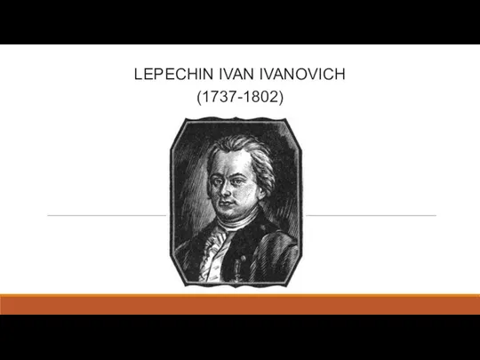 Lepechin Ivan Ivanovich
 СНК кафедры общей хирургии лечебного факультета РГМУ Современные аспекты панкреатодуоденальной резекции (ПДР) Докладчик студе
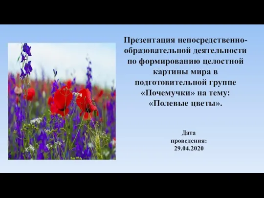 Полевые цветы
 Классная сказка Автор: Паплинская Владлена ученица 4 «А» класса школа № 5 г. Шелехов
 Организм человека
 Факты о тиграх
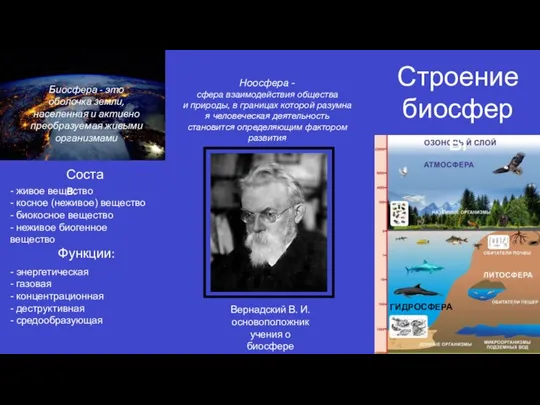 Биосфера. Строение биосферы
 Тихоходка
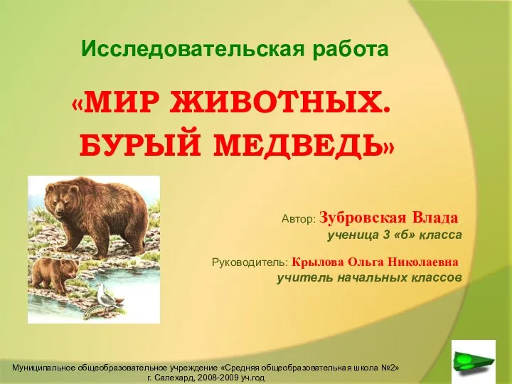 Презентация на тему "Мир животных. Бурый медведь" - скачать бесплатно презентации по Биологии
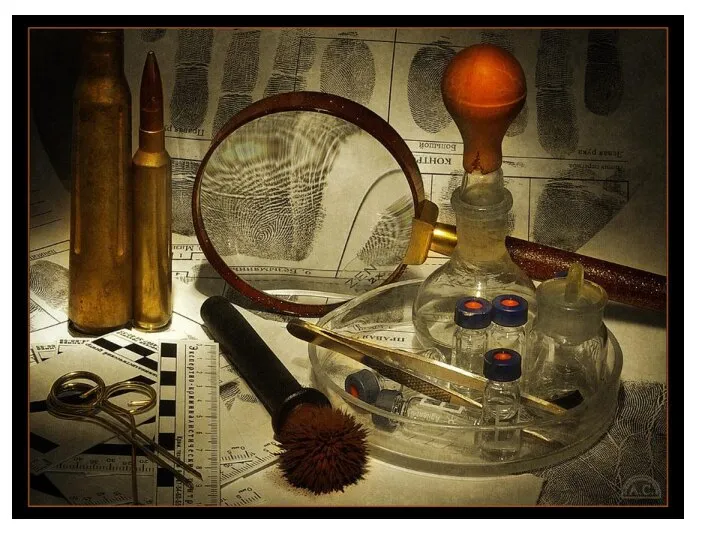 Строение кожи
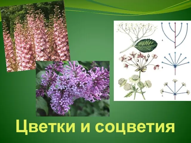 Презентация по биологии Цветки и соцветия
  Риби наших водойм!
  Методы исследования
 Общая характеристика царства Грибов
 Характеристика отряда Насекомоядные и отдельных его представителей
 Психофизиология. Сон
 Macaques. Introduction to macaques
 Основные направления эволюции
 Семейство пасленовые solanaceae. Подкласс lamiidae
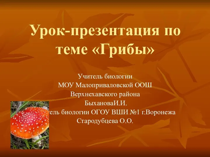 Урок-презентация по теме «Грибы» Учитель биологии МОУ Малоприваловской ООШ Верхнехавского района БыхановаИ.И. Учитель биолог
 Основные направления эволюционного процесса Зачетная работа по биологии учениц 11 класса «А» лицея им.В.Г. Сизова Заглада Юлии
 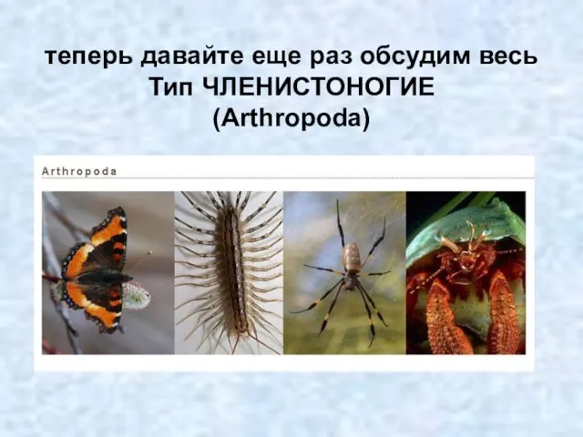 Тип членистоногие
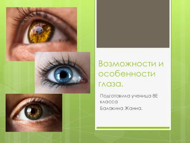 Возможности и особенности глаза
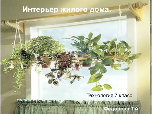 Роль комнатных растений. Уход за растениями