- Презентация к уроку английского языка "The Alimentary tract" - скачать

Содержание
- 2. The Alimentary Tract A long muscular tube with many sections and areas. Begins with the mouth
- 3. The Alimentary tract Mouth Pharynx Esophagus Stomach Small Intestine Large Intestine Anus
- 4. Accessory Parts Organs that are not in the Alimentary tract but helps in the digestion Teeth
- 5. Mouth Functions: Food enters in the mouth or oral cavity Tasting Mechanical breakdown of food Secretion
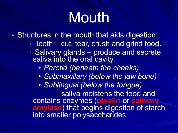
- 6. Mouth Structures in the mouth that aids digestion: Teeth – cut, tear, crush and grind food.
- 7. Mouth Tongue Mixes and rolls food into tiny mashed up bits (Bolus) Pushes the bolus toward
- 8. Anatomy of the Mouth and Throat
- 9. Human Deciduous and Permanent Teeth
- 10. Mechanism of Swallowing Swallowing is a coordinated activity of the tongue, soft palate, pharynx and esophagus.
- 12. Esophagus A straight muscular tube that is about 10 inches (25 cm) long which connects the
- 13. Peristalsis
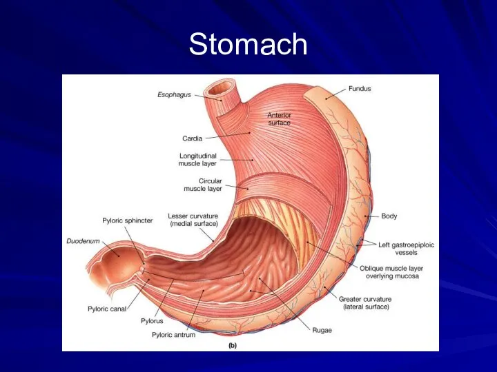
- 14. Stomach J-shaped muscular sac Has inner folds (rugae) that increases the surface area of the stomach.
- 15. Stomach
- 16. Stomach Pepsin – major enzyme; converts proteins into peptides in the presence of HCL. Mucus –
- 17. Movements in Stomach
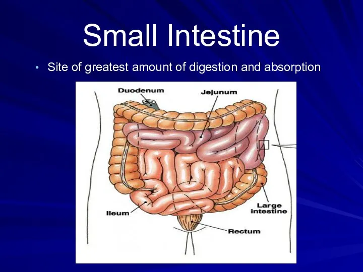
- 18. Small Intestine Long (20 ft), coiled tube beneath the stomach. Has three parts: Duodenum – upper
- 19. Small Intestine Site of greatest amount of digestion and absorption
- 20. Small Intestine Takes about 4 – 8 hrs to complete its journey. Mucosa (inner wall) –
- 21. Small Intestine Has folded inner walls covered with fingerlike projections (villi; sing. – villus) Each villus
- 22. Movement in small intestine: Mixing: Segmental contraction that occurs in small intestine Secretion: Lubricate, liquefy, digest
- 23. Large Intestine a.k.a. Colon larger diameter, but shorter (5 ft) Water is absorbed from the undigested
- 24. Large Intestine
- 25. Large Intestine Waste is pushed into the expanded portion (rectum) of the large intestine. Solid waste
- 26. Accessory Organs Produce or store enzymes that helps in digestion. Liver Largest gland of the body
- 27. Accessory Organs Gall bladder Stores bile in between meals Secretes bile to the duodenum through the
- 28. Accessory Organs Pancreas Produces a juice that contains enzymes (amylase and insulin) to break down carbohydrates,
- 30. Скачать презентацию
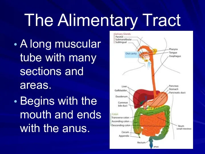
The Alimentary Tract
A long muscular tube with many sections and areas.
Begins
The Alimentary Tract
A long muscular tube with many sections and areas.
Begins
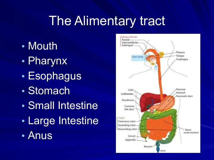
The Alimentary tract
Mouth
Pharynx
Esophagus
Stomach
Small Intestine
Large Intestine
Anus
The Alimentary tract
Mouth
Pharynx
Esophagus
Stomach
Small Intestine
Large Intestine
Anus
Accessory Parts
Organs that are not in the Alimentary tract but helps
Accessory Parts
Organs that are not in the Alimentary tract but helps
Mouth
Functions:
Food enters in the mouth or oral cavity
Tasting
Mechanical
Mouth
Functions:
Food enters in the mouth or oral cavity
Tasting
Mechanical
Mouth
Structures in the mouth that aids digestion:
Teeth – cut, tear,
Mouth
Structures in the mouth that aids digestion:
Teeth – cut, tear,
Mouth
Tongue
Mixes and rolls food into tiny mashed up bits
Mouth
Tongue
Mixes and rolls food into tiny mashed up bits
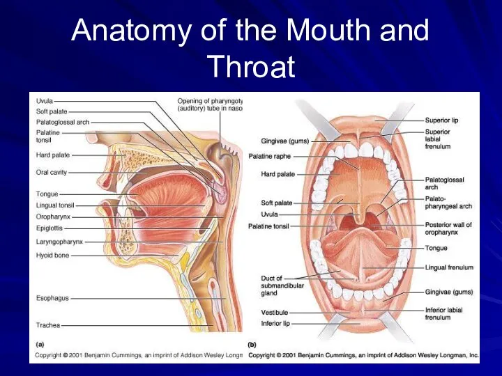
Anatomy of the Mouth and Throat
Anatomy of the Mouth and Throat
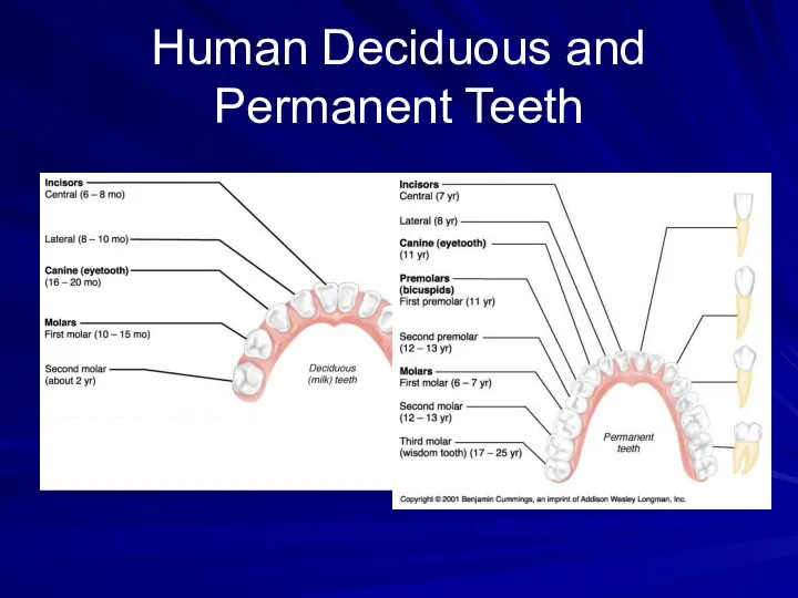
Human Deciduous and Permanent Teeth
Human Deciduous and Permanent Teeth

Mechanism of Swallowing
Swallowing is a coordinated activity of the tongue, soft
Mechanism of Swallowing
Swallowing is a coordinated activity of the tongue, soft


Esophagus
A straight muscular tube that is about 10 inches (25 cm)
Esophagus
A straight muscular tube that is about 10 inches (25 cm)
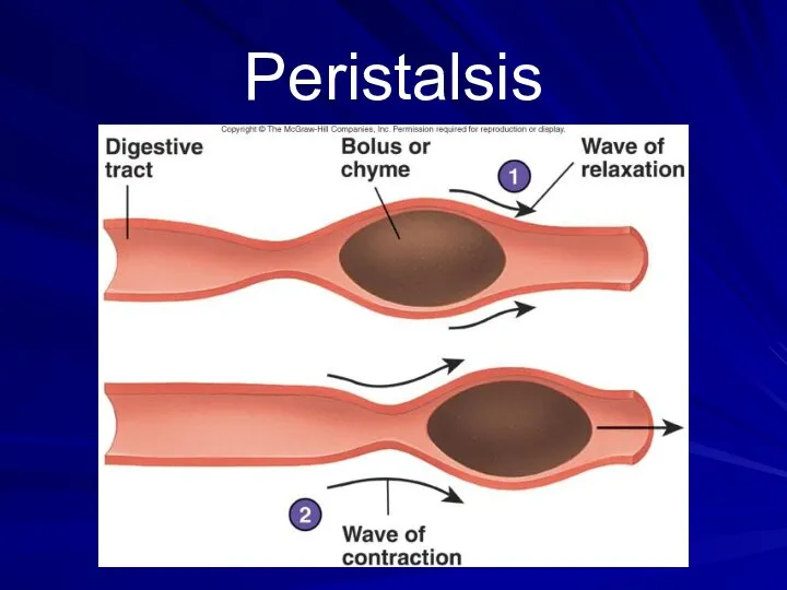
Peristalsis
Peristalsis

Stomach
J-shaped muscular sac
Has inner folds (rugae) that increases the surface area
Stomach
J-shaped muscular sac
Has inner folds (rugae) that increases the surface area
Stomach
Stomach
Stomach
Pepsin – major enzyme; converts proteins into peptides in the presence
Stomach
Pepsin – major enzyme; converts proteins into peptides in the presence

Movements in Stomach
Movements in Stomach

Small Intestine
Long (20 ft), coiled tube beneath the stomach.
Has three parts:
Duodenum
Small Intestine
Long (20 ft), coiled tube beneath the stomach.
Has three parts:
Duodenum
Small Intestine
Site of greatest amount of digestion and absorption
Small Intestine
Site of greatest amount of digestion and absorption

Small Intestine
Takes about 4 – 8 hrs to complete its journey.
Mucosa
Small Intestine
Takes about 4 – 8 hrs to complete its journey.
Mucosa

Small Intestine
Has folded inner walls covered with fingerlike projections (villi; sing.
Small Intestine
Has folded inner walls covered with fingerlike projections (villi; sing.
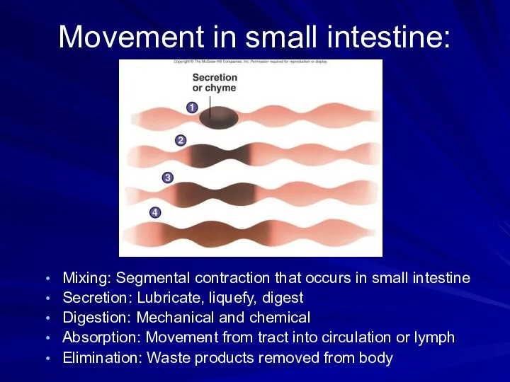
Movement in small intestine:
Mixing: Segmental contraction that occurs in small intestine
Secretion:
Movement in small intestine:
Mixing: Segmental contraction that occurs in small intestine
Secretion:
Large Intestine
a.k.a. Colon
larger diameter, but shorter (5 ft)
Water is absorbed from
Large Intestine
a.k.a. Colon
larger diameter, but shorter (5 ft)
Water is absorbed from

Large Intestine
Large Intestine

Large Intestine
Waste is pushed into the expanded portion (rectum) of the
Large Intestine
Waste is pushed into the expanded portion (rectum) of the
Accessory Organs
Produce or store enzymes that helps in digestion.
Liver
Largest
Accessory Organs
Produce or store enzymes that helps in digestion.
Liver
Largest

Accessory Organs
Gall bladder
Stores bile in between meals
Secretes bile
Accessory Organs
Gall bladder
Stores bile in between meals
Secretes bile
Accessory Organs
Pancreas
Produces a juice that contains enzymes (amylase and insulin)
Accessory Organs
Pancreas
Produces a juice that contains enzymes (amylase and insulin)
 Игра: страны
 Modal Verbs
 Flashcards. Things I can do
 Королевская семья Великобритании Отношения, семейное древо, тайны и многое другое...
  My perfect day
 Hotel “Golden Crown”
 Алгоритм перевода
 Snow White and the Seven Dwarfs
 Презентация к уроку английского языка "День святого патрика" - скачать бесплатно
 Present Perfect Tense
 Песни и стихи на уроках английского языка Гончаренко Анна 2338-2А, 303А
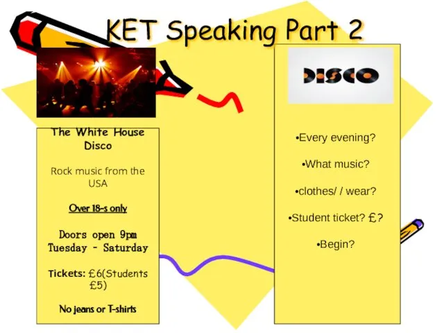 Ket. Speaking. (Рart 2)
 Have you re-gifted presents?
 Скачать Going to the Doctor
 My university KazNU
 Ecological problems. What you can do to protect our planet
 Borad or how the film can affect the flow of tourists
 Есть два друга - he и it. Кто из них и где стоит?!
 Benjamin Franklin 1706-1790
  Презентация по английскому языку Особенности фразеологии современного английского языка
 Australia
 Игрушки. Toys
 Cuba officially the Republic of Cuba
 Russian holidays It is a tradition to give greeting cards on these holidays. Here you can see some of them. Работу подготовила Игнатьева Е.Т. Учитель английского языка
 Make a sentence
 Скачать Exercise for eyes
 TRUE FRIENDS…
 Student recognition at BCC