- Muscle Tissue

Содержание
- 2. Muscle Tissue Muscles tissue distributed almost everywhere Some functions of muscular tissue Propels food we eat
- 3. Muscle Tissue Three types of muscle tissue: Skeletal muscle, cardiac muscle, smooth muscle Composes 40-50% of
- 4. 3 Types of Muscle Tissue Skeletal Smooth Cardiac
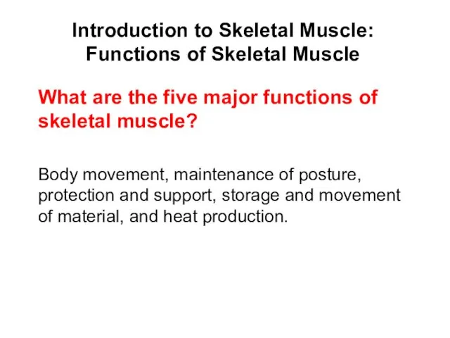
- 5. Introduction to Skeletal Muscle: Functions of Skeletal Muscle Functions of Skeletal Muscle Body movement Maintenance of
- 6. Introduction to Skeletal Muscle: Functions of Skeletal Muscle Body movement, maintenance of posture, protection and support,
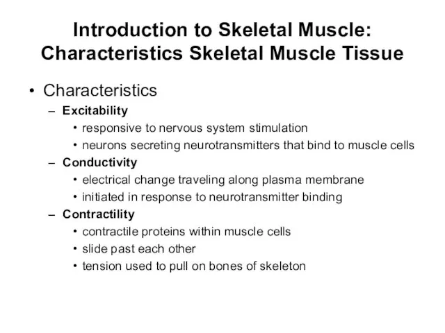
- 7. Introduction to Skeletal Muscle: Characteristics Skeletal Muscle Tissue Characteristics Excitability responsive to nervous system stimulation neurons
- 8. Introduction to Skeletal Muscle: Characteristics Skeletal Muscle Tissue Characteristics (continued) Elasticity due to protein fibers acting
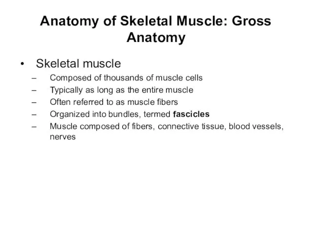
- 9. Anatomy of Skeletal Muscle: Gross Anatomy Skeletal muscle Composed of thousands of muscle cells Typically as
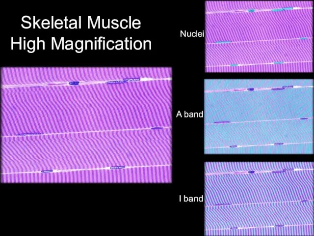
- 10. Skeletal Muscle High Magnification Nuclei A band I band
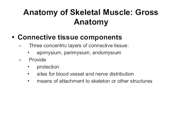
- 11. Anatomy of Skeletal Muscle: Gross Anatomy Connective tissue components Three concentric layers of connective tissue: epimysium,
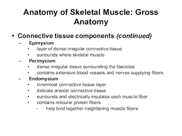
- 12. Anatomy of Skeletal Muscle: Gross Anatomy Connective tissue components (continued) Epimysium layer of dense irregular connective
- 13. Connective Tissue and Fascicles Epimysium Perimysium Fascicle Endomysium Epimysium + Perimysium + Endomysium = Tendon
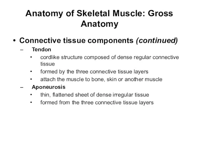
- 14. Anatomy of Skeletal Muscle: Gross Anatomy Connective tissue components (continued) Tendon cordlike structure composed of dense
- 15. Tendon and Aponeurosis of Palmaris Longus muscle
- 16. Anatomy of Skeletal Muscle: Gross Anatomy Connective tissue components (continued) Deep fascia additional sheet of dense
- 17. Superficial and Deep Fasciae 10- Superficial Deep
- 18. Anatomy of Skeletal Muscle: Gross Anatomy Connective tissue components (continued) Superficial fascia superficial to deep fascia
- 19. Skin
- 20. Superficial Fascia
- 21. Deep Fascia
- 22. Superficial Muscles
- 23. Deeper Muscles
- 24. Even Deeper Muscles
- 25. Yet Even Deeper Muscles
- 26. Soft Tissue and Bone
- 27. Bone
- 28. Anatomy of Skeletal Muscle: Gross Anatomy Blood vessels and nerves Skeletal muscles vascularized by extensive blood
- 29. Structural Organization of Skeletal Muscle (Figure 10.1) Copyright © The McGraw-Hill Companies, Inc. Permission required for
- 30. Anatomy of Skeletal Muscle: Gross Anatomy The endomysium is the layer of connective tissue surrounding the
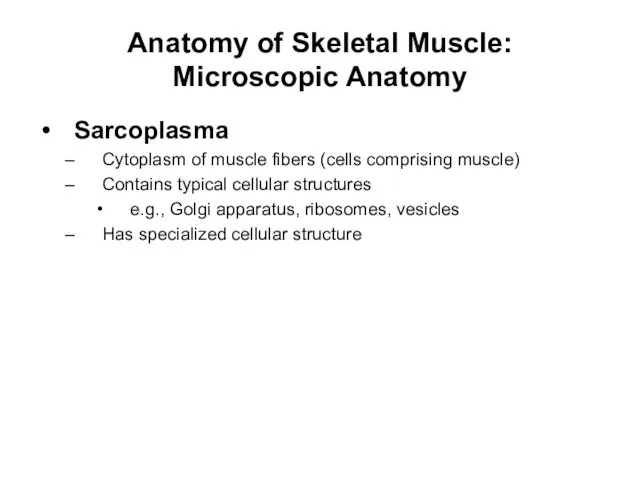
- 31. Anatomy of Skeletal Muscle: Microscopic Anatomy Sarcoplasma Cytoplasm of muscle fibers (cells comprising muscle) Contains typical
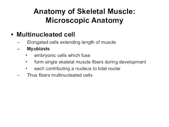
- 32. Anatomy of Skeletal Muscle: Microscopic Anatomy Multinucleated cell Elongated cells extending length of muscle Myoblasts embryonic
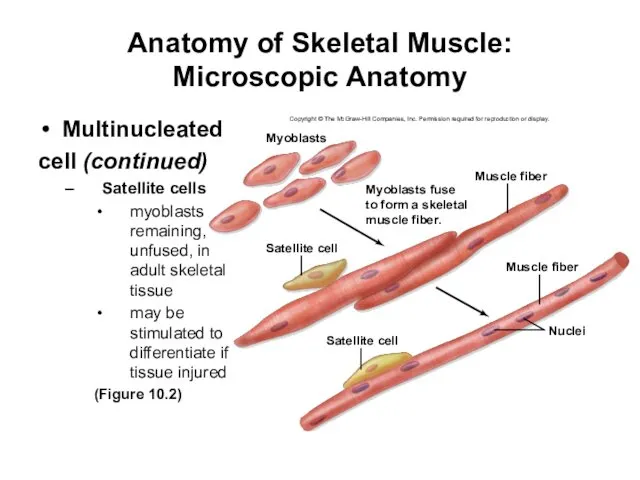
- 33. Anatomy of Skeletal Muscle: Microscopic Anatomy Multinucleated cell (continued) Satellite cells myoblasts remaining, unfused, in adult
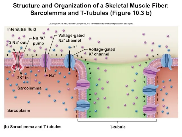
- 34. Anatomy of Skeletal Muscle: Microscopic Anatomy Sarcolemma and T-tubules Plasma membrane of a skeletal muscle fiber
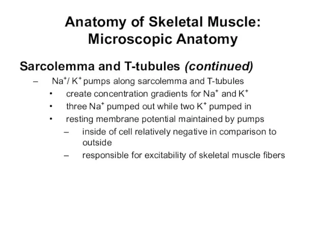
- 35. Anatomy of Skeletal Muscle: Microscopic Anatomy Sarcolemma and T-tubules (continued) Na+/ K+ pumps along sarcolemma and
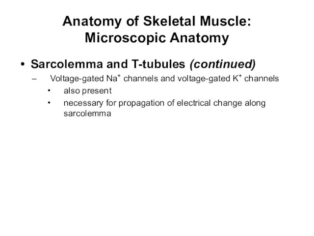
- 36. Anatomy of Skeletal Muscle: Microscopic Anatomy Sarcolemma and T-tubules (continued) Voltage-gated Na+ channels and voltage-gated K+
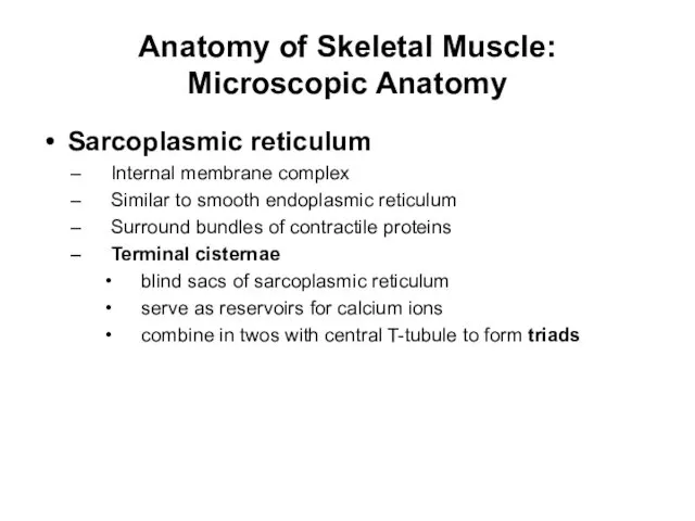
- 37. Anatomy of Skeletal Muscle: Microscopic Anatomy Sarcoplasmic reticulum Internal membrane complex Similar to smooth endoplasmic reticulum
- 38. Structure and Organization of a Skeletal Muscle Fiber: Sarcolemma and T-Tubules (Figure 10.3 b) Copyright ©
- 39. Sarcoplasmic reticulum (continued) Ca2+ pumps embedded in sarcoplasmic reticulum move Ca2+ into sarcoplasmic reticulum stored bound
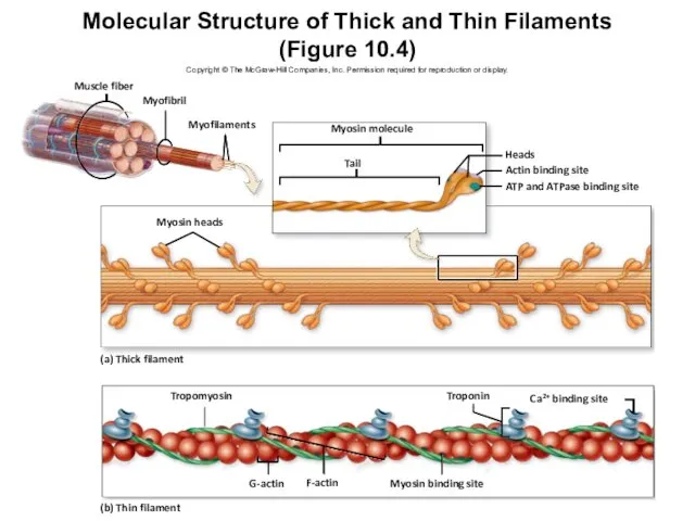
- 40. Anatomy of Skeletal Muscle: Microscopic Anatomy Muscle fibers and myofibrils Myofibrils long cylindrical structures extend length
- 41. Structure and Organization of a Skeletal Muscle Fiber (Figure 10.3 a) Copyright © The McGraw-Hill Companies,
- 42. Anatomy of Skeletal Muscle: Microscopic Anatomy Muscle fibers and myofibrils (continued) Thick filaments Assembled from bundles
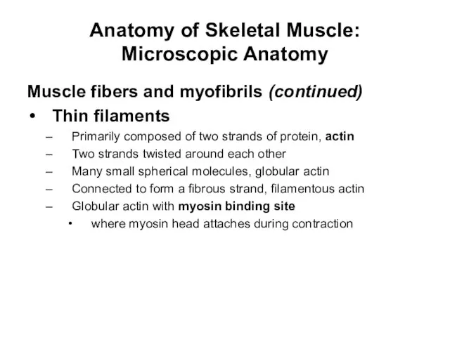
- 43. Anatomy of Skeletal Muscle: Microscopic Anatomy Muscle fibers and myofibrils (continued) Thin filaments Primarily composed of
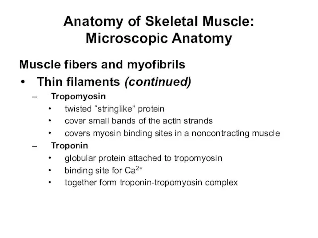
- 44. Anatomy of Skeletal Muscle: Microscopic Anatomy Muscle fibers and myofibrils Thin filaments (continued) Tropomyosin twisted “stringlike”
- 45. Molecular Structure of Thick and Thin Filaments (Figure 10.4) Copyright © The McGraw-Hill Companies, Inc. Permission
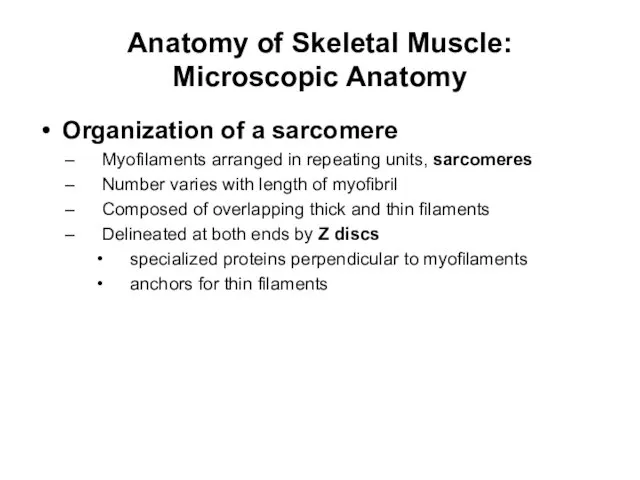
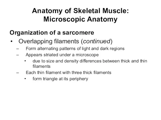
- 46. Anatomy of Skeletal Muscle: Microscopic Anatomy Organization of a sarcomere Myofilaments arranged in repeating units, sarcomeres
- 47. Anatomy of Skeletal Muscle: Microscopic Anatomy Organization of a sarcomere Overlapping filaments (continued) Form alternating patterns
- 48. Skeletal Muscle (striations) Skeletal muscle fiber A band I band Nuclei
- 49. Sarcomere A band H band I band M line Myofibril Sarcomere Z disc
- 50. Structure of a Sarcomere (Figure 10.5 a) Copyright © The McGraw-Hill Companies, Inc. Permission required for
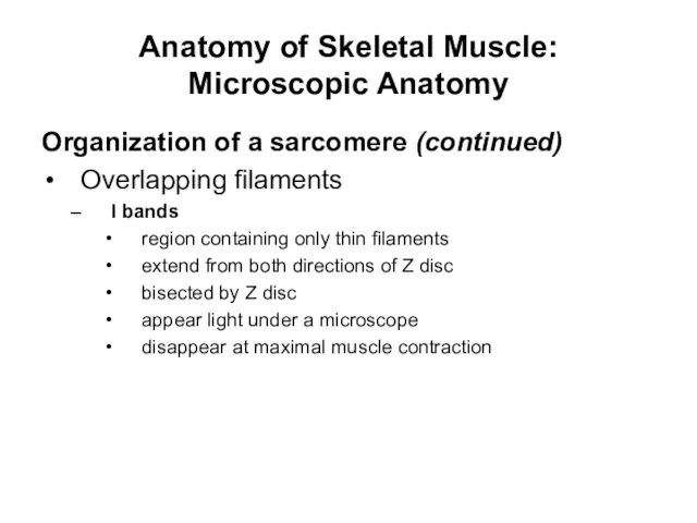
- 51. Anatomy of Skeletal Muscle: Microscopic Anatomy Organization of a sarcomere (continued) Overlapping filaments I bands region
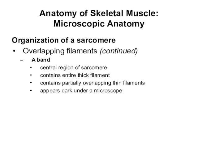
- 52. Anatomy of Skeletal Muscle: Microscopic Anatomy Organization of a sarcomere Overlapping filaments (continued) A band central
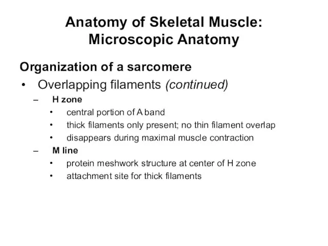
- 53. Anatomy of Skeletal Muscle: Microscopic Anatomy Organization of a sarcomere Overlapping filaments (continued) H zone central
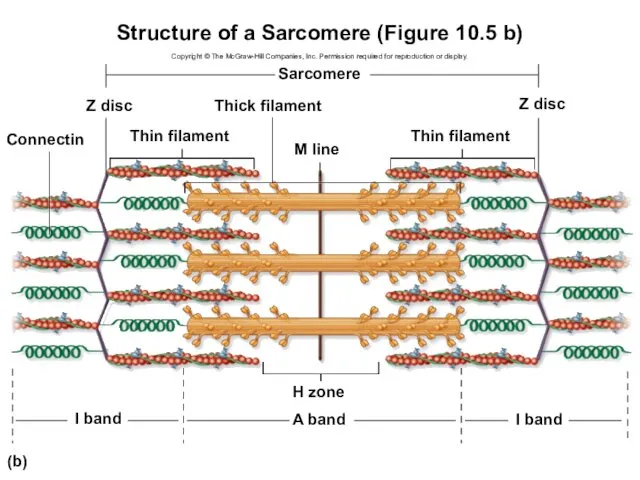
- 54. Structure of a Sarcomere (Figure 10.5 b) Copyright © The McGraw-Hill Companies, Inc. Permission required for
- 55. Structure of a Sarcomere (Figure 10.5 c) Copyright © The McGraw-Hill Companies, Inc. Permission required for
- 56. Anatomy of Skeletal Muscle: Microscopic Anatomy Organization of a sarcomere Other structural and functional proteins Connectin
- 57. Anatomy of Skeletal Muscle: Microscopic Anatomy Organization of a sarcomere Other structural and functional proteins (continued)
- 58. Anatomy of Skeletal Muscle: Microscopic Anatomy Mitochondria and other structures associated with energy production Muscle with
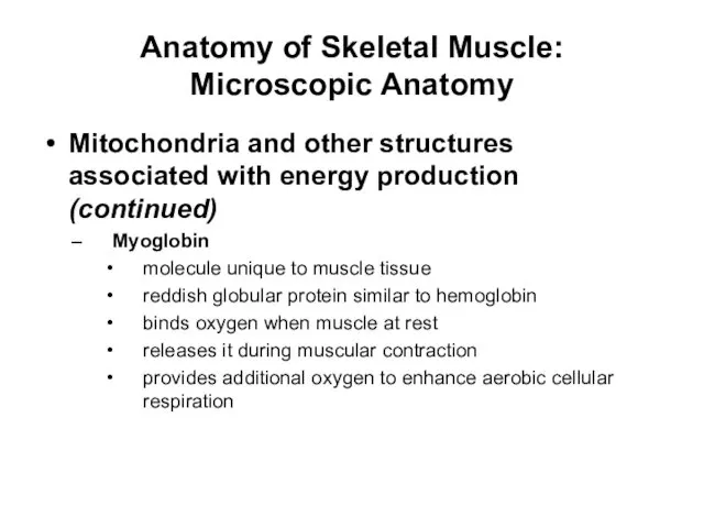
- 59. Anatomy of Skeletal Muscle: Microscopic Anatomy Mitochondria and other structures associated with energy production (continued) Myoglobin
- 60. Anatomy of Skeletal Muscle: Microscopic Anatomy Thick filaments are composed of myosin protein. What are the
- 61. Anatomy of Skeletal Muscle: Microscopic Anatomy H zone In which band are there thick filaments only,
- 62. Anatomy of Skeletal Muscle: Innervation of Skeletal Muscle Fibers Motor unit Motor neuron nerve cells transmit
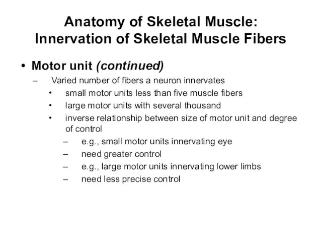
- 63. Anatomy of Skeletal Muscle: Innervation of Skeletal Muscle Fibers Motor unit (continued) Varied number of fibers
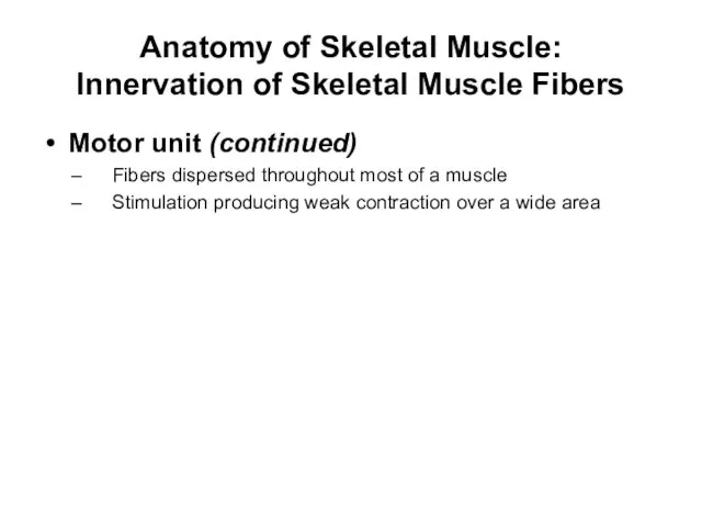
- 64. Anatomy of Skeletal Muscle: Innervation of Skeletal Muscle Fibers Motor unit (continued) Fibers dispersed throughout most
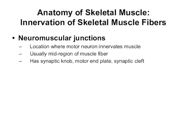
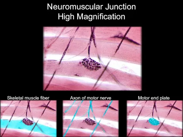
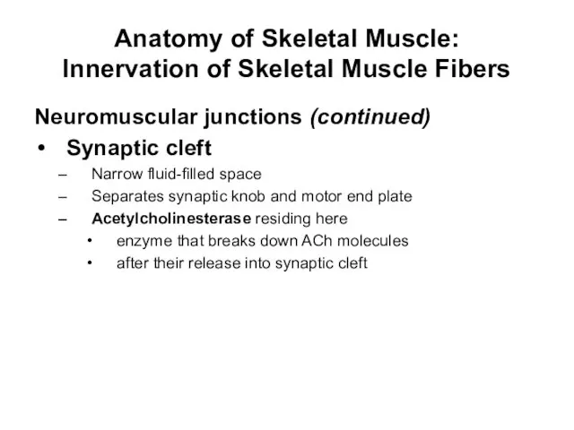
- 65. Anatomy of Skeletal Muscle: Innervation of Skeletal Muscle Fibers Neuromuscular junctions Location where motor neuron innervates
- 66. Neuromuscular Junction High Magnification Skeletal muscle fiber Axon of motor nerve Motor end plate
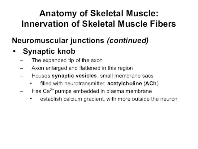
- 67. Anatomy of Skeletal Muscle: Innervation of Skeletal Muscle Fibers Neuromuscular junctions (continued) Synaptic knob The expanded
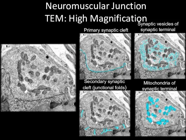
- 68. Neuromuscular Junction TEM: High Magnification Primary synaptic cleft Synaptic vesicles of synaptic terminal Secondary synaptic cleft
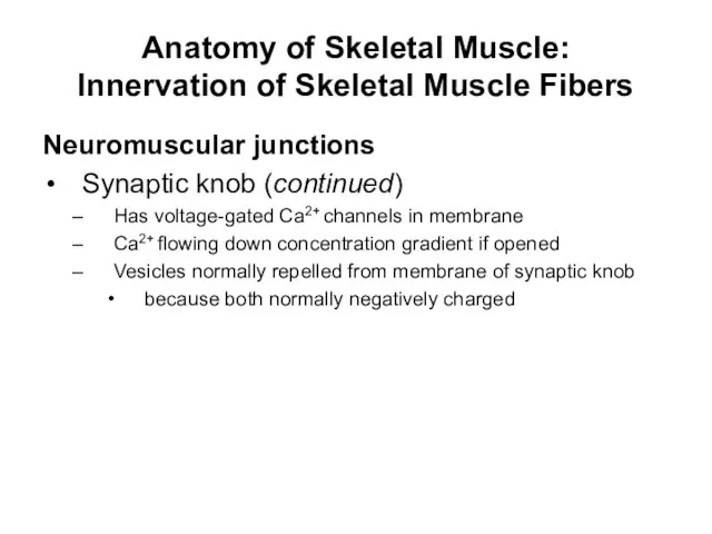
- 69. Anatomy of Skeletal Muscle: Innervation of Skeletal Muscle Fibers Neuromuscular junctions Synaptic knob (continued) Has voltage-gated
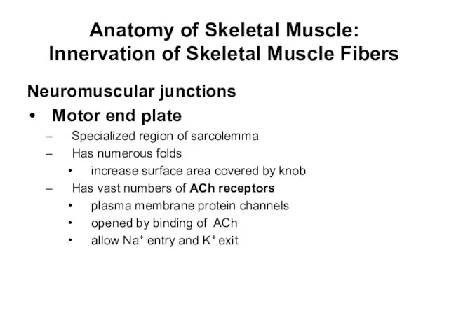
- 70. Anatomy of Skeletal Muscle: Innervation of Skeletal Muscle Fibers Neuromuscular junctions Motor end plate Specialized region
- 71. Anatomy of Skeletal Muscle: Innervation of Skeletal Muscle Fibers Neuromuscular junctions (continued) Synaptic cleft Narrow fluid-filled
- 72. Structure and Organization of a Neuromuscular Junction (Figure 10.7a) Neuromuscular junction Nerve signal Synaptic cleft Endomysium
- 73. Structure and Organization of a Neuromuscular Junction (Figure 10.7b) K+ (b) Interstitial fluid Ca2+ pump Voltage-gated
- 74. Anatomy of Skeletal Muscle: Innervation of Skeletal Muscle Fibers A motor unit is a single motor
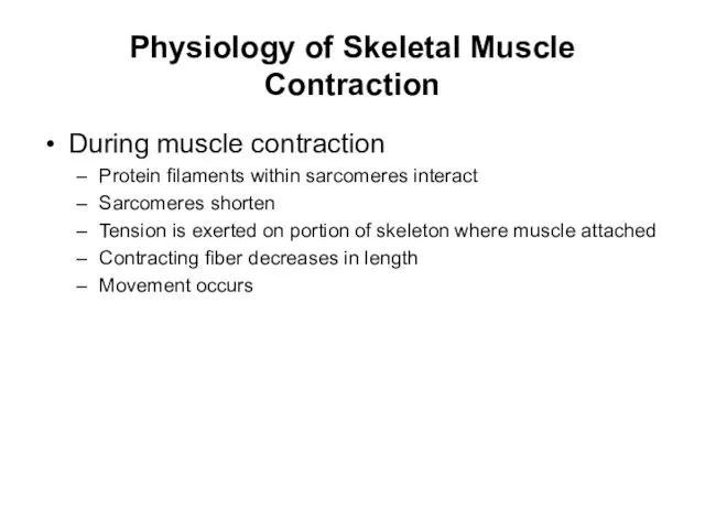
- 75. Physiology of Skeletal Muscle Contraction During muscle contraction Protein filaments within sarcomeres interact Sarcomeres shorten Tension
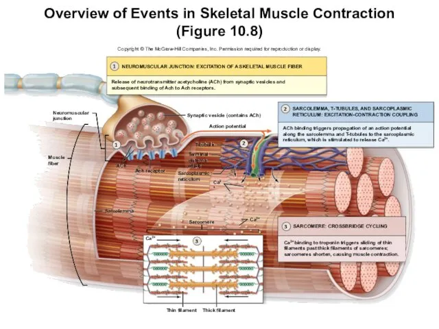
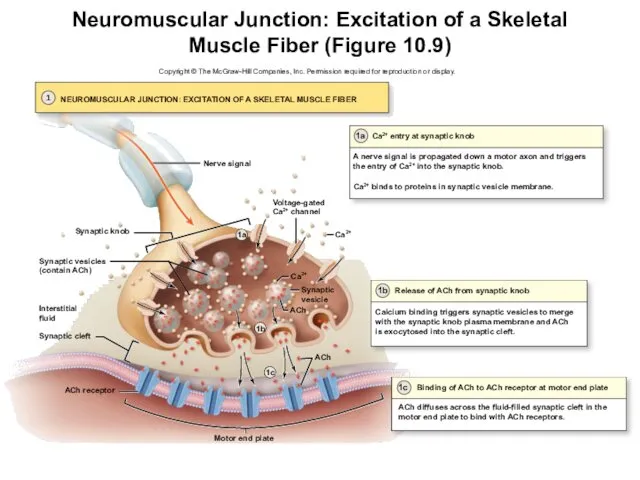
- 76. Overview of Events in Skeletal Muscle Contraction (Figure 10.8) 1 2 3 NEUROMUSCULAR JUNCTION: EXCITATION OF
- 77. Skeletal Muscle Contraction—Neuromuscular Junction: Excitation of a Skeletal Muscle Fiber First physiological event Muscular fiber excitation
- 78. Skeletal Muscle Contraction—Neuromuscular Junction: Excitation of a Skeletal Muscle Fiber Calcium entry at synaptic knob Nerve
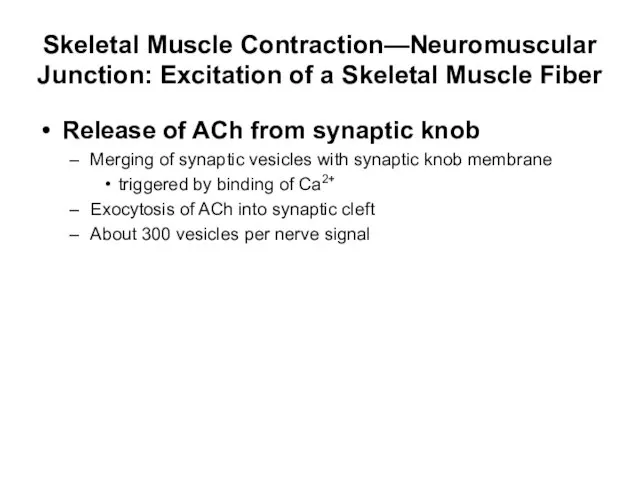
- 79. Skeletal Muscle Contraction—Neuromuscular Junction: Excitation of a Skeletal Muscle Fiber Release of ACh from synaptic knob
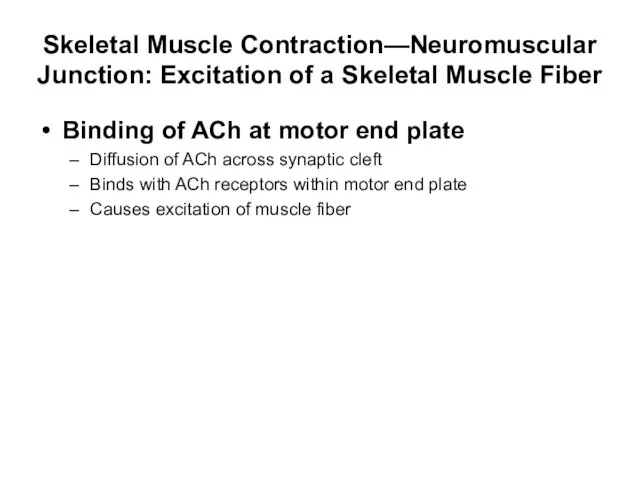
- 80. Skeletal Muscle Contraction—Neuromuscular Junction: Excitation of a Skeletal Muscle Fiber Binding of ACh at motor end
- 81. Neuromuscular Junction: Excitation of a Skeletal Muscle Fiber (Figure 10.9) Motor end plate 1 NEUROMUSCULAR JUNCTION:
- 82. Skeletal Muscle Contraction—Neuromuscular Junction: Excitation of a Skeletal Muscle Fiber Nerve signal triggers the entry of
- 83. Skeletal Muscle Contraction—Neuromuscular Junction: Excitation of a Skeletal Muscle Fiber Clinical View: Myasthenia Gravis Autoimmune disease,
- 84. Skeletal Muscle Contraction—Sarcolemma, T-Tubules, Sarcoplasmic Reticulum: Excitation-Contraction Coupling Second physiological event Excitation-contraction coupling Links skeletal muscle
- 85. Skeletal Muscle Contraction—Sarcolemma, T-Tubules, Sarcoplasmic Reticulum: Excitation-Contraction Coupling Development of an end-plate potential at the motor
- 86. Skeletal Muscle Contraction—Sarcolemma, T-Tubules, Sarcoplasmic Reticulum: Excitation-Contraction Coupling Development of an end-plate potential at the motor
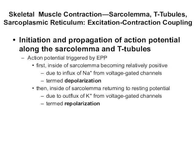
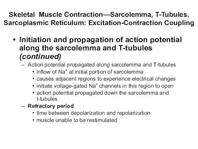
- 87. Skeletal Muscle Contraction—Sarcolemma, T-Tubules, Sarcoplasmic Reticulum: Excitation-Contraction Coupling Initiation and propagation of action potential along the
- 88. Skeletal Muscle Contraction—Sarcolemma, T-Tubules, Sarcoplasmic Reticulum: Excitation-Contraction Coupling Initiation and propagation of action potential along the
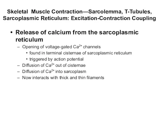
- 89. Skeletal Muscle Contraction—Sarcolemma, T-Tubules, Sarcoplasmic Reticulum: Excitation-Contraction Coupling Release of calcium from the sarcoplasmic reticulum Opening
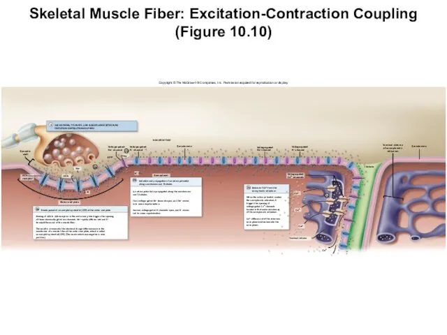
- 90. Skeletal Muscle Fiber: Excitation-Contraction Coupling (Figure 10.10) Copyright © The McGraw-Hill Companies, Inc. Permission required for
- 92. Скачать презентацию
Muscle Tissue
Muscles tissue distributed almost everywhere
Some functions of muscular tissue
Propels food
Muscle Tissue
Muscles tissue distributed almost everywhere
Some functions of muscular tissue
Propels food
Muscle Tissue
Three types of muscle tissue:
Skeletal muscle, cardiac muscle, smooth muscle
Composes
Muscle Tissue
Three types of muscle tissue:
Skeletal muscle, cardiac muscle, smooth muscle
Composes
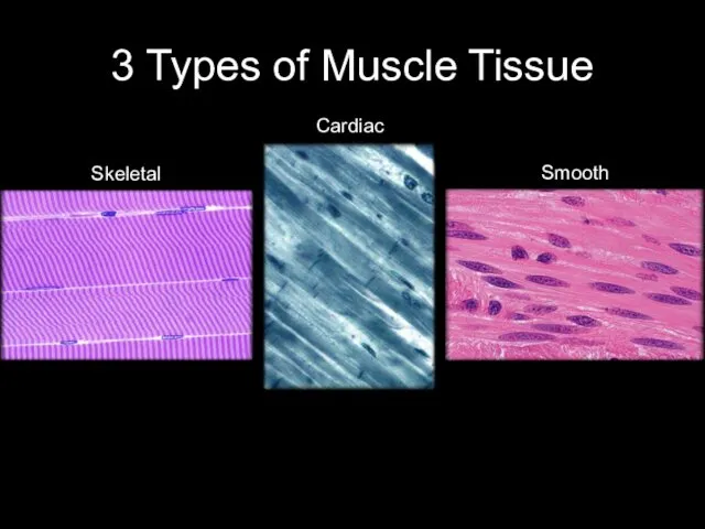
3 Types of Muscle Tissue
Skeletal
Smooth
Cardiac
3 Types of Muscle Tissue
Skeletal
Smooth
Cardiac
Introduction to Skeletal Muscle:
Functions of Skeletal Muscle
Functions of Skeletal Muscle
Introduction to Skeletal Muscle:
Functions of Skeletal Muscle
Functions of Skeletal Muscle
Introduction to Skeletal Muscle:
Functions of Skeletal Muscle
Body movement, maintenance of
Introduction to Skeletal Muscle:
Functions of Skeletal Muscle
Body movement, maintenance of
Introduction to Skeletal Muscle:
Characteristics Skeletal Muscle Tissue
Characteristics
Excitability
responsive to nervous system
Introduction to Skeletal Muscle:
Characteristics Skeletal Muscle Tissue
Characteristics
Excitability
responsive to nervous system

Introduction to Skeletal Muscle:
Characteristics Skeletal Muscle Tissue
Characteristics (continued)
Elasticity
due to protein
Introduction to Skeletal Muscle:
Characteristics Skeletal Muscle Tissue
Characteristics (continued)
Elasticity
due to protein
Anatomy of Skeletal Muscle: Gross Anatomy
Skeletal muscle
Composed of thousands of muscle
Anatomy of Skeletal Muscle: Gross Anatomy
Skeletal muscle
Composed of thousands of muscle
Skeletal Muscle
High Magnification
Nuclei
A band
I band
Skeletal Muscle
High Magnification
Nuclei
A band
I band
Anatomy of Skeletal Muscle: Gross Anatomy
Connective tissue components
Three concentric layers of
Anatomy of Skeletal Muscle: Gross Anatomy
Connective tissue components
Three concentric layers of
Anatomy of Skeletal Muscle: Gross Anatomy
Connective tissue components (continued)
Epimysium
layer of dense
Anatomy of Skeletal Muscle: Gross Anatomy
Connective tissue components (continued)
Epimysium
layer of dense
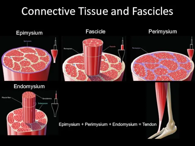
Connective Tissue and Fascicles
Epimysium
Perimysium
Fascicle
Endomysium
Epimysium + Perimysium + Endomysium = Tendon
Connective Tissue and Fascicles
Epimysium
Perimysium
Fascicle
Endomysium
Epimysium + Perimysium + Endomysium = Tendon
Anatomy of Skeletal Muscle: Gross Anatomy
Connective tissue components (continued)
Tendon
cordlike structure composed
Anatomy of Skeletal Muscle: Gross Anatomy
Connective tissue components (continued)
Tendon
cordlike structure composed
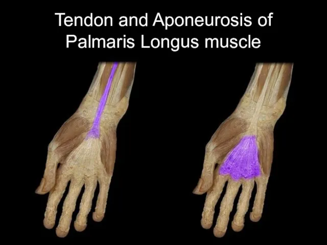
Tendon and Aponeurosis of Palmaris Longus muscle
Tendon and Aponeurosis of Palmaris Longus muscle
Anatomy of Skeletal Muscle: Gross Anatomy
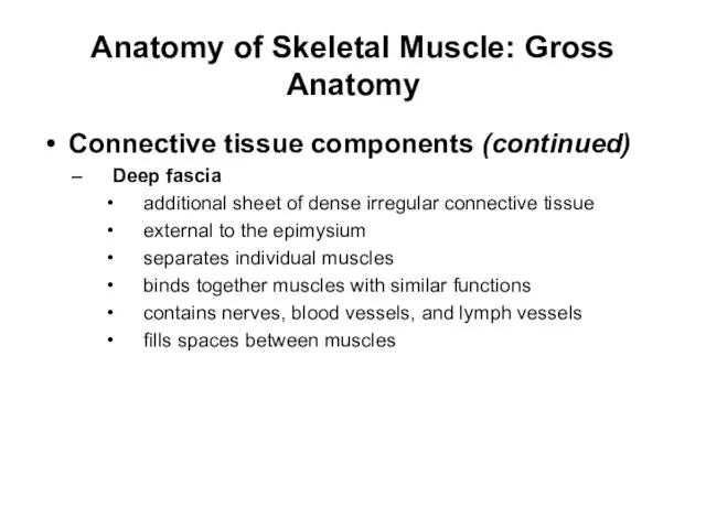
Connective tissue components (continued)
Deep fascia
additional sheet
Anatomy of Skeletal Muscle: Gross Anatomy
Connective tissue components (continued)
Deep fascia
additional sheet
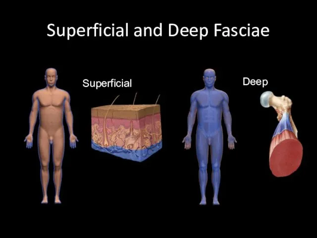
Superficial and Deep Fasciae
10-
Superficial
Deep
Superficial and Deep Fasciae
10-
Superficial
Deep
Anatomy of Skeletal Muscle: Gross Anatomy
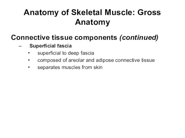
Connective tissue components (continued)
Superficial fascia
superficial to
Anatomy of Skeletal Muscle: Gross Anatomy
Connective tissue components (continued)
Superficial fascia
superficial to
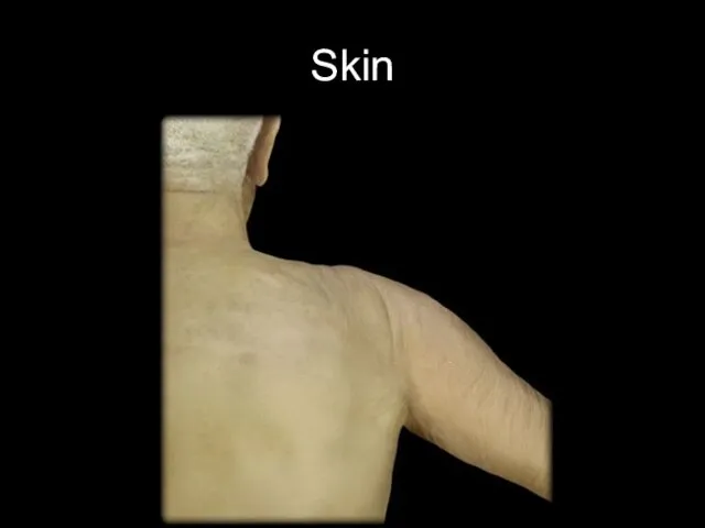
Skin
Skin
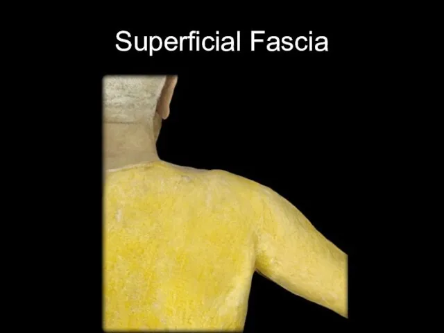
Superficial Fascia
Superficial Fascia
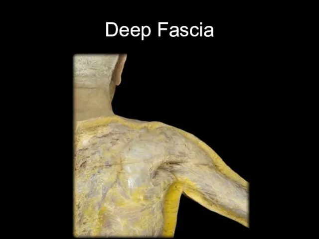
Deep Fascia
Deep Fascia
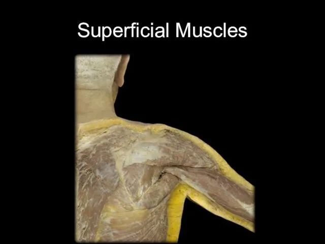
Superficial Muscles
Superficial Muscles
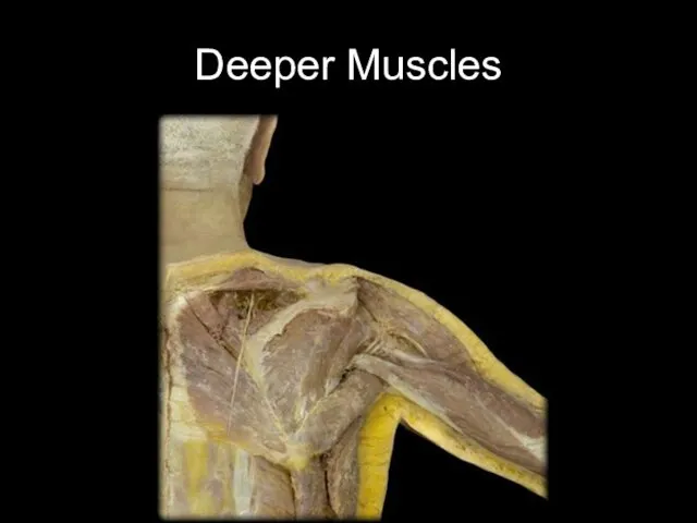
Deeper Muscles
Deeper Muscles
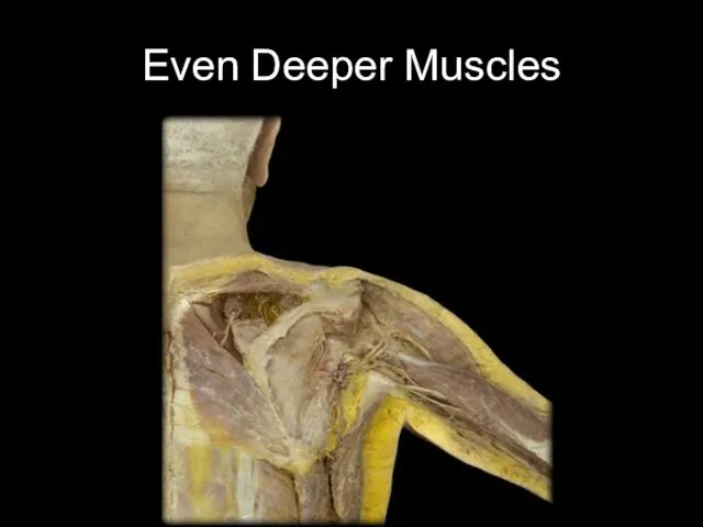
Even Deeper Muscles
Even Deeper Muscles
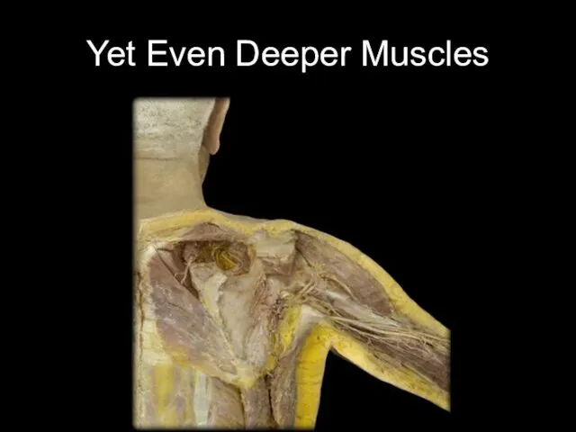
Yet Even Deeper Muscles
Yet Even Deeper Muscles
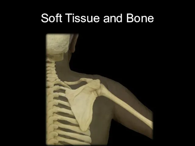
Soft Tissue and Bone
Soft Tissue and Bone
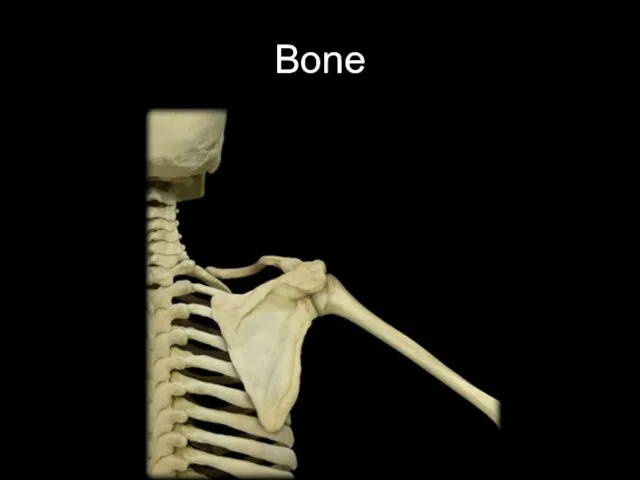
Bone
Bone
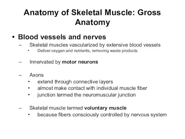
Anatomy of Skeletal Muscle: Gross Anatomy
Blood vessels and nerves
Skeletal muscles vascularized
Anatomy of Skeletal Muscle: Gross Anatomy
Blood vessels and nerves
Skeletal muscles vascularized
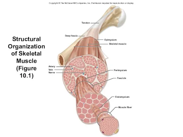
Structural Organization of Skeletal Muscle
(Figure
10.1)
Copyright © The McGraw-Hill Companies, Inc.
Structural Organization of Skeletal Muscle
(Figure
10.1)
Copyright © The McGraw-Hill Companies, Inc.
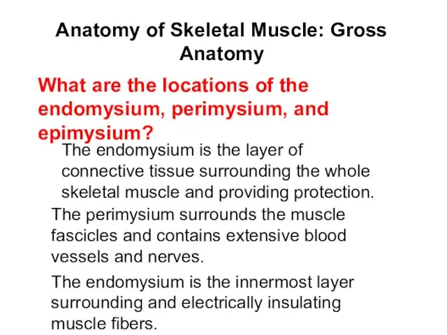
Anatomy of Skeletal Muscle: Gross Anatomy
The endomysium is the layer of
Anatomy of Skeletal Muscle: Gross Anatomy
The endomysium is the layer of
Anatomy of Skeletal Muscle:
Microscopic Anatomy
Sarcoplasma
Cytoplasm of muscle fibers (cells comprising
Anatomy of Skeletal Muscle:
Microscopic Anatomy
Sarcoplasma
Cytoplasm of muscle fibers (cells comprising
Anatomy of Skeletal Muscle:
Microscopic Anatomy
Multinucleated cell
Elongated cells extending length of
Anatomy of Skeletal Muscle:
Microscopic Anatomy
Multinucleated cell
Elongated cells extending length of
Anatomy of Skeletal Muscle:
Microscopic Anatomy
Multinucleated
cell (continued)
Satellite cells
myoblasts remaining, unfused, in
Anatomy of Skeletal Muscle:
Microscopic Anatomy
Multinucleated
cell (continued)
Satellite cells
myoblasts remaining, unfused, in
Anatomy of Skeletal Muscle:
Microscopic Anatomy
Sarcolemma and T-tubules
Plasma membrane of a
Anatomy of Skeletal Muscle:
Microscopic Anatomy
Sarcolemma and T-tubules
Plasma membrane of a
Anatomy of Skeletal Muscle:
Microscopic Anatomy
Sarcolemma and T-tubules (continued)
Na+/ K+ pumps
Anatomy of Skeletal Muscle:
Microscopic Anatomy
Sarcolemma and T-tubules (continued)
Na+/ K+ pumps
Anatomy of Skeletal Muscle:
Microscopic Anatomy
Sarcolemma and T-tubules (continued)
Voltage-gated Na+ channels
Anatomy of Skeletal Muscle:
Microscopic Anatomy
Sarcolemma and T-tubules (continued)
Voltage-gated Na+ channels
Anatomy of Skeletal Muscle:
Microscopic Anatomy
Sarcoplasmic reticulum
Internal membrane complex
Similar to
Anatomy of Skeletal Muscle:
Microscopic Anatomy
Sarcoplasmic reticulum
Internal membrane complex
Similar to
Structure and Organization of a Skeletal Muscle Fiber: Sarcolemma and T-Tubules
Structure and Organization of a Skeletal Muscle Fiber: Sarcolemma and T-Tubules
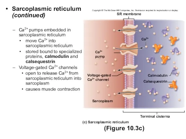
Sarcoplasmic reticulum (continued)
Ca2+ pumps embedded in sarcoplasmic reticulum
move Ca2+ into sarcoplasmic
Sarcoplasmic reticulum (continued)
Ca2+ pumps embedded in sarcoplasmic reticulum
move Ca2+ into sarcoplasmic
Anatomy of Skeletal Muscle:
Microscopic Anatomy
Muscle fibers and myofibrils
Myofibrils
long cylindrical structures
Anatomy of Skeletal Muscle:
Microscopic Anatomy
Muscle fibers and myofibrils
Myofibrils
long cylindrical structures
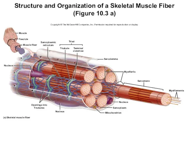
Structure and Organization of a Skeletal Muscle Fiber
(Figure 10.3 a)
Copyright
Structure and Organization of a Skeletal Muscle Fiber
(Figure 10.3 a)
Copyright
Anatomy of Skeletal Muscle:
Microscopic Anatomy
Muscle fibers and myofibrils (continued)
Thick filaments
Anatomy of Skeletal Muscle:
Microscopic Anatomy
Muscle fibers and myofibrils (continued)
Thick filaments
Anatomy of Skeletal Muscle:
Microscopic Anatomy
Muscle fibers and myofibrils (continued)
Thin filaments
Primarily
Anatomy of Skeletal Muscle:
Microscopic Anatomy
Muscle fibers and myofibrils (continued)
Thin filaments
Primarily
Anatomy of Skeletal Muscle:
Microscopic Anatomy
Muscle fibers and myofibrils
Thin filaments
Anatomy of Skeletal Muscle:
Microscopic Anatomy
Muscle fibers and myofibrils
Thin filaments
Molecular Structure of Thick and Thin Filaments
(Figure 10.4)
Copyright © The McGraw-Hill
Molecular Structure of Thick and Thin Filaments
(Figure 10.4)
Copyright © The McGraw-Hill
Anatomy of Skeletal Muscle:
Microscopic Anatomy
Organization of a sarcomere
Myofilaments arranged in
Anatomy of Skeletal Muscle:
Microscopic Anatomy
Organization of a sarcomere
Myofilaments arranged in
Anatomy of Skeletal Muscle:
Microscopic Anatomy
Organization of a sarcomere
Overlapping filaments (continued)
Form
Anatomy of Skeletal Muscle:
Microscopic Anatomy
Organization of a sarcomere
Overlapping filaments (continued)
Form
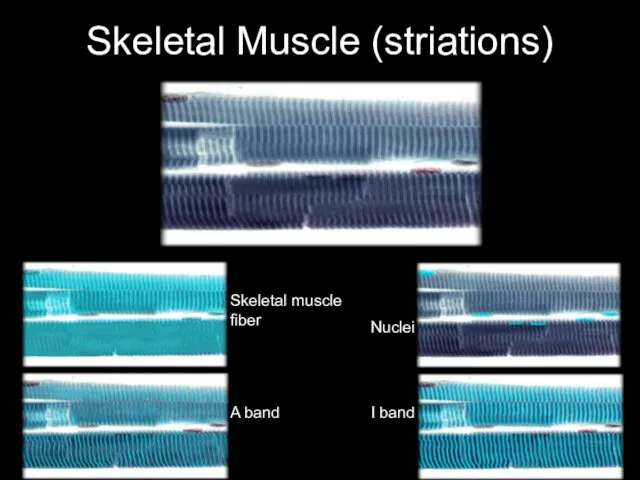
Skeletal Muscle (striations)
Skeletal muscle fiber
A band
I band
Nuclei
Skeletal Muscle (striations)
Skeletal muscle fiber
A band
I band
Nuclei
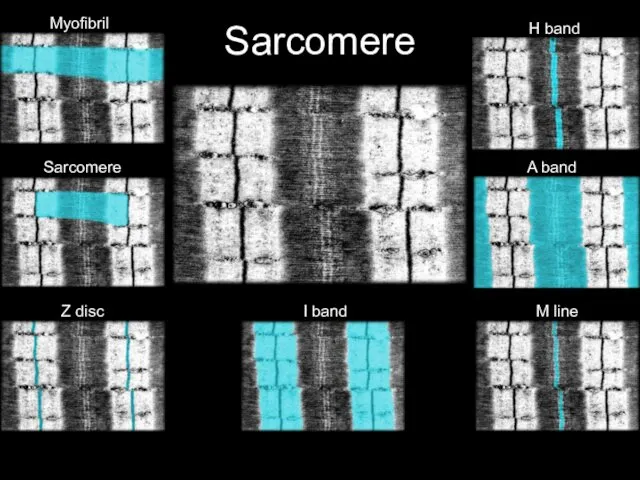
Sarcomere
A band
H band
I band
M line
Myofibril
Sarcomere
Z disc
Sarcomere
A band
H band
I band
M line
Myofibril
Sarcomere
Z disc
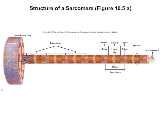
Structure of a Sarcomere (Figure 10.5 a)
Copyright © The McGraw-Hill Companies,
Structure of a Sarcomere (Figure 10.5 a)
Copyright © The McGraw-Hill Companies,
Anatomy of Skeletal Muscle:
Microscopic Anatomy
Organization of a sarcomere (continued)
Overlapping filaments
I
Anatomy of Skeletal Muscle:
Microscopic Anatomy
Organization of a sarcomere (continued)
Overlapping filaments
I
Anatomy of Skeletal Muscle:
Microscopic Anatomy
Organization of a sarcomere
Overlapping filaments (continued)
A
Anatomy of Skeletal Muscle:
Microscopic Anatomy
Organization of a sarcomere
Overlapping filaments (continued)
A
Anatomy of Skeletal Muscle:
Microscopic Anatomy
Organization of a sarcomere
Overlapping filaments (continued)
H
Anatomy of Skeletal Muscle:
Microscopic Anatomy
Organization of a sarcomere
Overlapping filaments (continued)
H
Structure of a Sarcomere (Figure 10.5 b)
Copyright © The McGraw-Hill Companies,
Structure of a Sarcomere (Figure 10.5 b)
Copyright © The McGraw-Hill Companies,
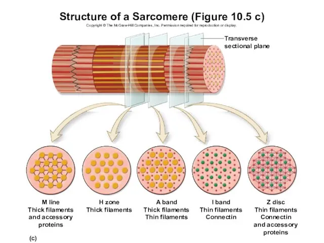
Structure of a Sarcomere (Figure 10.5 c)
Copyright © The McGraw-Hill Companies,
Structure of a Sarcomere (Figure 10.5 c)
Copyright © The McGraw-Hill Companies,
Anatomy of Skeletal Muscle:
Microscopic Anatomy
Organization of a sarcomere
Other structural and
Anatomy of Skeletal Muscle:
Microscopic Anatomy
Organization of a sarcomere
Other structural and
Anatomy of Skeletal Muscle:
Microscopic Anatomy
Organization of a sarcomere
Other structural and
Anatomy of Skeletal Muscle:
Microscopic Anatomy
Organization of a sarcomere
Other structural and
Anatomy of Skeletal Muscle:
Microscopic Anatomy
Mitochondria and other structures associated with
Anatomy of Skeletal Muscle:
Microscopic Anatomy
Mitochondria and other structures associated with
Anatomy of Skeletal Muscle:
Microscopic Anatomy
Mitochondria and other structures associated with
Anatomy of Skeletal Muscle:
Microscopic Anatomy
Mitochondria and other structures associated with
Anatomy of Skeletal Muscle:
Microscopic Anatomy
Thick filaments are composed of myosin
Anatomy of Skeletal Muscle:
Microscopic Anatomy
Thick filaments are composed of myosin
Anatomy of Skeletal Muscle:
Microscopic Anatomy
H zone
In which band are there
Anatomy of Skeletal Muscle:
Microscopic Anatomy
H zone
In which band are there
Anatomy of Skeletal Muscle:
Innervation of Skeletal Muscle Fibers
Motor unit
Motor neuron
Anatomy of Skeletal Muscle:
Innervation of Skeletal Muscle Fibers
Motor unit
Motor neuron
Anatomy of Skeletal Muscle:
Innervation of Skeletal Muscle Fibers
Motor unit (continued)
Varied
Anatomy of Skeletal Muscle:
Innervation of Skeletal Muscle Fibers
Motor unit (continued)
Varied
Anatomy of Skeletal Muscle:
Innervation of Skeletal Muscle Fibers
Motor unit (continued)
Fibers
Anatomy of Skeletal Muscle:
Innervation of Skeletal Muscle Fibers
Motor unit (continued)
Fibers
Anatomy of Skeletal Muscle:
Innervation of Skeletal Muscle Fibers
Neuromuscular junctions
Location where
Anatomy of Skeletal Muscle:
Innervation of Skeletal Muscle Fibers
Neuromuscular junctions
Location where
Neuromuscular Junction
High Magnification
Skeletal muscle fiber
Axon of motor nerve
Motor end plate
Neuromuscular Junction
High Magnification
Skeletal muscle fiber
Axon of motor nerve
Motor end plate
Anatomy of Skeletal Muscle:
Innervation of Skeletal Muscle Fibers
Neuromuscular junctions (continued)
Synaptic
Anatomy of Skeletal Muscle:
Innervation of Skeletal Muscle Fibers
Neuromuscular junctions (continued)
Synaptic
Neuromuscular Junction
TEM: High Magnification
Primary synaptic cleft
Synaptic vesicles of synaptic terminal
Secondary
Neuromuscular Junction
TEM: High Magnification
Primary synaptic cleft
Synaptic vesicles of synaptic terminal
Secondary
Anatomy of Skeletal Muscle:
Innervation of Skeletal Muscle Fibers
Neuromuscular junctions
Synaptic knob
Anatomy of Skeletal Muscle:
Innervation of Skeletal Muscle Fibers
Neuromuscular junctions
Synaptic knob
Anatomy of Skeletal Muscle:
Innervation of Skeletal Muscle Fibers
Neuromuscular junctions
Motor end
Anatomy of Skeletal Muscle:
Innervation of Skeletal Muscle Fibers
Neuromuscular junctions
Motor end
Anatomy of Skeletal Muscle:
Innervation of Skeletal Muscle Fibers
Neuromuscular junctions (continued)
Synaptic
Anatomy of Skeletal Muscle:
Innervation of Skeletal Muscle Fibers
Neuromuscular junctions (continued)
Synaptic
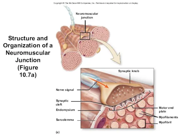
Structure and Organization of a Neuromuscular Junction
(Figure
10.7a)
Neuromuscular
junction
Nerve signal
Synaptic
cleft
Endomysium
Sarcolemma
(a)
Synaptic knob
Myofibril
Myofilaments
Motor end
plate
Copyright
Structure and Organization of a Neuromuscular Junction
(Figure
10.7a)
Neuromuscular
junction
Nerve signal
Synaptic
cleft
Endomysium
Sarcolemma
(a)
Synaptic knob
Myofibril
Myofilaments
Motor end
plate
Copyright
Structure and Organization of a Neuromuscular Junction
(Figure
10.7b)
K+
(b)
Interstitial fluid
Ca2+ pump
Voltage-gated
Ca2+ channels
Sarcolemma
Synaptic
Structure and Organization of a Neuromuscular Junction
(Figure
10.7b)
K+
(b)
Interstitial fluid
Ca2+ pump
Voltage-gated
Ca2+ channels
Sarcolemma
Synaptic
Anatomy of Skeletal Muscle:
Innervation of Skeletal Muscle Fibers
A motor unit
Anatomy of Skeletal Muscle:
Innervation of Skeletal Muscle Fibers
A motor unit
Physiology of Skeletal Muscle Contraction
During muscle contraction
Protein filaments within sarcomeres interact
Sarcomeres
Physiology of Skeletal Muscle Contraction
During muscle contraction
Protein filaments within sarcomeres interact
Sarcomeres
Overview of Events in Skeletal Muscle Contraction
(Figure 10.8)
1
2
3
NEUROMUSCULAR JUNCTION: EXCITATION
Overview of Events in Skeletal Muscle Contraction
(Figure 10.8)
1
2
3
NEUROMUSCULAR JUNCTION: EXCITATION
Skeletal Muscle Contraction—Neuromuscular Junction: Excitation of a Skeletal Muscle Fiber
First
Skeletal Muscle Contraction—Neuromuscular Junction: Excitation of a Skeletal Muscle Fiber
First
Skeletal Muscle Contraction—Neuromuscular Junction: Excitation of a Skeletal Muscle Fiber
Calcium
Skeletal Muscle Contraction—Neuromuscular Junction: Excitation of a Skeletal Muscle Fiber
Calcium
Skeletal Muscle Contraction—Neuromuscular Junction: Excitation of a Skeletal Muscle Fiber
Release
Skeletal Muscle Contraction—Neuromuscular Junction: Excitation of a Skeletal Muscle Fiber
Release
Skeletal Muscle Contraction—Neuromuscular Junction: Excitation of a Skeletal Muscle Fiber
Binding
Skeletal Muscle Contraction—Neuromuscular Junction: Excitation of a Skeletal Muscle Fiber
Binding
Neuromuscular Junction: Excitation of a Skeletal Muscle Fiber (Figure 10.9)
Motor end
Neuromuscular Junction: Excitation of a Skeletal Muscle Fiber (Figure 10.9)
Motor end
Skeletal Muscle Contraction—Neuromuscular Junction: Excitation of a Skeletal Muscle Fiber
Nerve
Skeletal Muscle Contraction—Neuromuscular Junction: Excitation of a Skeletal Muscle Fiber
Nerve
Skeletal Muscle Contraction—Neuromuscular Junction: Excitation of a Skeletal Muscle Fiber
Clinical
Skeletal Muscle Contraction—Neuromuscular Junction: Excitation of a Skeletal Muscle Fiber
Clinical
Skeletal Muscle Contraction—Sarcolemma, T-Tubules, Sarcoplasmic Reticulum: Excitation-Contraction Coupling
Second physiological event
Excitation-contraction coupling
Links
Skeletal Muscle Contraction—Sarcolemma, T-Tubules, Sarcoplasmic Reticulum: Excitation-Contraction Coupling
Second physiological event
Excitation-contraction coupling
Links
Skeletal Muscle Contraction—Sarcolemma, T-Tubules, Sarcoplasmic Reticulum: Excitation-Contraction Coupling
Development of an end-plate
Skeletal Muscle Contraction—Sarcolemma, T-Tubules, Sarcoplasmic Reticulum: Excitation-Contraction Coupling
Development of an end-plate
Skeletal Muscle Contraction—Sarcolemma, T-Tubules, Sarcoplasmic Reticulum: Excitation-Contraction Coupling
Development of an end-plate
Skeletal Muscle Contraction—Sarcolemma, T-Tubules, Sarcoplasmic Reticulum: Excitation-Contraction Coupling
Development of an end-plate
Skeletal Muscle Contraction—Sarcolemma, T-Tubules, Sarcoplasmic Reticulum: Excitation-Contraction Coupling
Initiation and propagation of
Skeletal Muscle Contraction—Sarcolemma, T-Tubules, Sarcoplasmic Reticulum: Excitation-Contraction Coupling
Initiation and propagation of
Skeletal Muscle Contraction—Sarcolemma, T-Tubules, Sarcoplasmic Reticulum: Excitation-Contraction Coupling
Initiation and propagation of
Skeletal Muscle Contraction—Sarcolemma, T-Tubules, Sarcoplasmic Reticulum: Excitation-Contraction Coupling
Initiation and propagation of
Skeletal Muscle Contraction—Sarcolemma, T-Tubules, Sarcoplasmic Reticulum: Excitation-Contraction Coupling
Release of calcium from
Skeletal Muscle Contraction—Sarcolemma, T-Tubules, Sarcoplasmic Reticulum: Excitation-Contraction Coupling
Release of calcium from
Skeletal Muscle Fiber: Excitation-Contraction Coupling (Figure 10.10)
Copyright © The McGraw-Hill Companies,
Skeletal Muscle Fiber: Excitation-Contraction Coupling (Figure 10.10)
Copyright © The McGraw-Hill Companies,
 Тип Моллюски
 Введение в курс анатомии
 Анализатор вкуса
 Презентация на тему Как не поссориться с медведем!?
 Презентация на тему "В мире растений и животных" - скачать презентации по Биологии
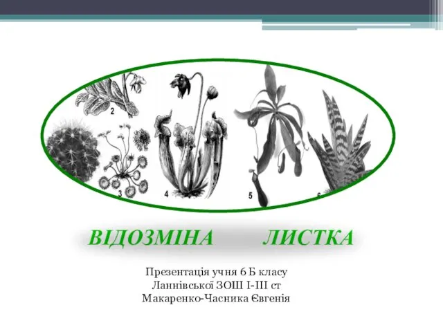 Видозміна листка
 Происхождение человека. Человеческие расы. Стадии антропосоциогенеза. Адаптация человека.
 Плесневые грибы
 КЛАСС ЛЕНТОЧНЫЕ ЧЕРВИ
 Класс Двудольные, характерные особенности растений семейства Крестоцветные или Капустные
 Основы физиологии растений. Дыхание растений
 1. Аминокислоты и белки В состав природных полипептидов и белков входят -аминокислоты, в молекулах которых амино- и карбоксильная группы связаны с одним и тем же атомом углерода. H2N–СН–СООН R В зависимости от строения угле
 Витамины. Их влияние на здоровье человека. Подготовила: Садовникова Александра Евгеньевна
 Практическая работа: Мои фенологические наблюдения
 Зейін. Зейіннің физиологиялық механизмдері
 Презентация на тему "Влияние алкоголя на беременную женщину" - скачать бесплатно презентации по Биологии
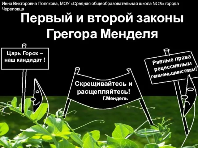 Первый и второй законы Грегора Менделя
 Значение птиц в природе и жизни человека
 Витамины. Классификация витаминов
 Северный олень
 Ayurveda cosmetics. Отражение внутренней гармонии
 Основы генетики
 Микроорганизмы, характерные для микрофлоры человека
 Презентация на тему "Питание и пищеварение 6 класс" - скачать презентации по Биологии
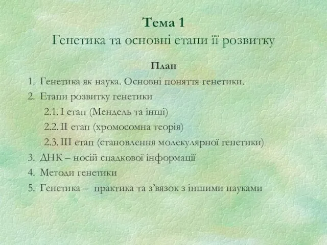 Генетика та основні етапи її розвитку. Тема 1
 Внутриклеточные паразиты вирусы
 КЛАСС ЛЕНТОЧНЫЕ ЧЕРВИ
  Нервная система. Спинной мозг