- Pathologic Protozoa

Содержание
- 2. CHARACTERISTICS OF PROTOZOA 1. Unicellular 2. Chemoheterotrophs (get their energy by breaking down organic matter). 3.
- 3. CHARACTERISTICS OF PROTOZOA 4. The vegetative form is the TROPHOZOA (tropho = movement; zoite = animal;
- 4. CHARACTERISTICS OF PROTOZOA 6. Some produce cysts. These are not tissue cysts like a human gets
- 5. PROTOZOA CYSTS Cysts are not as resistant as a bacterial endospore. You can kill cysts by
- 6. Classification Domain: Eukaryotes Kingdom: Protista
- 7. Classification Traditional classification of protozoa phylae was based on mode of locomotion. MASTIGOPHORA (flagella) CILIOPHORA (cilia)
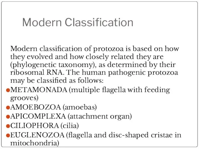
- 8. Modern Classification Modern classification of protozoa is based on how they evolved and how closely related
- 9. EUGLENOZOA EUGLENOZOA (older classification = Mastigophora): has flagella and its mitochondria have disc-shaped cristae Organisms Trypanosoma
- 10. MASTIGOPHORA DISEASES Trypanosomiasis Leishmaniasis
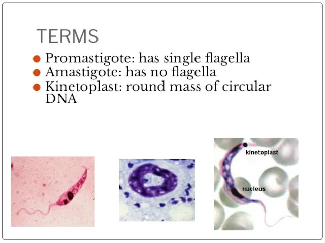
- 11. TERMS Promastigote: has single flagella Amastigote: has no flagella Kinetoplast: round mass of circular DNA
- 12. Leishmania donovani Domain: Eukaryota Kingdom: Protista Phylum: Mastigophora Class: Kinetoplastida Order: Trypanosomatida Genus: Leishmania Species: donovani
- 13. Leishmania donovani Disease: Leishmaniasis Vector-borne disease transmitted by sandflies.
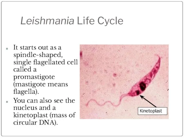
- 14. Leishmania Life Cycle Kinetoplast It starts out as a spindle-shaped, single flagellated cell called a promastigote
- 15. Leishmania rosette In prepared slides you can see promastigotes align their nose in a circle, called
- 16. Leishmaniasis rosette
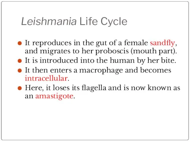
- 17. Leishmania Life Cycle It reproduces in the gut of a female sandfly, and migrates to her
- 18. Leishmaniasis These amastigotes multiply in various organs including the spleen, liver, and lymph nodes. Symptoms include
- 19. Leishmania Life Cycle The female sandflies inject the infective stage, promastigotes, during blood meals. Macrophages phagocytize
- 20. Leishmaniasis Life Cycle
- 21. Leishmania donovani (Promastigote) Single flagellum found in sand flies
- 22. Leishmaniasis Amastogotes Amastogotes with nucleus and kinetoplast Macrophage rupturing
- 23. Leishmania Amastigotes
- 24. Sandfly This looks like a mosquito, except its body is hairy and the wings are feathery.
- 25. Leishmaniasis Geographic Distribution: More than 90 percent of the world's cases of visceral leishmaniasis are in
- 26. Leishmaniasis There are three forms of Leishmaniasis: Cutaneous Mucocutaneus Visceral
- 27. Cutaneous Leishmaniasis The disease is only at the site of the bite. This form is seen
- 28. Leishmaniasis (cutaneous)
- 29. Leishmaniasis (cutaneous)
- 30. Leishmaniasis (cutaneous)
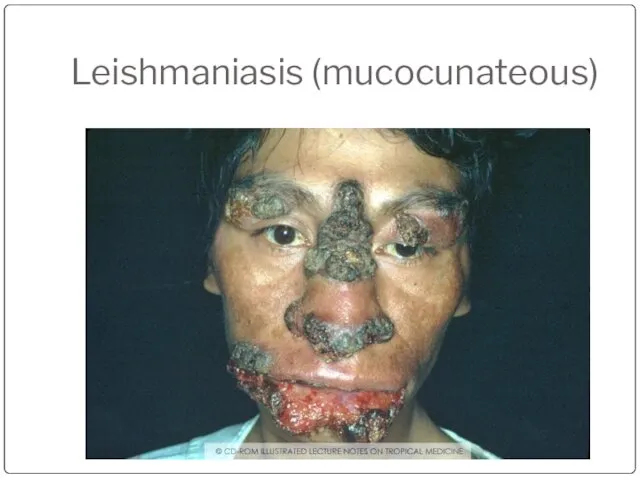
- 31. Leishmaniasis (mucocunateous) This is when the disease located in the mucous membranes of the nose and
- 32. Leishmaniasis (mucocunateous)
- 33. Leishmaniasis (visceral) This is the most serious form. It occurs especially in immunocompromised people, especially HIV
- 34. Kala Azar Hepatosplenomegaly
- 35. Kala Azar (duodenum)
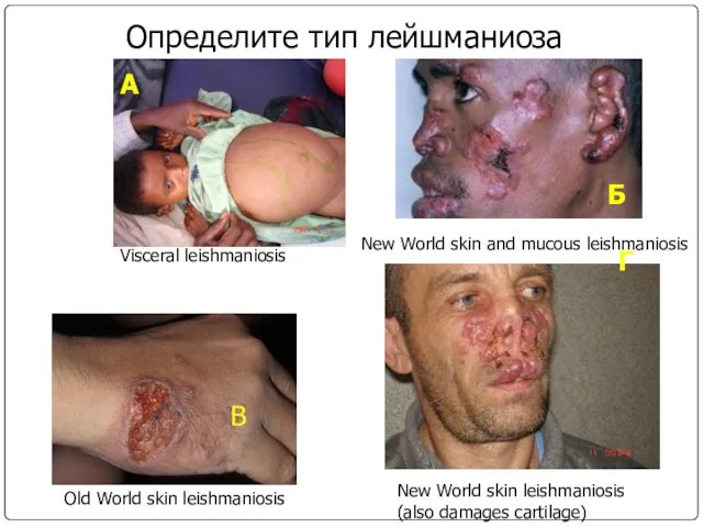
- 36. Определите тип лейшманиоза А Б В Г Visceral leishmaniosis Old World skin leishmaniosis New World skin
- 37. Leishmania life cycle
- 38. TERMS Mastigote = flagella Promastigote: has single flagella Amastigote: has no flagella Kinetoplast: round mass of
- 39. Trypanosomiasis African Trypanosomiasis (African Sleeping Sickness) American Trypanosomiasis (Chaga’s Disease)
- 40. “African Sleeping Sickness” Disease: African Tryptanosomiasis Causal Agents: Trypanosoma brucei gambiense Trypanosoma brucei rhodesiense
- 42. Geographic Distribution T. b. gambiense is found in foci in large areas of West and Central
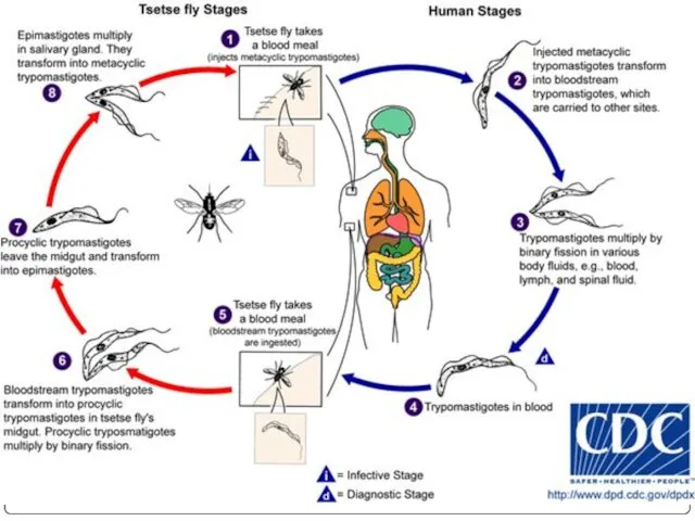
- 43. Trypanosomiasis Trypanosomiasis has a biological vector, the tsetse (pronounced “set-see”) fly. Wild animals may also be
- 44. Trypanosomiasis The tsetse fly bites a human and injects the trypanomastigotes into the skin. This causes
- 45. Trypanosomiasis It is characterized by Winterbottom’s Sign: swelling of the cervical lymph nodes in the head
- 46. Trypanosomiasis CNS symptoms Shuffling gait Slurred speech Malaise (sleeping all day) Treatment Melarsoprol: which has dangerous
- 47. Trypanosoma brucei Trypomastigote stages are the only form found in patients. Posterior kinetoplast Centrally located nucleus
- 48. Trypanosoma brucei
- 49. Trypanosoma brucei gambiense trypomastigote
- 50. Trypanosoma brucei rhodesiense
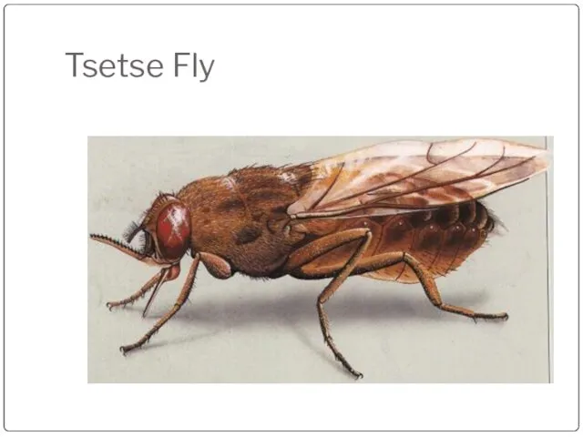
- 51. Tsetse Fly
- 52. “Chaga’s Disease” Disease: American Tryptanosomiasis A zoonotic disease (can infect animals) that can be transmitted to
- 53. “Chaga’s Disease” This disease is NOT found in Africa. This disease is also zoonotic; it can
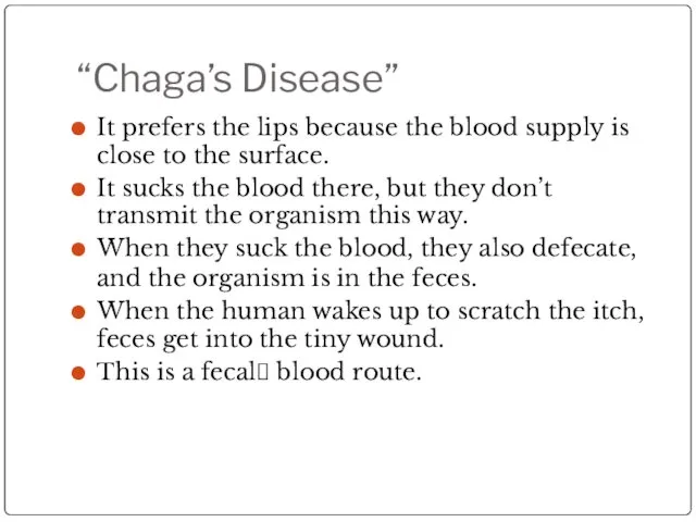
- 54. “Chaga’s Disease” It prefers the lips because the blood supply is close to the surface. It
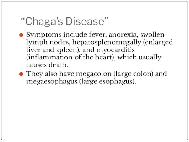
- 55. “Chaga’s Disease” Symptoms include fever, anorexia, swollen lymph nodes, hepatosplenomegally (enlarged liver and spleen), and myocarditis
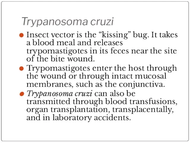
- 57. Trypanosoma cruzi Insect vector is the “kissing” bug. It takes a blood meal and releases trypomastigotes
- 58. Trypanosoma cruzi Geographic Distribution: The Americas from the southern United States to southern Argentina. Mostly in
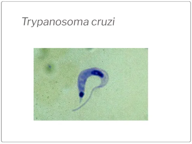
- 59. Trypanosoma cruzi
- 60. Trypanosoma cruzi
- 61. Trypanosoma cruzi large kinetoplast
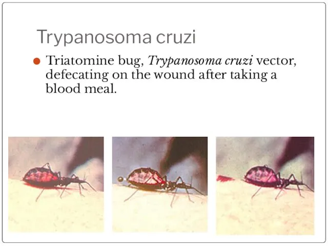
- 62. Trypanosoma cruzi Triatomine bug, Trypanosoma cruzi vector, defecating on the wound after taking a blood meal.
- 63. Kissing Bug
- 64. Romana’s sign Swollen eye, seen in Chagra’s disease.
- 66. Скачать презентацию
CHARACTERISTICS OF PROTOZOA
1. Unicellular
2. Chemoheterotrophs (get their energy by breaking down
CHARACTERISTICS OF PROTOZOA
1. Unicellular
2. Chemoheterotrophs (get their energy by breaking down

CHARACTERISTICS OF PROTOZOA
4. The vegetative form is the TROPHOZOA (tropho =
CHARACTERISTICS OF PROTOZOA
4. The vegetative form is the TROPHOZOA (tropho =
CHARACTERISTICS OF PROTOZOA
6. Some produce cysts.
These are not tissue cysts
CHARACTERISTICS OF PROTOZOA
6. Some produce cysts.
These are not tissue cysts

PROTOZOA CYSTS
Cysts are not as resistant as a bacterial endospore.
You
PROTOZOA CYSTS
Cysts are not as resistant as a bacterial endospore.
You
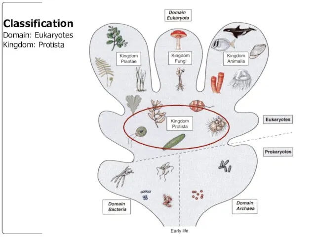
Classification
Domain: Eukaryotes
Kingdom: Protista
Classification
Domain: Eukaryotes
Kingdom: Protista
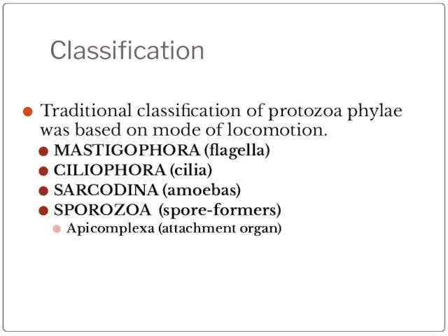
Classification
Traditional classification of protozoa phylae was based on mode of locomotion.
MASTIGOPHORA
Classification
Traditional classification of protozoa phylae was based on mode of locomotion.
MASTIGOPHORA
Modern Classification
Modern classification of protozoa is based on how they evolved
Modern Classification
Modern classification of protozoa is based on how they evolved

EUGLENOZOA
EUGLENOZOA (older classification = Mastigophora): has flagella and its mitochondria have
EUGLENOZOA
EUGLENOZOA (older classification = Mastigophora): has flagella and its mitochondria have

MASTIGOPHORA DISEASES
Trypanosomiasis
Leishmaniasis
MASTIGOPHORA DISEASES
Trypanosomiasis
Leishmaniasis
TERMS
Promastigote: has single flagella
Amastigote: has no flagella
Kinetoplast: round mass of circular
TERMS
Promastigote: has single flagella
Amastigote: has no flagella
Kinetoplast: round mass of circular
Leishmania donovani
Domain: Eukaryota
Kingdom: Protista
Phylum: Mastigophora
Class: Kinetoplastida
Order: Trypanosomatida
Genus: Leishmania
Species: donovani
Leishmania donovani
Domain: Eukaryota
Kingdom: Protista
Phylum: Mastigophora
Class: Kinetoplastida
Order: Trypanosomatida
Genus: Leishmania
Species: donovani

Leishmania donovani
Disease: Leishmaniasis
Vector-borne disease transmitted by sandflies.
Leishmania donovani
Disease: Leishmaniasis
Vector-borne disease transmitted by sandflies.
Leishmania Life Cycle
Kinetoplast
It starts out as a spindle-shaped, single flagellated cell
Leishmania Life Cycle
Kinetoplast
It starts out as a spindle-shaped, single flagellated cell

Leishmania rosette
In prepared slides you can see promastigotes align their nose
Leishmania rosette
In prepared slides you can see promastigotes align their nose
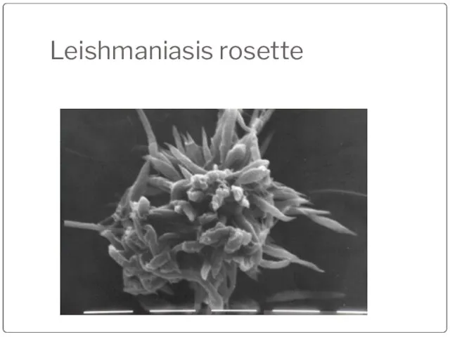
Leishmaniasis rosette
Leishmaniasis rosette
Leishmania Life Cycle
It reproduces in the gut of a female sandfly,
Leishmania Life Cycle
It reproduces in the gut of a female sandfly,
Leishmaniasis
These amastigotes multiply in various organs including the spleen, liver, and
Leishmaniasis
These amastigotes multiply in various organs including the spleen, liver, and
Leishmania Life Cycle
The female sandflies inject the infective stage, promastigotes, during
Leishmania Life Cycle
The female sandflies inject the infective stage, promastigotes, during

Leishmaniasis Life Cycle
Leishmaniasis Life Cycle

Leishmania donovani (Promastigote)
Single flagellum found in sand flies
Leishmania donovani (Promastigote)
Single flagellum found in sand flies

Leishmaniasis
Amastogotes
Amastogotes with nucleus and kinetoplast
Macrophage rupturing
Leishmaniasis
Amastogotes
Amastogotes with nucleus and kinetoplast
Macrophage rupturing

Leishmania
Amastigotes
Leishmania
Amastigotes

Sandfly
This looks like a mosquito, except its body is hairy and
Sandfly
This looks like a mosquito, except its body is hairy and
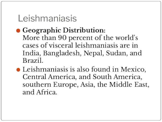
Leishmaniasis
Geographic Distribution:
More than 90 percent of the world's cases of visceral
Leishmaniasis
Geographic Distribution: More than 90 percent of the world's cases of visceral
Leishmaniasis
There are three forms of Leishmaniasis:
Cutaneous
Mucocutaneus
Visceral
Leishmaniasis
There are three forms of Leishmaniasis:
Cutaneous
Mucocutaneus
Visceral

Cutaneous Leishmaniasis
The disease is only at the site of the bite.
Cutaneous Leishmaniasis
The disease is only at the site of the bite.
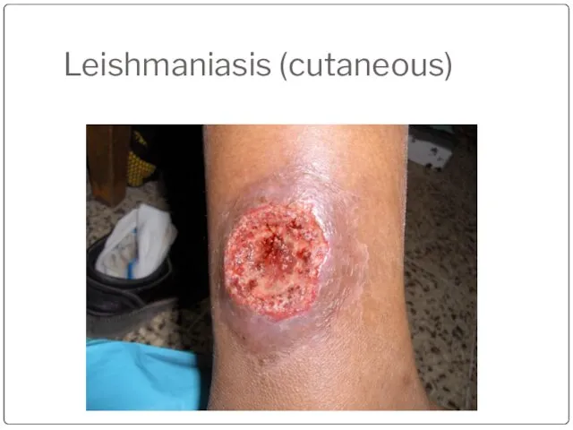
Leishmaniasis (cutaneous)
Leishmaniasis (cutaneous)

Leishmaniasis (cutaneous)
Leishmaniasis (cutaneous)

Leishmaniasis (cutaneous)
Leishmaniasis (cutaneous)
Leishmaniasis (mucocunateous)
This is when the disease located in the mucous membranes
Leishmaniasis (mucocunateous)
This is when the disease located in the mucous membranes
Leishmaniasis (mucocunateous)
Leishmaniasis (mucocunateous)
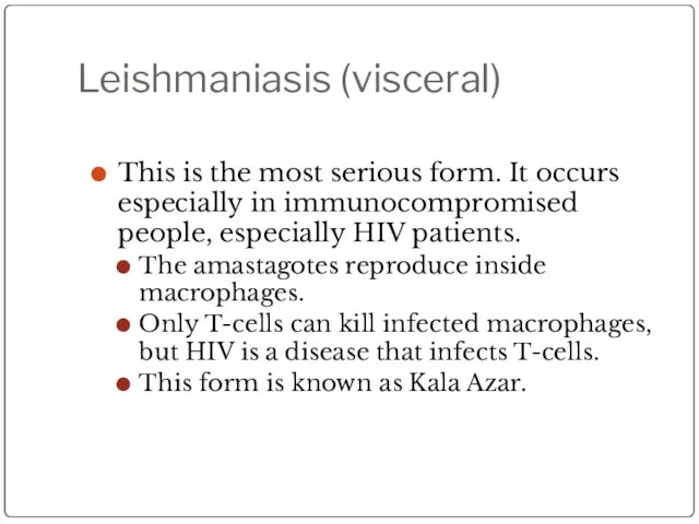
Leishmaniasis (visceral)
This is the most serious form. It occurs especially in
Leishmaniasis (visceral)
This is the most serious form. It occurs especially in
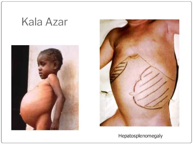
Kala Azar
Hepatosplenomegaly
Kala Azar
Hepatosplenomegaly

Kala Azar (duodenum)
Kala Azar (duodenum)
Определите тип лейшманиоза
А
Б
В
Г
Visceral leishmaniosis
Old World skin leishmaniosis
New World skin and mucous
Определите тип лейшманиоза
А
Б
В
Г
Visceral leishmaniosis
Old World skin leishmaniosis
New World skin and mucous

Leishmania life cycle
Leishmania life cycle

TERMS
Mastigote = flagella
Promastigote: has single flagella
Amastigote: has no flagella
Kinetoplast: round mass
TERMS
Mastigote = flagella
Promastigote: has single flagella
Amastigote: has no flagella
Kinetoplast: round mass

Trypanosomiasis
African Trypanosomiasis
(African Sleeping Sickness)
American Trypanosomiasis
(Chaga’s Disease)
Trypanosomiasis
African Trypanosomiasis
(African Sleeping Sickness)
American Trypanosomiasis
(Chaga’s Disease)

“African Sleeping Sickness”
Disease: African Tryptanosomiasis
Causal Agents:
Trypanosoma brucei gambiense
Trypanosoma brucei rhodesiense
“African Sleeping Sickness”
Disease: African Tryptanosomiasis
Causal Agents:
Trypanosoma brucei gambiense
Trypanosoma brucei rhodesiense

Geographic Distribution
T. b. gambiense is found in foci in large areas
Geographic Distribution
T. b. gambiense is found in foci in large areas

Trypanosomiasis
Trypanosomiasis has a biological vector, the tsetse (pronounced “set-see”) fly.
Wild animals
Trypanosomiasis
Trypanosomiasis has a biological vector, the tsetse (pronounced “set-see”) fly.
Wild animals

Trypanosomiasis
The tsetse fly bites a human and injects the trypanomastigotes into
Trypanosomiasis
The tsetse fly bites a human and injects the trypanomastigotes into

Trypanosomiasis
It is characterized by Winterbottom’s Sign: swelling of the cervical lymph
Trypanosomiasis
It is characterized by Winterbottom’s Sign: swelling of the cervical lymph
Trypanosomiasis
CNS symptoms
Shuffling gait
Slurred speech
Malaise (sleeping all day)
Treatment
Melarsoprol: which has dangerous side-effects
Trypanosomiasis
CNS symptoms
Shuffling gait
Slurred speech
Malaise (sleeping all day)
Treatment
Melarsoprol: which has dangerous side-effects

Trypanosoma brucei
Trypomastigote stages are the only form found in patients.
Posterior kinetoplast
Centrally
Trypanosoma brucei
Trypomastigote stages are the only form found in patients.
Posterior kinetoplast
Centrally

Trypanosoma brucei
Trypanosoma brucei

Trypanosoma brucei gambiense
trypomastigote
Trypanosoma brucei gambiense
trypomastigote

Trypanosoma brucei rhodesiense
Trypanosoma brucei rhodesiense
Tsetse Fly
Tsetse Fly
“Chaga’s Disease”
Disease: American Tryptanosomiasis
A zoonotic disease (can infect animals) that can
“Chaga’s Disease”
Disease: American Tryptanosomiasis
A zoonotic disease (can infect animals) that can

“Chaga’s Disease”
This disease is NOT found in Africa.
This disease is
“Chaga’s Disease”
This disease is NOT found in Africa.
This disease is
“Chaga’s Disease”
It prefers the lips because the blood supply is close
“Chaga’s Disease”
It prefers the lips because the blood supply is close
“Chaga’s Disease”
Symptoms include fever, anorexia, swollen lymph nodes, hepatosplenomegally (enlarged liver
“Chaga’s Disease”
Symptoms include fever, anorexia, swollen lymph nodes, hepatosplenomegally (enlarged liver

Trypanosoma cruzi
Insect vector is the “kissing” bug. It takes a blood
Trypanosoma cruzi
Insect vector is the “kissing” bug. It takes a blood

Trypanosoma cruzi
Geographic Distribution:
The Americas from the southern United States to southern
Trypanosoma cruzi
Geographic Distribution: The Americas from the southern United States to southern
Trypanosoma cruzi
Trypanosoma cruzi

Trypanosoma cruzi
Trypanosoma cruzi

Trypanosoma cruzi
large kinetoplast
Trypanosoma cruzi
large kinetoplast
Trypanosoma cruzi
Triatomine bug, Trypanosoma cruzi vector, defecating on the wound after
Trypanosoma cruzi
Triatomine bug, Trypanosoma cruzi vector, defecating on the wound after

Kissing Bug
Kissing Bug

Romana’s sign
Swollen eye, seen in Chagra’s disease.
Romana’s sign
Swollen eye, seen in Chagra’s disease.
 Презентация на тему "Клетка – структурная единица всего живого" - скачать презентации по Биологии
 Анатомо-физиологические особенности сердечно-сосудистой системы
 Интересное животное Африки - викунья
 Гипофиз. Надпочечники. Поджелудочная железа. Половые железы
 Всасывание продуктов гидролиза липидов в тонком кишечнике. Ресинтез жиров. Образование смешанных мицелл
 Покровы тела. Сравнительная характеристика.
  Прионный ген
 Фотосинтез. Открытие процесса фотосинтеза
 Иммунитет
 Самые большие животные планеты Несколько миллионов лет назад динозавры были самыми крупными животными планеты. Некоторые из н
 Корни растений - Живые якоря
 Презентация на тему Весеннее пробуждение растений
  Химический состав бактериальной клетки
 Презентация на тему "Где зимуют птицы?" - скачать бесплатно презентации по Биологии
 ПТИЦЫ
ПТИЦЫ  Горох Менделя
 Caracteristică succintă a angiospermelor
 Органические вещества клетки Общая биология 10 класс
 ЦИТРУСОВОЕ ХОББИ, или как вырастить лимон в домашних условиях Автор Логунова Наталья, учащаяся 3 "А" класса МОУ гимнази
 ВИРУСЫ – НЕКЛЕТОЧНАЯ ФОРМА ЖИЗНИ…
 Қой ценурозы
 ИССЛЕДОВАТЕЛЬСКАЯ РАБОТА НА ТЕМУ: ВКЛАД ДМИТРИЯ ИВАНОВИЧА МЕНДЕЛЕЕВА В РАЗВИТИЕ МОЛОЧНОЙ ПРОМЫШЛЕННОСТИ И СЫРОВАРЕНИЯ Выпол
 Физиологические адаптации
 Агератум. Агротенхнические мероприятия по выращиванию растения агератум
 Загальна характеристики Царства Рослини
  Законы конкурентных отношений в природе
 Анатомия половой системы человека
 Биохимический анализ крови в диагностике болезней животных. Часть 1