- The Integumentary System

Содержание
- 2. Integumentary System Only 1.5 to 4 mm thick but is body’s largest organ 7% of body
- 3. Integument Composition Two basic layers Epidermis External Stratified squamous epithelium Dermis Deeper Mostly dense irregular connective
- 4. Integument Epidermis Dermis Subcutaneous layer
- 5. Layers of the Integument (Figure 6.1) Copyright © The McGraw-Hill Companies, Inc. Permission required for reproduction
- 6. Epidermis Epithelial tissue arranged in 5 layers or strata Cells divide in only in base layer
- 7. Epidermal Strata From deep to superficial Stratum basale (base layer) Stratum spinosum (spiny layer) Stratum granulosum
- 8. Thick Skin High Magnification Epidermis Dermis Stratum basale Stratum spinosum Stratum granulosum Stratum lucidum Stratum corneum
- 9. Stratum Basale Single layer of cuboidal to low columnar cells attached to a basement membrane Include
- 10. Stratum Spinosum Cells are pushed up from below and become “squished” and look spiny on cross
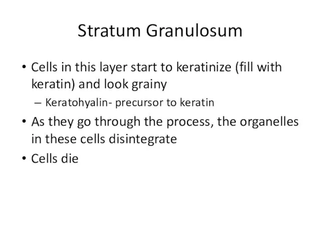
- 11. Stratum Granulosum Cells in this layer start to keratinize (fill with keratin) and look grainy Keratohyalin-
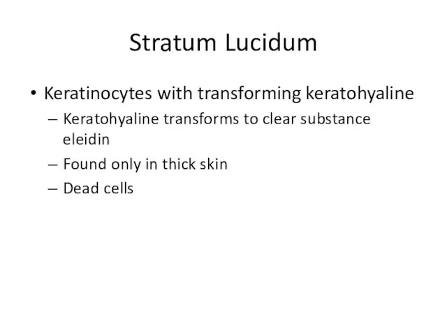
- 12. Stratum Lucidum Keratinocytes with transforming keratohyaline Keratohyaline transforms to clear substance eleidin Found only in thick
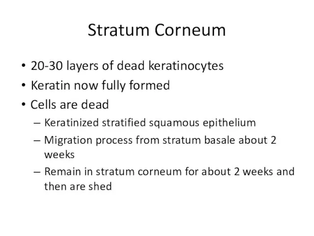
- 13. Stratum Corneum 20-30 layers of dead keratinocytes Keratin now fully formed Cells are dead Keratinized stratified
- 14. Epidermal Strata (Figure 6.2) Copyright © The McGraw-Hill Companies, Inc. Permission required for reproduction or display.
- 15. Variations in the Epidermis Epidermis is not the same over all portions of the body or
- 16. Thick Skin Thin Skin
- 17. Thick Skin High Magnification Epidermis Dermis Stratum basale Stratum spinosum Stratum granulosum Stratum lucidum Stratum corneum
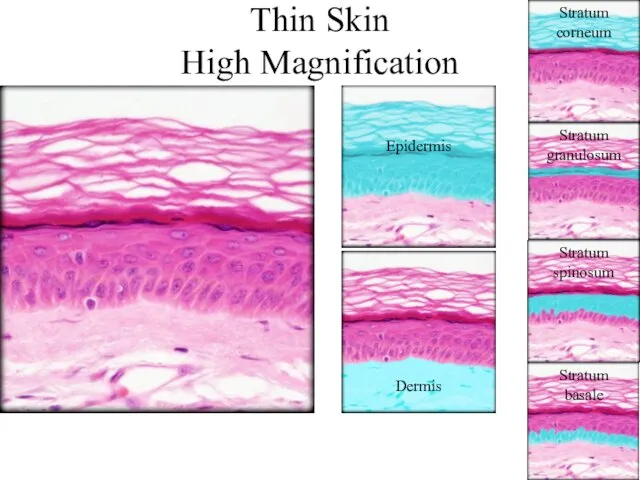
- 18. Thin Skin High Magnification Epidermis Dermis Stratum basale Stratum spinosum Stratum granulosum Stratum corneum
- 19. Thin Skin Low Magnification Epidermis Dermis Dermal papillae Duct of merocrine sweat gland Stratum corneum Stratum
- 21. Скачать презентацию
Integumentary System
Only 1.5 to 4 mm thick but is body’s largest
Integumentary System
Only 1.5 to 4 mm thick but is body’s largest
Integument Composition
Two basic layers
Epidermis
External
Stratified squamous epithelium
Dermis
Deeper
Mostly dense irregular connective tissue
Additional
Integument Composition
Two basic layers
Epidermis
External
Stratified squamous epithelium
Dermis
Deeper
Mostly dense irregular connective tissue
Additional
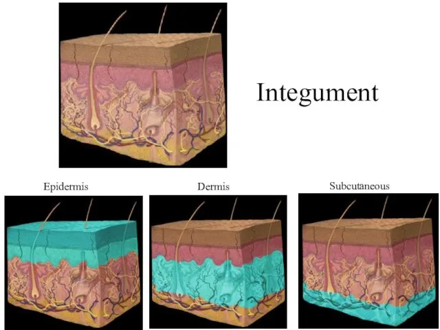
Integument
Epidermis
Dermis
Subcutaneous layer
Integument
Epidermis
Dermis
Subcutaneous layer
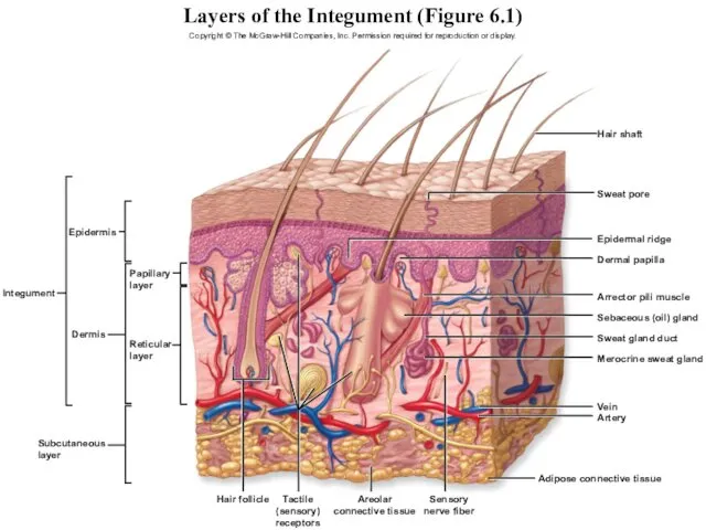
Layers of the Integument (Figure 6.1)
Copyright © The McGraw-Hill Companies, Inc.
Layers of the Integument (Figure 6.1)
Copyright © The McGraw-Hill Companies, Inc.
Epidermis
Epithelial tissue arranged in 5 layers or strata
Cells divide in only
Epidermis
Epithelial tissue arranged in 5 layers or strata
Cells divide in only
Epidermal Strata
From deep to superficial
Stratum basale (base layer)
Stratum spinosum (spiny layer)
Stratum
Epidermal Strata
From deep to superficial
Stratum basale (base layer)
Stratum spinosum (spiny layer)
Stratum
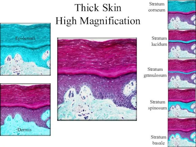
Thick Skin
High Magnification
Epidermis
Dermis
Stratum
basale
Stratum
spinosum
Stratum
granulosum
Stratum
lucidum
Stratum
corneum
Thick Skin
High Magnification
Epidermis
Dermis
Stratum
basale
Stratum
spinosum
Stratum
granulosum
Stratum
lucidum
Stratum
corneum

Stratum Basale
Single layer of cuboidal to low columnar cells attached to
Stratum Basale
Single layer of cuboidal to low columnar cells attached to
Stratum Spinosum
Cells are pushed up from below and become “squished” and
Stratum Spinosum
Cells are pushed up from below and become “squished” and
Stratum Granulosum
Cells in this layer start to keratinize (fill with keratin)
Stratum Granulosum
Cells in this layer start to keratinize (fill with keratin)
Stratum Lucidum
Keratinocytes with transforming keratohyaline
Keratohyaline transforms to clear substance eleidin
Found
Stratum Lucidum
Keratinocytes with transforming keratohyaline
Keratohyaline transforms to clear substance eleidin
Found
Stratum Corneum
20-30 layers of dead keratinocytes
Keratin now fully formed
Cells are dead
Keratinized
Stratum Corneum
20-30 layers of dead keratinocytes
Keratin now fully formed
Cells are dead
Keratinized
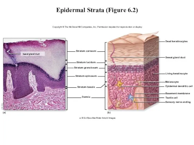
Epidermal Strata (Figure 6.2)
Copyright © The McGraw-Hill Companies, Inc. Permission required
Epidermal Strata (Figure 6.2)
Copyright © The McGraw-Hill Companies, Inc. Permission required
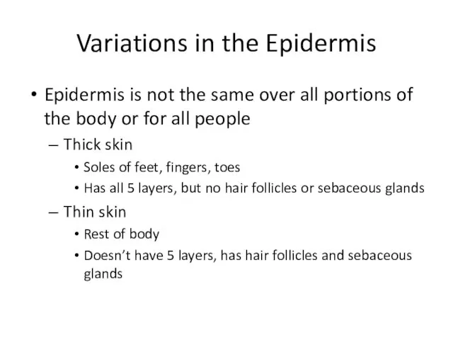
Variations in the Epidermis
Epidermis is not the same over all portions
Variations in the Epidermis
Epidermis is not the same over all portions
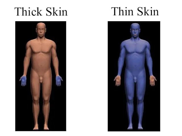
Thick Skin
Thin Skin
Thick Skin
Thin Skin

Thick Skin
High Magnification
Epidermis
Dermis
Stratum
basale
Stratum
spinosum
Stratum
granulosum
Stratum
lucidum
Stratum
corneum
Thick Skin
High Magnification
Epidermis
Dermis
Stratum
basale
Stratum
spinosum
Stratum
granulosum
Stratum
lucidum
Stratum
corneum
Thin Skin
High Magnification
Epidermis
Dermis
Stratum
basale
Stratum
spinosum
Stratum
granulosum
Stratum
corneum
Thin Skin
High Magnification
Epidermis
Dermis
Stratum
basale
Stratum
spinosum
Stratum
granulosum
Stratum
corneum
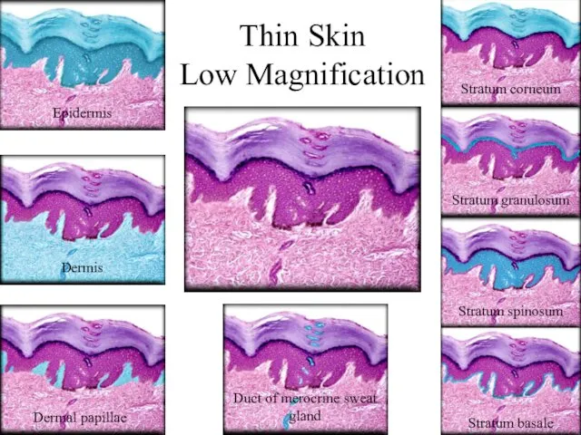
Thin Skin
Low Magnification
Epidermis
Dermis
Dermal papillae
Duct of merocrine sweat gland
Stratum corneum
Stratum granulosum
Stratum spinosum
Stratum
Thin Skin
Low Magnification
Epidermis
Dermis
Dermal papillae
Duct of merocrine sweat gland
Stratum corneum
Stratum granulosum
Stratum spinosum
Stratum
 Презентация Цветок
  Биоэкология растений
 Презентация на тему Неправильное питание
  Гипотеза происхождения жизни
 Дыхательная система человека
 Обитатели Тайги
 Видовое разнообразие флоры в Казахстане
 Презентация на тему О птицах зимой
 Ученица 9 «Б» класса Нежинского лицея КИЛЯЗОВА ЕКАТЕРИНА
  Тигровая викторина
 Тупорылая Акула
 День моржа
 Биохимия, как наука. Элементарный и молекулярный состав живых организмов
 Тест по гистологии. Практика №1
 Кровь и остальные компоненты внутренней среды организма
 Задачи по семеноводству
 Концепции организации живых систем. Популяционно-видовой уровень живого. (Лекция 15)
 Органы чувств
 Теории возникновения жизни Теории возникновения жизни на Земле.
 Передача наследственной информации от ДНК к и-РНК и к белку
 Экологическое загрязнение Брянской области _
 Понятие о микроорганизмах. Тема № 7/1
 СТРУКТУРА И ФУНКЦИИ ХРОМОСОМ Автор Долгорукова С.В., учитель высшей категории МОУ гимназия № 2 Г.Екатеринбурга
  Презентация на тему Решение проблем с отходами
  Томаты
 Основные виды питательных веществ и их значение в питании человека
 Клеточный цикл. Митоз. Стволовые клетки. Понятие о детерминации и дифференцировке
 Иерархия в табуне!