Cross-section sample preparation using focused ion beam system (FIB) for transmission electron microscopy (TEM)
- Cross-section sample preparation using focused ion beam system (FIB) for transmission electron microscopy (TEM)

Содержание
- 2. What is TEM? TEM Hitachi HT 7700 What can be observed by TEM: Thin films and
- 3. Supporting grid for TEM specimens TEM specimen holder
- 4. Examples Ni nanoparticles (catalysts) and carbon nanotubes Ni-Ti thin foil Neonothopanus nambi (lat.) biological specimen Co-Al2O3
- 5. What is “cross-section”? cross-section is basic method of observation multi layers structures cross-section method allows to
- 6. Classic method of cross-section sample preparation Four pieces of specimens on the silicon substrate are glued
- 7. 3 sawing disc of 100 um thickness 4 Thinning the disk up to 10 microns by
- 8. 5 Thinning the disk up to 10 nm by Precision Ion Polishing System (PIPS)
- 9. Modern method of cross-section sample preparation Focused Ion Beam System (FIB) Hitachi FB 2100
- 10. Basic steps of cross-section sample preparation by FIB 1. Deposition of protective tungsten coating on the
- 11. 2. Cutting half-finished (lamella)
- 12. 2. Cutting lamella (continuation)
- 13. 3. Fixing microprobe
- 14. 4. Cutting left side and removing lamella
- 15. 5. Fixing lamella on the toothed semicircle
- 16. 6. Cutting and removing microprobe
- 17. 7. Thinning specimen to 50-100 nm
- 18. Finish result Область просмотра в ПЭМ Disadvantages of FIB method Damaging top layer during deposition tungsten
- 19. Some features sample preparation with thin layers (thickness less than 300 nm)
- 22. Pre-sputtering of Ge protective layer
- 23. substrate (Si) Ge (protection layer) Mn (epitaxial layers) Examples
- 24. substrate (Si) Ge (protection layer) Co Pt
- 25. comparison of the two methods Classic method FIB method
- 27. Скачать презентацию
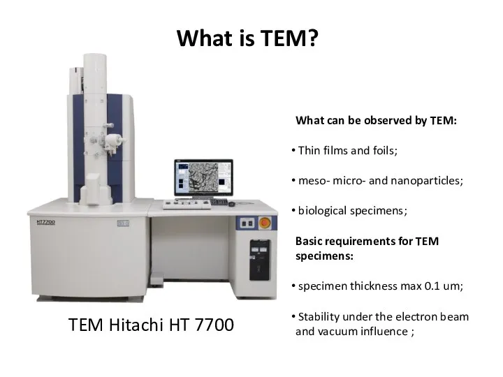
What is TEM?
TEM Hitachi HT 7700
What can be observed by TEM:
What is TEM?
TEM Hitachi HT 7700
What can be observed by TEM:
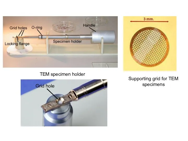
Supporting grid for TEM specimens
TEM specimen holder
Supporting grid for TEM specimens
TEM specimen holder
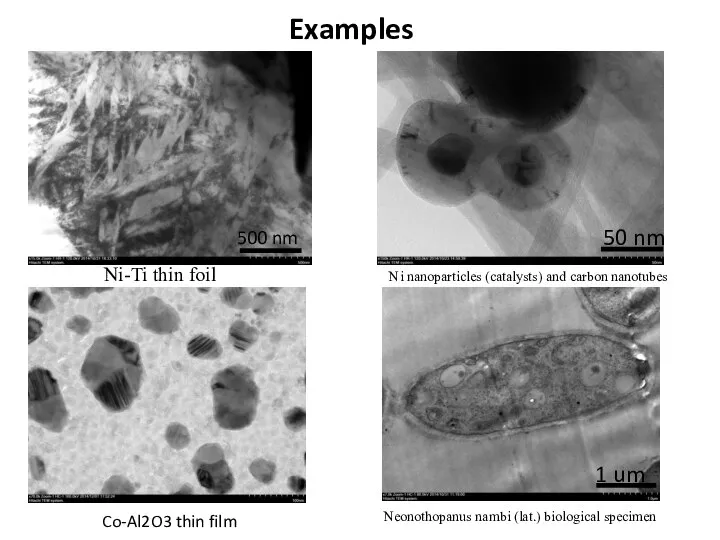
Examples
Ni nanoparticles (catalysts) and carbon nanotubes
Ni-Ti thin foil
Neonothopanus nambi (lat.) biological
Examples
Ni nanoparticles (catalysts) and carbon nanotubes
Ni-Ti thin foil
Neonothopanus nambi (lat.) biological
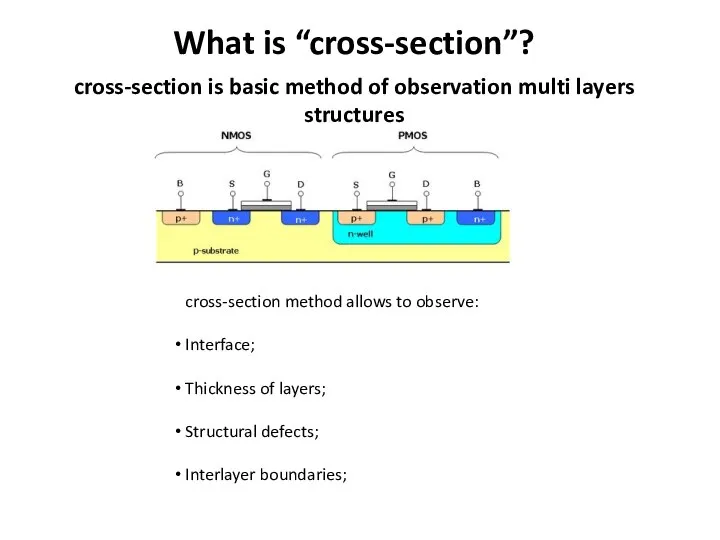
What is “cross-section”?
cross-section is basic method of observation multi layers structures
What is “cross-section”?
cross-section is basic method of observation multi layers structures
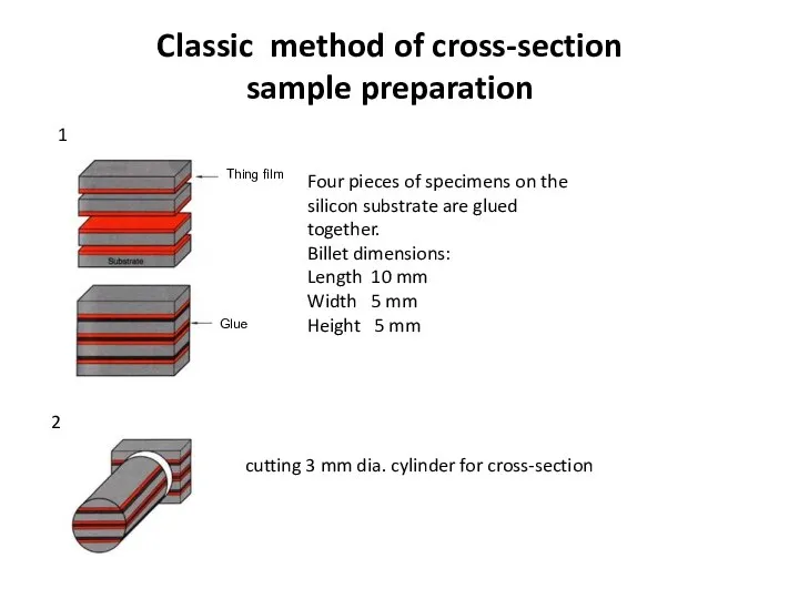
Classic method of cross-section sample preparation
Four pieces of specimens on
Classic method of cross-section sample preparation
Four pieces of specimens on
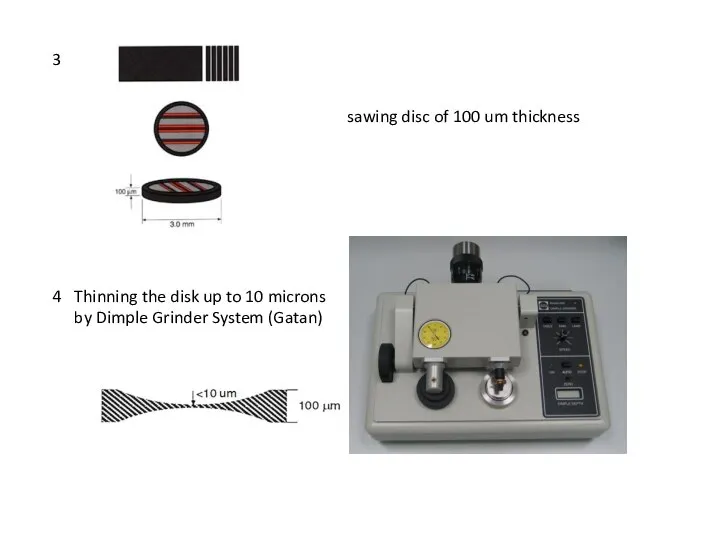
3
sawing disc of 100 um thickness
4
Thinning the disk up to 10
3
sawing disc of 100 um thickness
4
Thinning the disk up to 10
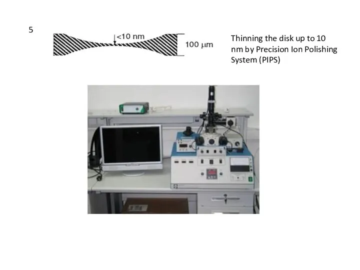
5
Thinning the disk up to 10 nm by Precision Ion Polishing
5
Thinning the disk up to 10 nm by Precision Ion Polishing
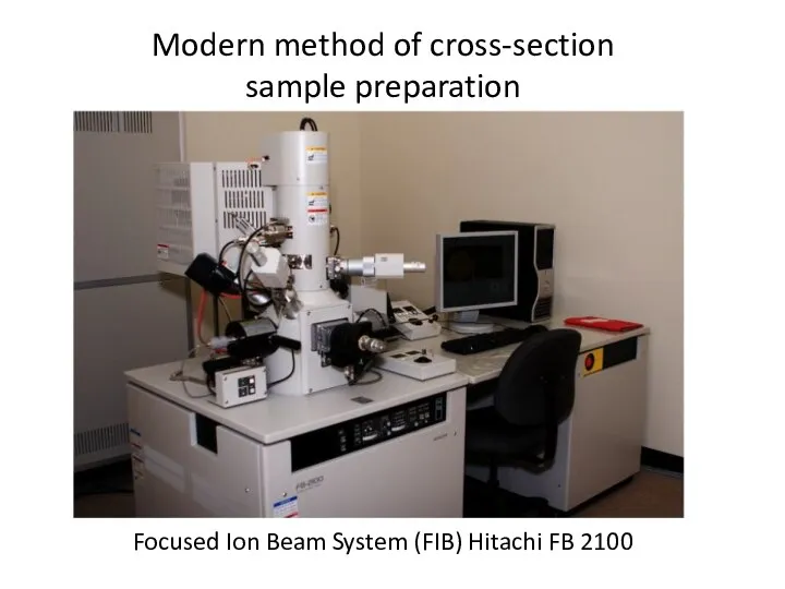
Modern method of cross-section sample preparation
Focused Ion Beam System (FIB)
Modern method of cross-section sample preparation
Focused Ion Beam System (FIB)

Basic steps of cross-section sample preparation by FIB
1. Deposition of protective
Basic steps of cross-section sample preparation by FIB
1. Deposition of protective
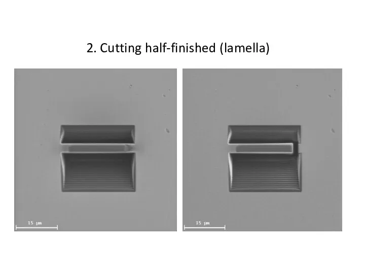
2. Cutting half-finished (lamella)
2. Cutting half-finished (lamella)

2. Cutting lamella (continuation)
2. Cutting lamella (continuation)

3. Fixing microprobe
3. Fixing microprobe

4. Cutting left side and removing lamella
4. Cutting left side and removing lamella
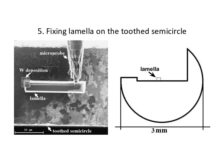
5. Fixing lamella on the toothed semicircle
5. Fixing lamella on the toothed semicircle

6. Cutting and removing microprobe
6. Cutting and removing microprobe

7. Thinning specimen to 50-100 nm
7. Thinning specimen to 50-100 nm

Finish result
Область просмотра в ПЭМ
Disadvantages of FIB method
Damaging top layer during
Finish result
Область просмотра в ПЭМ
Disadvantages of FIB method
Damaging top layer during

Some features sample preparation with thin layers (thickness less than 300
Some features sample preparation with thin layers (thickness less than 300


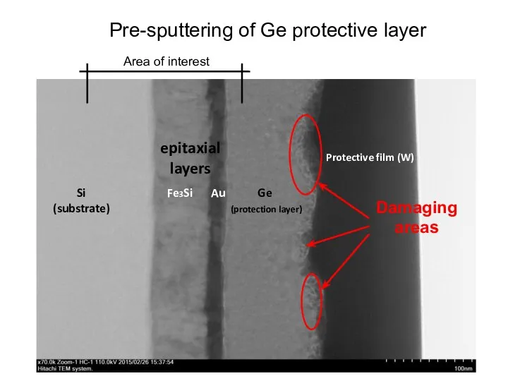
Pre-sputtering of Ge protective layer
Pre-sputtering of Ge protective layer

substrate (Si)
Ge (protection layer)
Mn
(epitaxial layers)
Examples
substrate (Si)
Ge (protection layer)
Mn
(epitaxial layers)
Examples

substrate (Si)
Ge
(protection layer)
Co
Pt
substrate (Si)
Ge
(protection layer)
Co
Pt

comparison of the two methods
Classic method
FIB method
comparison of the two methods
Classic method
FIB method
 Обзор основных методов исследования супрамолекулярных объектов
 Презентация по Химии "Многоатомные спирты" - скачать смотреть
  Родючість ґрунтів , можливості її регулювання. Учень 9-Г класу Юрчак Владислав Керівник Садовська Т.Я.
 Симметрия в химии. Кристаллы
 Количественное определение лекарственных средств
 Электронные конфигурации атомов
 Презентація З хімії На тему : ” Майбутнє альтернативного палива” Виконала учениця 11-А класу Твердохліб Анжеліка
  Кислоты органические и неорганические.
 Синтетические моющие средства.(СМС) Подготовила Живулько Елена.
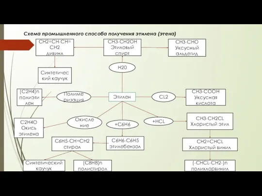 Промышленній способ получения этилена (этена)
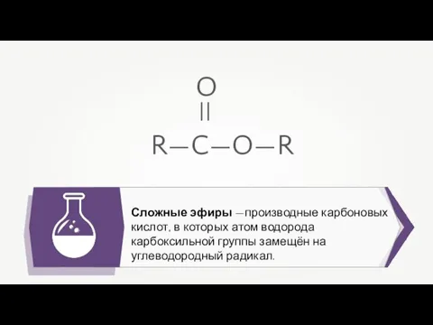 Сложные эфиры
 Растворы. Способы выражения концентрации растворенного вещества
 Титани органічної хімії
 Энергетика химических процессов
 Презентация по химии Химические уравнения Реакции обмена
 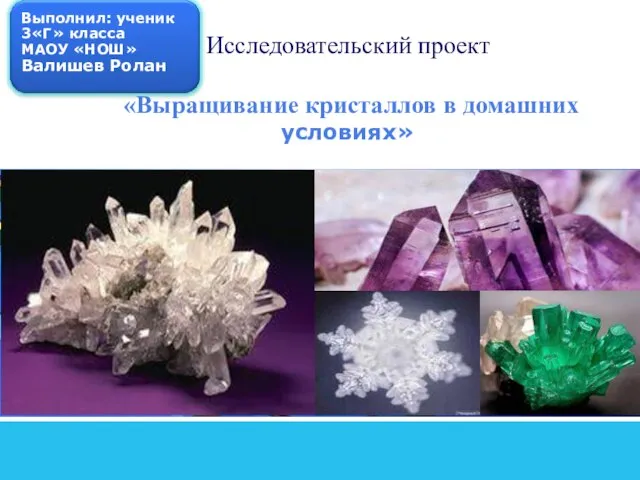 Выращивание кристаллов в домашних условиях
 Неметаллы. Обобщение. Открытый урок
 Окислительно-восстановительные реакции
 Биогеохимия Оренбургской области
 Химия элементов VIA группы. Сера
 Кремнийорганическая гипотеза
 Неметаллы. Положение неметаллов в ПСХЭ
 Полифункциональды (гетерофункциональды) биоорганикалық қосылыстар: оксиқышқылдар, альдегидо- және кетоқышқылдар
 - Это оружие массового поражения, действие которого основано на токсических свойствах химических веществ. - Это оружие массового
 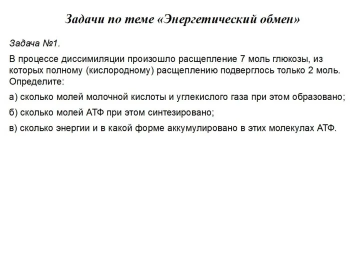 Задачи по теме Энергетический обмен
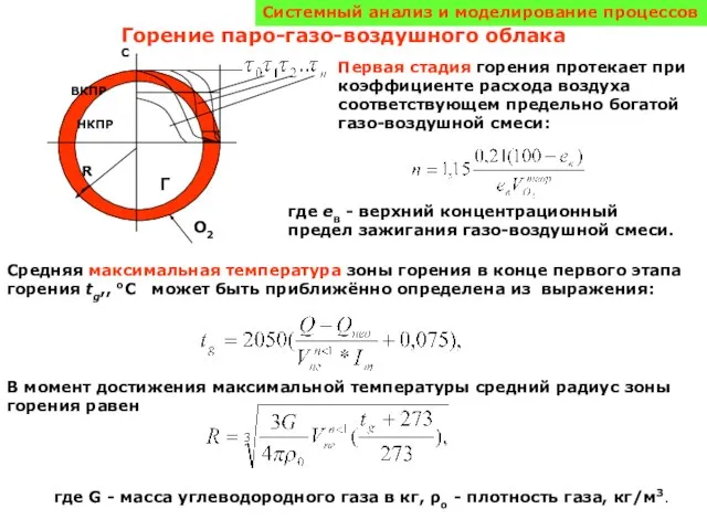 Горение паро-газо-воздушного облака
 Ковалентная полярная связь
 Нітратна кислота. Нітрати Підготувала:Рибальчук Вікторія