- Disease of Honeybees

Содержание
- 2. Introduction Honeybees are attacked both at brood and adult stages by microorganisms. The disease of honeybees
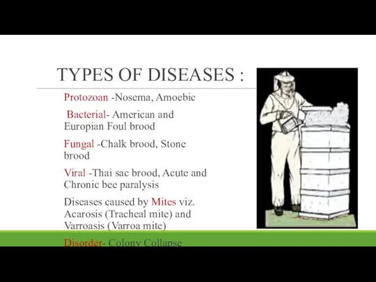
- 3. TYPES OF DISEASES : Protozoan -Nosema, Amoebic Bacterial- American and Europian Foul brood Fungal -Chalk brood,
- 4. Brood diseases American foul brood European foul brood Fungal brood diseases Chalk brood Stone brood Viral
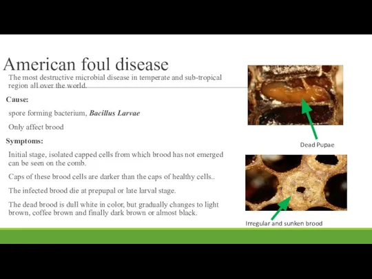
- 5. American foul disease The most destructive microbial disease in temperate and sub-tropical region all over the
- 6. Diagnostic procedure: The simple test for AFD is the “Stretch test” A match stick or tooth
- 7. Feeding streptomycine in sugar syrup @ 0.05-0.15g/litre. Dust Terramycin(TM50) in powdered sugar (1.20) @ 4 tea
- 8. European foul Brood In India, except Maharastra, EFB has not been recorded so for in Apis
- 9. The color of the larvae changes from shiny white to pale yellow and then brown, as
- 10. Fungal Brood Diseases Chalk brood Cause: causative agent is Ascophaer apis It is a heterothallic fungus
- 11. Treatment: Equipment's should be sterilized using formalin or carbolic acid. 0. 7% of thymol has been
- 12. Stone Brood Cause: it is caused by Aspergillus niger, Aspergillus fumigates, Aspergillus flavus. Symptoms: Mummification of
- 13. Viral Diseases So far, the world over, 18 viruses have been found to infect honeybees. Thai
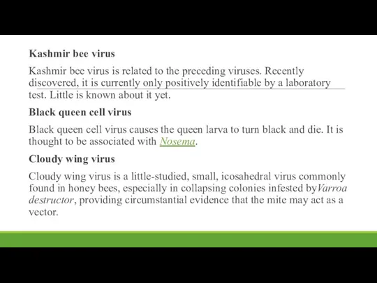
- 14. Kashmir bee virus Kashmir bee virus is related to the preceding viruses. Recently discovered, it is
- 15. Control measurements: use antibiotics such as Rifampin, Leavamisol, Amentidine along with vitamin B complex fed to
- 16. Adult Bee Diseases: The diseases of adult bees are caused by protozoa which are single celled
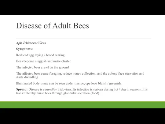
- 17. Disease of Adult Bees Apis Iridescent Virus Symptoms: Reduced egg laying / brood rearing. Bees become
- 18. Management of Viral Diseases: For viral pathogens, there is no chemical control. Affected colonies should be
- 19. Nosema Disease: This disease is caused by Nosema apis Zander. It is disease of adult bees.
- 20. Symptoms Bees start foraging at younger age. Bees feel fatigued, are less able to fly and
- 21. Mid intestine is swollen and if dissected, shows dull greyish white contents. Bees soil the hive
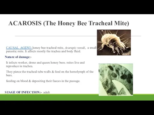
- 22. ACAROSIS (The Honey Bee Tracheal Mite) CAUSAL AGENT- honey bee tracheal mite, Acarapis woodi, a small
- 23. PLACE OF INFECTION: Trachea and body fluid MANAGEMENT:- Use of grease patties ( typically made from
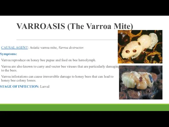
- 24. VARROASIS (The Varroa Mite) CAUSAL AGENT: Asiatic varroa mite, Varroa destructor. Symptoms: Varroa reproduce on honey
- 25. PLACE OF INFECTION: Body and body fluid i.e. haemolymph MANAGEMENT: Apivar: Apivar is effective against varroa
- 27. Скачать презентацию
Introduction
Honeybees are attacked both at brood and adult stages by
Introduction
Honeybees are attacked both at brood and adult stages by
TYPES OF DISEASES :
Protozoan -Nosema, Amoebic
Bacterial- American and Europian Foul
TYPES OF DISEASES :
Protozoan -Nosema, Amoebic
Bacterial- American and Europian Foul
Brood diseases
American foul brood
European foul brood
Fungal brood diseases
Brood diseases
American foul brood
European foul brood
Fungal brood diseases
American foul disease
The most destructive microbial disease in temperate and
American foul disease
The most destructive microbial disease in temperate and
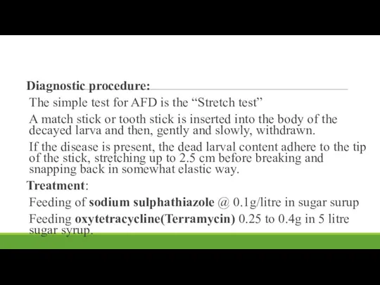
Diagnostic procedure:
The simple test for AFD is the “Stretch test”
A match
The simple test for AFD is the “Stretch test”
A match
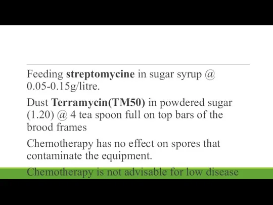
Feeding streptomycine in sugar syrup @ 0.05-0.15g/litre.
Dust Terramycin(TM50) in powdered sugar
Feeding streptomycine in sugar syrup @ 0.05-0.15g/litre.
Dust Terramycin(TM50) in powdered sugar
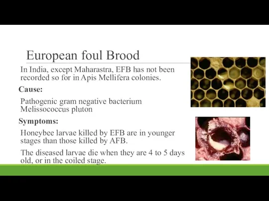
European foul Brood
In India, except Maharastra, EFB has not been recorded
European foul Brood
In India, except Maharastra, EFB has not been recorded
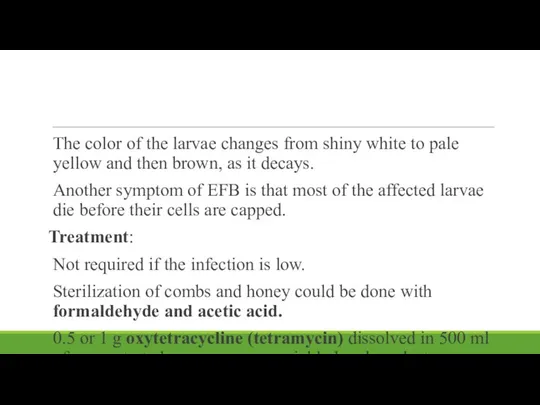
The color of the larvae changes from shiny white to pale
The color of the larvae changes from shiny white to pale
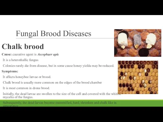
Fungal Brood Diseases
Chalk brood
Cause: causative agent is Ascophaer apis
It
Fungal Brood Diseases
Chalk brood
Cause: causative agent is Ascophaer apis
It
Treatment:
Equipment's should be sterilized using formalin or carbolic acid.
0. 7% of
Equipment's should be sterilized using formalin or carbolic acid.
0. 7% of
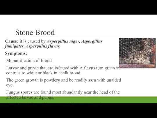
Stone Brood
Cause: it is caused by Aspergillus niger, Aspergillus fumigates,
Stone Brood
Cause: it is caused by Aspergillus niger, Aspergillus fumigates,
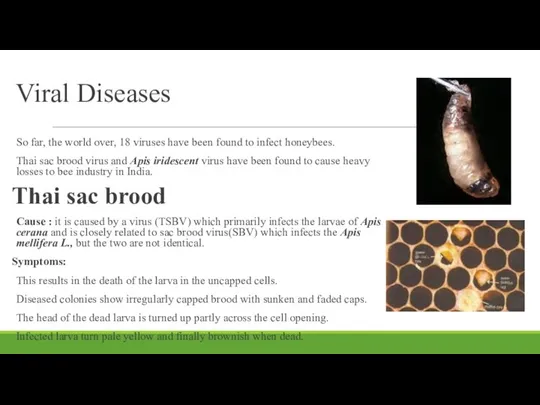
Viral Diseases
So far, the world over, 18 viruses have been
Viral Diseases
So far, the world over, 18 viruses have been
Kashmir bee virus
Kashmir bee virus is related to the preceding viruses. Recently discovered,
Kashmir bee virus
Kashmir bee virus is related to the preceding viruses. Recently discovered,
Control measurements:
use antibiotics such as Rifampin, Leavamisol, Amentidine along with
Control measurements: use antibiotics such as Rifampin, Leavamisol, Amentidine along with
Adult Bee Diseases:
The diseases of adult bees are caused by protozoa
Adult Bee Diseases:
The diseases of adult bees are caused by protozoa
Disease of Adult Bees
Apis Iridescent Virus
Symptoms:
Reduced egg laying / brood rearing.
Bees
Disease of Adult Bees
Apis Iridescent Virus
Symptoms:
Reduced egg laying / brood rearing.
Bees
Management of Viral Diseases:
For viral pathogens, there is no chemical control.
Affected
For viral pathogens, there is no chemical control.
Affected
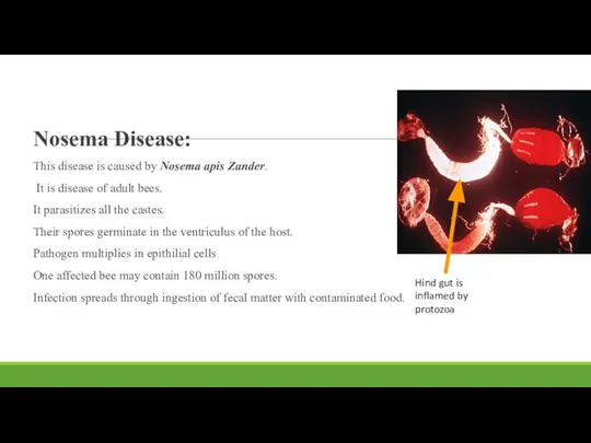
Nosema Disease:
This disease is caused by Nosema apis Zander.
It is
This disease is caused by Nosema apis Zander.
It is
Symptoms
Bees start foraging at younger age.
Bees feel fatigued, are less able
Symptoms
Bees start foraging at younger age.
Bees feel fatigued, are less able
Mid intestine is swollen and if dissected, shows dull greyish white
Mid intestine is swollen and if dissected, shows dull greyish white
ACAROSIS (The Honey Bee Tracheal Mite)
CAUSAL AGENT- honey bee tracheal mite,
ACAROSIS (The Honey Bee Tracheal Mite)
CAUSAL AGENT- honey bee tracheal mite,
PLACE OF INFECTION: Trachea and body fluid
MANAGEMENT:-
Use of grease patties (
MANAGEMENT:-
Use of grease patties (
VARROASIS (The Varroa Mite)
CAUSAL AGENT: Asiatic varroa mite, Varroa destructor.
Symptoms:
Varroa reproduce
VARROASIS (The Varroa Mite)
CAUSAL AGENT: Asiatic varroa mite, Varroa destructor.
Symptoms:
Varroa reproduce
PLACE OF INFECTION: Body and body fluid i.e. haemolymph
MANAGEMENT:
Apivar: Apivar is
PLACE OF INFECTION: Body and body fluid i.e. haemolymph
MANAGEMENT:
Apivar: Apivar is
 Бесполое размножение организмов – формы и значение в природе
 Презентация на тему Что такое кровь?
  Біологічний прогрес і біологічний регрес.
  Перелётные птицы Ульяновской области. Черный аист
 Презентация на тему Естественный отбор
  Чинники середовища життя на планеті Земля
 Лекция 7. Активный обмен. Энергобаланс
 Презентация на тему "Прогностическая эффективность биомаркеров" - скачать бесплатно презентации по Биологии
 Презентация на тему "Рациональное питание – залог здоровья" - скачать презентации по Биологии
 «Расширенное изучение анатомии человека» Руководитель курса Рогальский Аркадий Ильич, учитель биологии, высшая к/к, победитель
  Эндокринная система
 Отдел хвощевидные, или членистые
 Презентация на тему "Птиц" - скачать бесплатно презентации по Биологии
 Растения Тороповой дачи
 Царства органического мира
 Лесной комплекс - ресурсы и их эксплуатация
 Кругосветное путешествие Ч. Дарвина
 Сканирующий рентгенофлуоресцентный анализ биологических образцов
 Исследовательская работа. Чипсы, вред или польза
 Дене импульсі, импульстің сақталу заңы тақырыптарына есепрер шығару
 Сердце: его строение и работа
 Конечный мозг: полушария, борозды, извилины, базальные ядра. Локализация функций в коре полушарий
 Нейрогуморальная и эндокринная регуляция функций
  Устройство увеличительных приборов. 6 класс
 Витамины. Их влияние на здоровье человека. Подготовила: Садовникова Александра Евгеньевна
 Решение генетических задач. Анализ и составление родословных
 МУНИЦИПАЛЬНОЕ ОБЩЕОБРАЗОВАТЕЛЬНОЕ УЧРЕЖДЕНИЕ ОДИНЦОВСКАЯ ГИМНАЗИЯ № 11 ВИРУСЫ – НЕКЛЕТОЧНАЯ ФОРМА ЖИЗНИ…
  ДТ биотехнология