- The Structure and Function of Large Biological Molecules

Содержание
- 2. Overview: The Molecules of Life All living things are made up of four classes of large
- 3. Fig. 5-1
- 4. Concept 5.1: Macromolecules are polymers, built from monomers A polymer is a long molecule consisting of
- 5. A condensation reaction or more specifically a dehydration reaction occurs when two monomers bond together through
- 6. Fig. 5-2 Short polymer HO 1 2 3 H HO H Unlinked monomer Dehydration removes a
- 7. Fig. 5-2a Dehydration removes a water molecule, forming a new bond Short polymer Unlinked monomer Longer
- 8. Fig. 5-2b Hydrolysis adds a water molecule, breaking a bond Hydrolysis of a polymer HO HO
- 9. The Diversity of Polymers Each cell has thousands of different kinds of macromolecules Macromolecules vary among
- 10. Concept 5.2: Carbohydrates serve as fuel and building material Carbohydrates include sugars and the polymers of
- 11. Sugars Monosaccharides have molecular formulas that are usually multiples of CH2O Glucose (C6H12O6) is the most
- 12. Fig. 5-3 Dihydroxyacetone Ribulose Ketoses Aldoses Fructose Glyceraldehyde Ribose Glucose Galactose Hexoses (C6H12O6) Pentoses (C5H10O5) Trioses
- 13. Fig. 5-3a Aldoses Glyceraldehyde Ribose Glucose Galactose Hexoses (C6H12O6) Pentoses (C5H10O5) Trioses (C3H6O3)
- 14. Fig. 5-3b Ketoses Dihydroxyacetone Ribulose Fructose Hexoses (C6H12O6) Pentoses (C5H10O5) Trioses (C3H6O3)
- 15. Though often drawn as linear skeletons, in aqueous solutions many sugars form rings Monosaccharides serve as
- 16. Fig. 5-4 (a) Linear and ring forms (b) Abbreviated ring structure
- 17. Fig. 5-4a (a) Linear and ring forms
- 18. Fig. 5-4b (b) Abbreviated ring structure
- 19. A disaccharide is formed when a dehydration reaction joins two monosaccharides This covalent bond is called
- 20. Fig. 5-5 (b) Dehydration reaction in the synthesis of sucrose Glucose Fructose Sucrose Maltose Glucose Glucose
- 21. Polysaccharides Polysaccharides, the polymers of sugars, have storage and structural roles The structure and function of
- 22. Storage Polysaccharides Starch, a storage polysaccharide of plants, consists entirely of glucose monomers Plants store surplus
- 23. Fig. 5-6 (b) Glycogen: an animal polysaccharide Starch Glycogen Amylose Chloroplast (a) Starch: a plant polysaccharide
- 24. Glycogen is a storage polysaccharide in animals Humans and other vertebrates store glycogen mainly in liver
- 25. Structural Polysaccharides The polysaccharide cellulose is a major component of the tough wall of plant cells
- 26. Fig. 5-7 (a) and glucose ring structures Glucose Glucose (b) Starch: 1–4
- 27. Fig. 5-7a (a) and glucose ring structures Glucose Glucose
- 28. Fig. 5-7bc (b) Starch: 1–4 linkage of glucose monomers (c) Cellulose: 1–4 linkage of
- 29. Polymers with α glucose are helical Polymers with β glucose are straight In straight structures, H
- 30. Fig. 5-8 Glucose monomer Cellulose molecules Microfibril Cellulose microfibrils in a plant cell wall 0.5 µm
- 31. Enzymes that digest starch by hydrolyzing α linkages can’t hydrolyze β linkages in cellulose Cellulose in
- 32. Fig. 5-9
- 33. Chitin, another structural polysaccharide, is found in the exoskeleton of arthropods Chitin also provides structural support
- 34. Fig. 5-10 The structure of the chitin monomer. (a) (b) (c) Chitin forms the exoskeleton of
- 35. Concept 5.3: Lipids are a diverse group of hydrophobic molecules Lipids are the one class of
- 36. Fats Fats are constructed from two types of smaller molecules: glycerol and fatty acids Glycerol is
- 37. Fig. 5-11 Fatty acid (palmitic acid) Glycerol (a) Dehydration reaction in the synthesis of a fat
- 38. Fig. 5-11a Fatty acid (palmitic acid) (a) Dehydration reaction in the synthesis of a fat Glycerol
- 39. Fig. 5-11b (b) Fat molecule (triacylglycerol) Ester linkage
- 40. Fats separate from water because water molecules form hydrogen bonds with each other and exclude the
- 41. Fatty acids vary in length (number of carbons) and in the number and locations of double
- 42. Fig. 5-12 Structural formula of a saturated fat molecule Stearic acid, a saturated fatty acid (a)
- 43. Fig. 5-12a (a) Saturated fat Structural formula of a saturated fat molecule Stearic acid, a saturated
- 44. Fig. 5-12b (b) Unsaturated fat Structural formula of an unsaturated fat molecule Oleic acid, an unsaturated
- 45. Fats made from saturated fatty acids are called saturated fats, and are solid at room temperature
- 46. A diet rich in saturated fats may contribute to cardiovascular disease through plaque deposits Hydrogenation is
- 47. The major function of fats is energy storage Humans and other mammals store their fat in
- 48. Phospholipids In a phospholipid, two fatty acids and a phosphate group are attached to glycerol The
- 49. Fig. 5-13 (b) Space-filling model (a) (c) Structural formula Phospholipid symbol Fatty acids Hydrophilic head Hydrophobic
- 50. Fig. 5-13ab (b) Space-filling model (a) Structural formula Fatty acids Choline Phosphate Glycerol Hydrophobic tails Hydrophilic
- 51. When phospholipids are added to water, they self-assemble into a bilayer, with the hydrophobic tails pointing
- 52. Fig. 5-14 Hydrophilic head Hydrophobic tail WATER WATER
- 53. Steroids Steroids are lipids characterized by a carbon skeleton consisting of four fused rings Cholesterol, an
- 54. Fig. 5-15
- 55. Concept 5.4: Proteins have many structures, resulting in a wide range of functions Proteins account for
- 56. Table 5-1
- 57. Animation: Structural Proteins Animation: Storage Proteins Animation: Transport Proteins Animation: Receptor Proteins Animation: Contractile Proteins Animation:
- 58. Enzymes are a type of protein that acts as a catalyst to speed up chemical reactions
- 59. Fig. 5-16 Enzyme (sucrase) Substrate (sucrose) Fructose Glucose OH H O H2O
- 60. Polypeptides Polypeptides are polymers built from the same set of 20 amino acids A protein consists
- 61. Amino Acid Monomers Amino acids are organic molecules with carboxyl and amino groups Amino acids differ
- 62. Fig. 5-UN1 Amino group Carboxyl group carbon
- 63. Fig. 5-17 Nonpolar Glycine (Gly or G) Alanine (Ala or A) Valine (Val or V) Leucine
- 64. Fig. 5-17a Nonpolar Glycine (Gly or G) Alanine (Ala or A) Valine (Val or V) Leucine
- 65. Fig. 5-17b Polar Asparagine (Asn or N) Glutamine (Gln or Q) Serine (Ser or S) Threonine
- 66. Fig. 5-17c Acidic Arginine (Arg or R) Histidine (His or H) Aspartic acid (Asp or D)
- 67. Amino Acid Polymers Amino acids are linked by peptide bonds A polypeptide is a polymer of
- 68. Peptide bond Fig. 5-18 Amino end (N-terminus) Peptide bond Side chains Backbone Carboxyl end (C-terminus) (a)
- 69. Protein Structure and Function A functional protein consists of one or more polypeptides twisted, folded, and
- 70. Fig. 5-19 A ribbon model of lysozyme (a) (b) A space-filling model of lysozyme Groove Groove
- 71. Fig. 5-19a A ribbon model of lysozyme (a) Groove
- 72. Fig. 5-19b (b) A space-filling model of lysozyme Groove
- 73. The sequence of amino acids determines a protein’s three-dimensional structure A protein’s structure determines its function
- 74. Fig. 5-20 Antibody protein Protein from flu virus
- 75. Four Levels of Protein Structure The primary structure of a protein is its unique sequence of
- 76. Primary structure, the sequence of amino acids in a protein, is like the order of letters
- 77. Fig. 5-21 Primary Structure Secondary Structure Tertiary Structure pleated sheet Examples of amino acid subunits
- 78. Fig. 5-21a Amino acid subunits +H3N Amino end 25 20 15 10 5 1 Primary Structure
- 79. Fig. 5-21b Amino acid subunits +H3N Amino end Carboxyl end 125 120 115 110 105 100
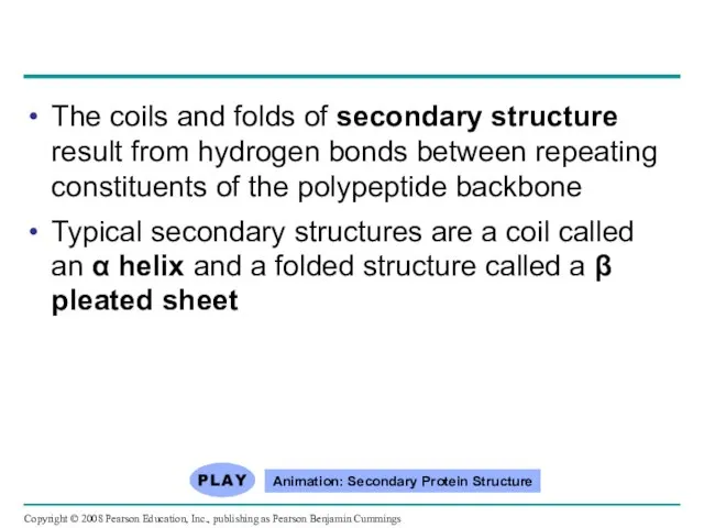
- 80. The coils and folds of secondary structure result from hydrogen bonds between repeating constituents of the
- 81. Fig. 5-21c Secondary Structure pleated sheet Examples of amino acid subunits helix
- 82. Fig. 5-21d Abdominal glands of the spider secrete silk fibers made of a structural protein containing
- 83. Tertiary structure is determined by interactions between R groups, rather than interactions between backbone constituents These
- 84. Fig. 5-21e Tertiary Structure Quaternary Structure
- 85. Fig. 5-21f Polypeptide backbone Hydrophobic interactions and van der Waals interactions Disulfide bridge Ionic bond Hydrogen
- 86. Fig. 5-21g Polypeptide chain Chains Heme Iron Chains Collagen Hemoglobin
- 87. Quaternary structure results when two or more polypeptide chains form one macromolecule Collagen is a fibrous
- 88. Sickle-Cell Disease: A Change in Primary Structure A slight change in primary structure can affect a
- 89. Fig. 5-22 Primary structure Secondary and tertiary structures Quaternary structure Normal hemoglobin (top view) Primary structure
- 90. Fig. 5-22a Primary structure Secondary and tertiary structures Function Quaternary structure Molecules do not associate with
- 91. Fig. 5-22b Primary structure Secondary and tertiary structures Function Quaternary structure Molecules interact with one another
- 92. Fig. 5-22c Normal red blood cells are full of individual hemoglobin molecules, each carrying oxygen. Fibers
- 93. What Determines Protein Structure? In addition to primary structure, physical and chemical conditions can affect structure
- 94. Fig. 5-23 Normal protein Denatured protein Denaturation Renaturation
- 95. Protein Folding in the Cell It is hard to predict a protein’s structure from its primary
- 96. Fig. 5-24 Hollow cylinder Cap Chaperonin (fully assembled) Polypeptide Steps of Chaperonin Action: An unfolded poly-
- 97. Fig. 5-24a Hollow cylinder Chaperonin (fully assembled) Cap
- 98. Fig. 5-24b Correctly folded protein Polypeptide Steps of Chaperonin Action: 1 2 An unfolded poly- peptide
- 99. Scientists use X-ray crystallography to determine a protein’s structure Another method is nuclear magnetic resonance (NMR)
- 100. Fig. 5-25 EXPERIMENT RESULTS X-ray source X-ray beam Diffracted X-rays Crystal Digital detector X-ray diffraction pattern
- 101. Fig. 5-25a Diffracted X-rays EXPERIMENT X-ray source X-ray beam Crystal Digital detector X-ray diffraction pattern
- 102. Fig. 5-25b RESULTS RNA RNA polymerase II DNA
- 103. Concept 5.5: Nucleic acids store and transmit hereditary information The amino acid sequence of a polypeptide
- 104. The Roles of Nucleic Acids There are two types of nucleic acids: Deoxyribonucleic acid (DNA) Ribonucleic
- 105. Fig. 5-26-1 mRNA Synthesis of mRNA in the nucleus DNA NUCLEUS CYTOPLASM 1
- 106. Fig. 5-26-2 mRNA Synthesis of mRNA in the nucleus DNA NUCLEUS mRNA CYTOPLASM Movement of mRNA
- 107. Fig. 5-26-3 mRNA Synthesis of mRNA in the nucleus DNA NUCLEUS mRNA CYTOPLASM Movement of mRNA
- 108. The Structure of Nucleic Acids Nucleic acids are polymers called polynucleotides Each polynucleotide is made of
- 109. Fig. 5-27 5 end Nucleoside Nitrogenous base Phosphate group Sugar (pentose) (b) Nucleotide (a) Polynucleotide, or
- 110. Fig. 5-27ab 5' end 5'C 3'C 5'C 3'C 3' end (a) Polynucleotide, or nucleic acid (b)
- 111. Fig. 5-27c-1 (c) Nucleoside components: nitrogenous bases Purines Guanine (G) Adenine (A) Cytosine (C) Thymine (T,
- 112. Fig. 5-27c-2 Ribose (in RNA) Deoxyribose (in DNA) Sugars (c) Nucleoside components: sugars
- 113. Nucleotide Monomers Nucleoside = nitrogenous base + sugar There are two families of nitrogenous bases: Pyrimidines
- 114. Nucleotide Polymers Nucleotide polymers are linked together to build a polynucleotide Adjacent nucleotides are joined by
- 115. The DNA Double Helix A DNA molecule has two polynucleotides spiraling around an imaginary axis, forming
- 116. Fig. 5-28 Sugar-phosphate backbones 3' end 3' end 3' end 3' end 5' end 5' end
- 117. DNA and Proteins as Tape Measures of Evolution The linear sequences of nucleotides in DNA molecules
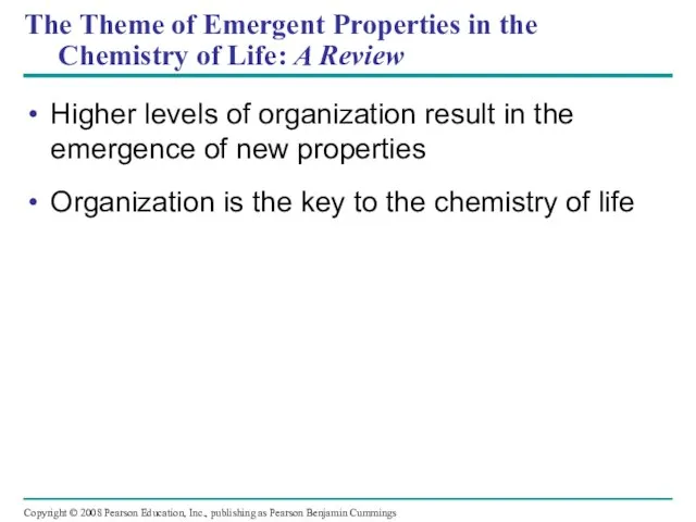
- 118. The Theme of Emergent Properties in the Chemistry of Life: A Review Higher levels of organization
- 119. Fig. 5-UN2
- 120. Fig. 5-UN2a
- 121. Fig. 5-UN2b
- 122. Fig. 5-UN3 % of glycosidic linkages broken 100 50 0 Time
- 123. Fig. 5-UN4
- 124. Fig. 5-UN5
- 125. Fig. 5-UN6
- 126. Fig. 5-UN7
- 127. Fig. 5-UN8
- 128. Fig. 5-UN9
- 129. Fig. 5-UN10
- 130. You should now be able to: List and describe the four major classes of molecules Describe
- 132. Скачать презентацию
Overview: The Molecules of Life
All living things are made up of
Overview: The Molecules of Life
All living things are made up of
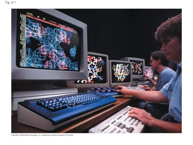
Fig. 5-1
Fig. 5-1
Concept 5.1: Macromolecules are polymers, built from monomers
A polymer is a
Concept 5.1: Macromolecules are polymers, built from monomers
A polymer is a
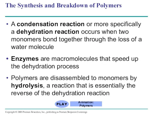
A condensation reaction or more specifically a dehydration reaction occurs when
A condensation reaction or more specifically a dehydration reaction occurs when
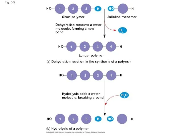
Fig. 5-2
Short polymer
HO
1
2
3
H
HO
H
Unlinked monomer
Dehydration removes a water
molecule, forming a new bond
HO
H2O
H
1
2
3
4
Longer
Fig. 5-2
Short polymer
HO
1
2
3
H
HO
H
Unlinked monomer
Dehydration removes a water
molecule, forming a new bond
HO
H2O
H
1
2
3
4
Longer
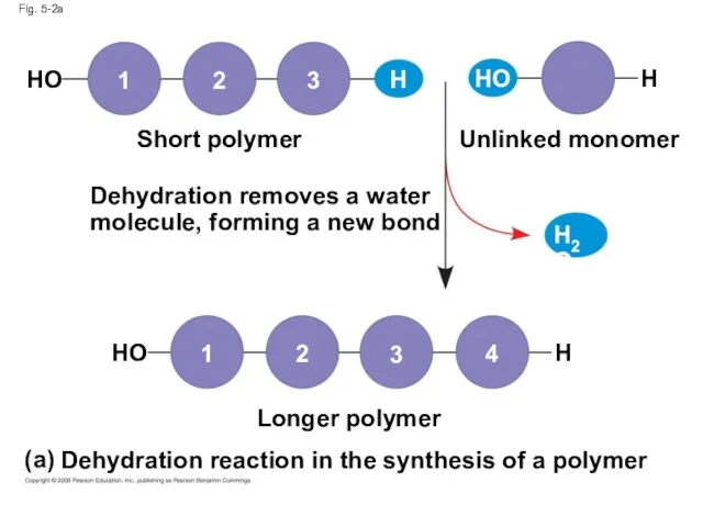
Fig. 5-2a
Dehydration removes a water
molecule, forming a new bond
Short polymer
Unlinked monomer
Longer
Fig. 5-2a
Dehydration removes a water
molecule, forming a new bond
Short polymer
Unlinked monomer
Longer
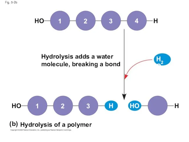
Fig. 5-2b
Hydrolysis adds a water
molecule, breaking a bond
Hydrolysis of a polymer
HO
HO
HO
H2O
H
H
H
3
2
1
1
2
3
4
(b)
Fig. 5-2b
Hydrolysis adds a water
molecule, breaking a bond
Hydrolysis of a polymer
HO
HO
HO
H2O
H
H
H
3
2
1
1
2
3
4
(b)
The Diversity of Polymers
Each cell has thousands of different kinds of
The Diversity of Polymers
Each cell has thousands of different kinds of
Concept 5.2: Carbohydrates serve as fuel and building material
Carbohydrates include sugars
Concept 5.2: Carbohydrates serve as fuel and building material
Carbohydrates include sugars
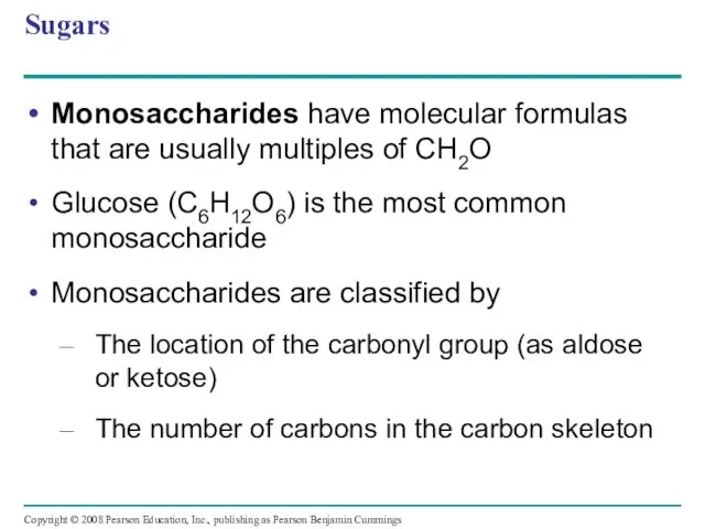
Sugars
Monosaccharides have molecular formulas that are usually multiples of CH2O
Glucose (C6H12O6)
Sugars
Monosaccharides have molecular formulas that are usually multiples of CH2O
Glucose (C6H12O6)
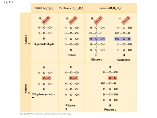
Fig. 5-3
Dihydroxyacetone
Ribulose
Ketoses
Aldoses
Fructose
Glyceraldehyde
Ribose
Glucose
Galactose
Hexoses (C6H12O6)
Pentoses (C5H10O5)
Trioses (C3H6O3)
Fig. 5-3
Dihydroxyacetone
Ribulose
Ketoses
Aldoses
Fructose
Glyceraldehyde
Ribose
Glucose
Galactose
Hexoses (C6H12O6)
Pentoses (C5H10O5)
Trioses (C3H6O3)
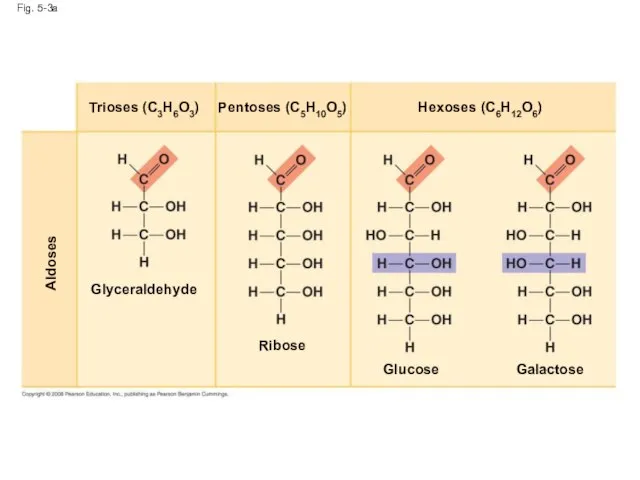
Fig. 5-3a
Aldoses
Glyceraldehyde
Ribose
Glucose
Galactose
Hexoses (C6H12O6)
Pentoses (C5H10O5)
Trioses (C3H6O3)
Fig. 5-3a
Aldoses
Glyceraldehyde
Ribose
Glucose
Galactose
Hexoses (C6H12O6)
Pentoses (C5H10O5)
Trioses (C3H6O3)
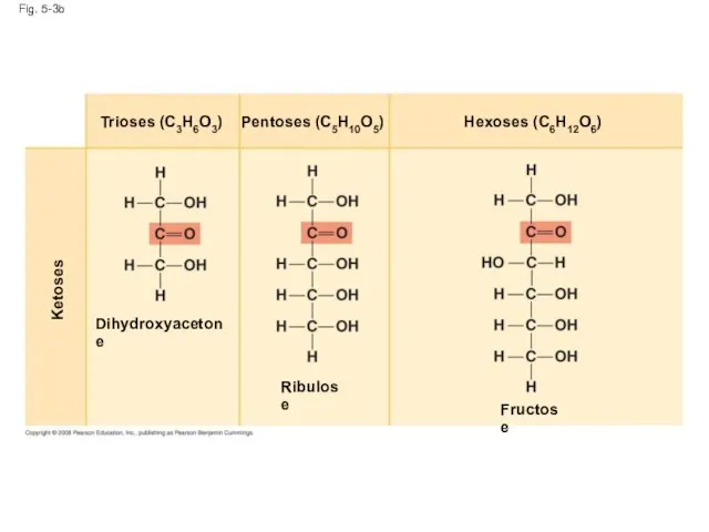
Fig. 5-3b
Ketoses
Dihydroxyacetone
Ribulose
Fructose
Hexoses (C6H12O6)
Pentoses (C5H10O5)
Trioses (C3H6O3)
Fig. 5-3b
Ketoses
Dihydroxyacetone
Ribulose
Fructose
Hexoses (C6H12O6)
Pentoses (C5H10O5)
Trioses (C3H6O3)
Though often drawn as linear skeletons, in aqueous solutions many sugars
Though often drawn as linear skeletons, in aqueous solutions many sugars
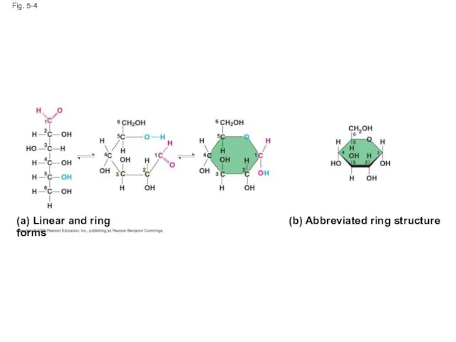
Fig. 5-4
(a) Linear and ring forms
(b) Abbreviated ring structure
Fig. 5-4
(a) Linear and ring forms
(b) Abbreviated ring structure
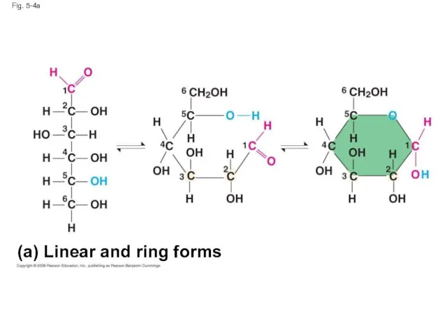
Fig. 5-4a
(a) Linear and ring forms
Fig. 5-4a
(a) Linear and ring forms
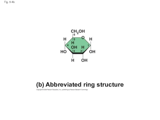
Fig. 5-4b
(b) Abbreviated ring structure
Fig. 5-4b
(b) Abbreviated ring structure
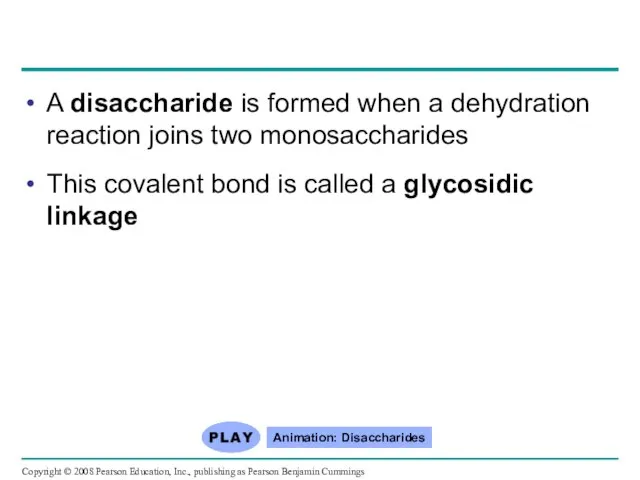
A disaccharide is formed when a dehydration reaction joins two monosaccharides
A disaccharide is formed when a dehydration reaction joins two monosaccharides
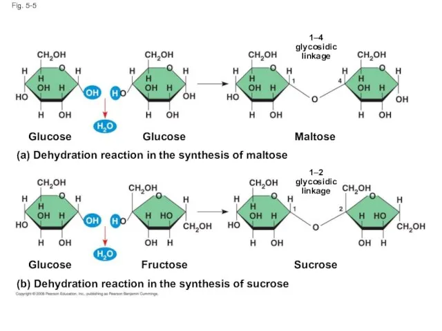
Fig. 5-5
(b) Dehydration reaction in the synthesis of sucrose
Glucose
Fructose
Sucrose
Maltose
Glucose
Glucose
(a) Dehydration reaction
Fig. 5-5
(b) Dehydration reaction in the synthesis of sucrose
Glucose
Fructose
Sucrose
Maltose
Glucose
Glucose
(a) Dehydration reaction
Polysaccharides
Polysaccharides, the polymers of sugars, have storage and structural roles
The structure
Polysaccharides
Polysaccharides, the polymers of sugars, have storage and structural roles
The structure
Storage Polysaccharides
Starch, a storage polysaccharide of plants, consists entirely of glucose
Storage Polysaccharides
Starch, a storage polysaccharide of plants, consists entirely of glucose
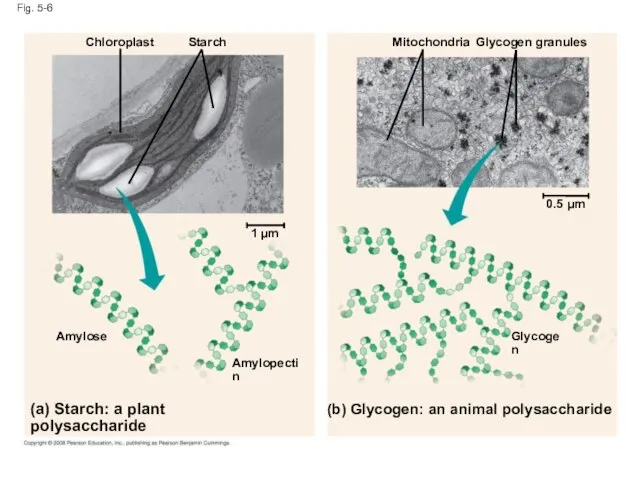
Fig. 5-6
(b) Glycogen: an animal polysaccharide
Starch
Glycogen
Amylose
Chloroplast
(a) Starch: a plant polysaccharide
Amylopectin
Mitochondria
Glycogen granules
0.5
Fig. 5-6
(b) Glycogen: an animal polysaccharide
Starch
Glycogen
Amylose
Chloroplast
(a) Starch: a plant polysaccharide
Amylopectin
Mitochondria
Glycogen granules
0.5
Glycogen is a storage polysaccharide in animals
Humans and other vertebrates store
Glycogen is a storage polysaccharide in animals
Humans and other vertebrates store
Structural Polysaccharides
The polysaccharide cellulose is a major component of the tough
Structural Polysaccharides
The polysaccharide cellulose is a major component of the tough
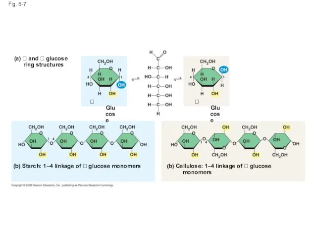
Fig. 5-7
(a) and glucose
ring structures
Glucose
Glucose
(b) Starch:
Fig. 5-7
(a) and glucose
ring structures
Glucose
Glucose
(b) Starch:
Fig. 5-7a
(a) and glucose ring structures
Glucose
Fig. 5-7a
(a) and glucose ring structures
Glucose
Fig. 5-7bc
(b) Starch: 1–4 linkage of glucose monomers
(c) Cellulose: 1–4
Fig. 5-7bc
(b) Starch: 1–4 linkage of glucose monomers
(c) Cellulose: 1–4
Polymers with α glucose are helical
Polymers with β glucose are straight
In
Polymers with α glucose are helical
Polymers with β glucose are straight
In
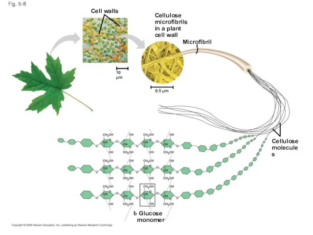
Fig. 5-8
Glucose
monomer
Cellulose
molecules
Microfibril
Cellulose
microfibrils
in a plant
cell wall
0.5 µm
10 µm
Cell walls
Fig. 5-8
Glucose
monomer
Cellulose
molecules
Microfibril
Cellulose
microfibrils
in a plant
cell wall
0.5 µm
10 µm
Cell walls
Enzymes that digest starch by hydrolyzing α linkages can’t hydrolyze β
Enzymes that digest starch by hydrolyzing α linkages can’t hydrolyze β
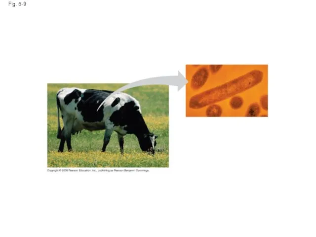
Fig. 5-9
Fig. 5-9
Chitin, another structural polysaccharide, is found in the exoskeleton of arthropods
Chitin
Chitin, another structural polysaccharide, is found in the exoskeleton of arthropods
Chitin
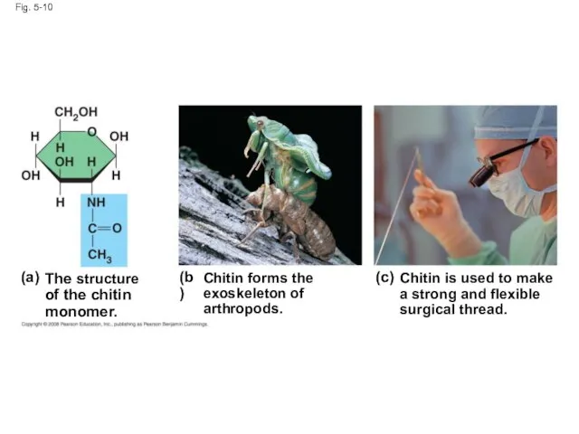
Fig. 5-10
The structure
of the chitin
monomer.
(a)
(b)
(c)
Chitin forms the
exoskeleton of
arthropods.
Chitin is used to
Fig. 5-10
The structure
of the chitin
monomer.
(a)
(b)
(c)
Chitin forms the
exoskeleton of
arthropods.
Chitin is used to
Concept 5.3: Lipids are a diverse group of hydrophobic molecules
Lipids are
Concept 5.3: Lipids are a diverse group of hydrophobic molecules
Lipids are
Fats
Fats are constructed from two types of smaller molecules: glycerol and
Fats
Fats are constructed from two types of smaller molecules: glycerol and
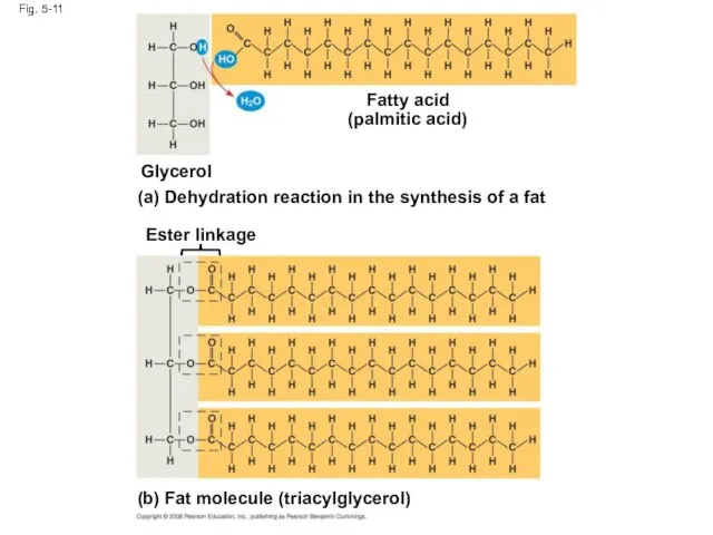
Fig. 5-11
Fatty acid
(palmitic acid)
Glycerol
(a) Dehydration reaction in the synthesis of a
Fig. 5-11
Fatty acid
(palmitic acid)
Glycerol
(a) Dehydration reaction in the synthesis of a
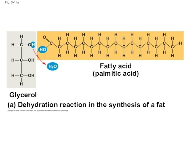
Fig. 5-11a
Fatty acid
(palmitic acid)
(a)
Dehydration reaction in the synthesis of a fat
Glycerol
Fig. 5-11a
Fatty acid
(palmitic acid)
(a)
Dehydration reaction in the synthesis of a fat
Glycerol
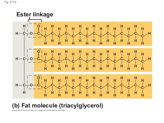
Fig. 5-11b
(b)
Fat molecule (triacylglycerol)
Ester linkage
Fig. 5-11b
(b)
Fat molecule (triacylglycerol)
Ester linkage
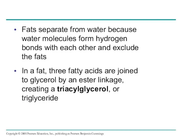
Fats separate from water because water molecules form hydrogen bonds with
Fats separate from water because water molecules form hydrogen bonds with
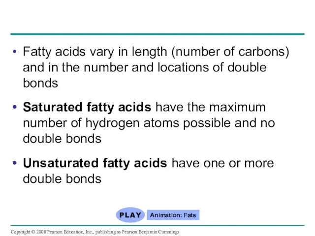
Fatty acids vary in length (number of carbons) and in the
Fatty acids vary in length (number of carbons) and in the
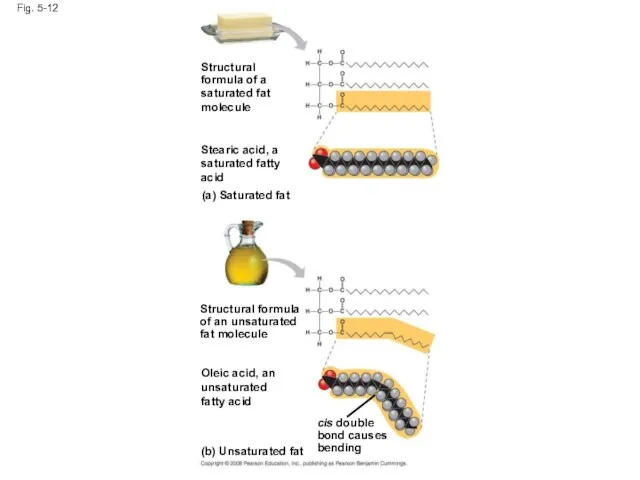
Fig. 5-12
Structural
formula of a
saturated fat
molecule
Stearic acid, a
saturated fatty
acid
(a) Saturated fat
Structural formula
of
Fig. 5-12
Structural
formula of a
saturated fat
molecule
Stearic acid, a
saturated fatty
acid
(a) Saturated fat
Structural formula
of
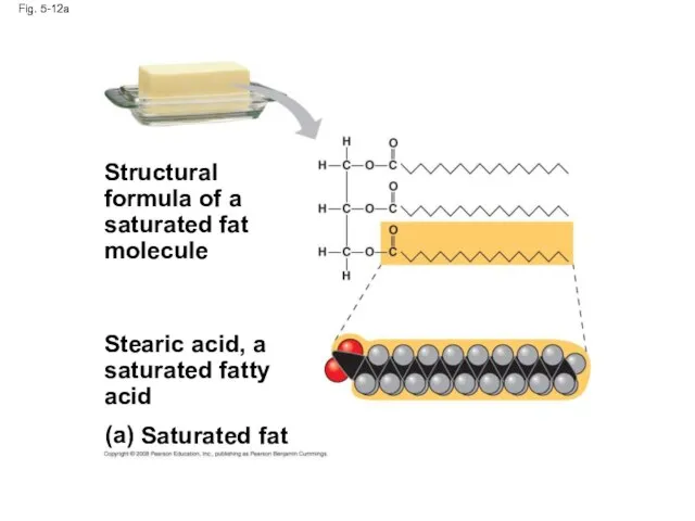
Fig. 5-12a
(a)
Saturated fat
Structural
formula of a
saturated fat
molecule
Stearic acid, a
saturated fatty
acid
Fig. 5-12a
(a)
Saturated fat
Structural
formula of a
saturated fat
molecule
Stearic acid, a
saturated fatty
acid
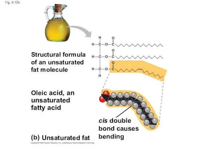
Fig. 5-12b
(b)
Unsaturated fat
Structural formula
of an unsaturated
fat molecule
Oleic acid, an
unsaturated
fatty acid
cis double
bond
Fig. 5-12b
(b)
Unsaturated fat
Structural formula
of an unsaturated
fat molecule
Oleic acid, an
unsaturated
fatty acid
cis double
bond
Fats made from saturated fatty acids are called saturated fats, and
Fats made from saturated fatty acids are called saturated fats, and
A diet rich in saturated fats may contribute to cardiovascular disease
A diet rich in saturated fats may contribute to cardiovascular disease
The major function of fats is energy storage
Humans and other mammals
The major function of fats is energy storage
Humans and other mammals
Phospholipids
In a phospholipid, two fatty acids and a phosphate group are
Phospholipids
In a phospholipid, two fatty acids and a phosphate group are
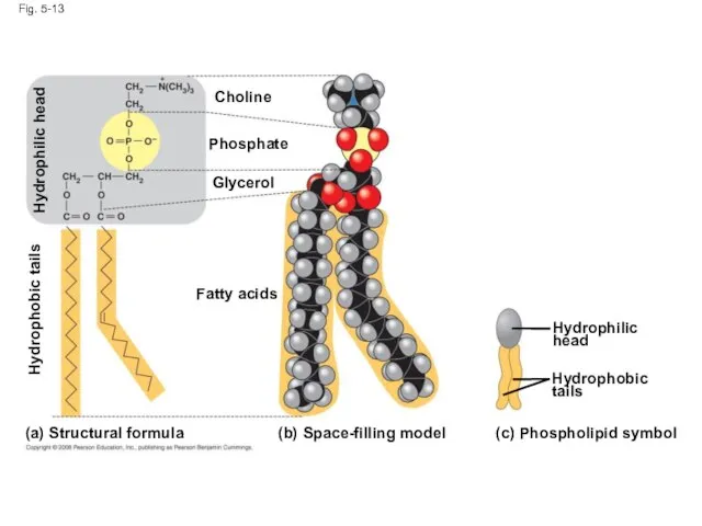
Fig. 5-13
(b)
Space-filling model
(a)
(c)
Structural formula
Phospholipid symbol
Fatty acids
Hydrophilic
head
Hydrophobic
tails
Choline
Phosphate
Glycerol
Hydrophobic tails
Hydrophilic head
Fig. 5-13
(b)
Space-filling model
(a)
(c)
Structural formula
Phospholipid symbol
Fatty acids
Hydrophilic
head
Hydrophobic
tails
Choline
Phosphate
Glycerol
Hydrophobic tails
Hydrophilic head
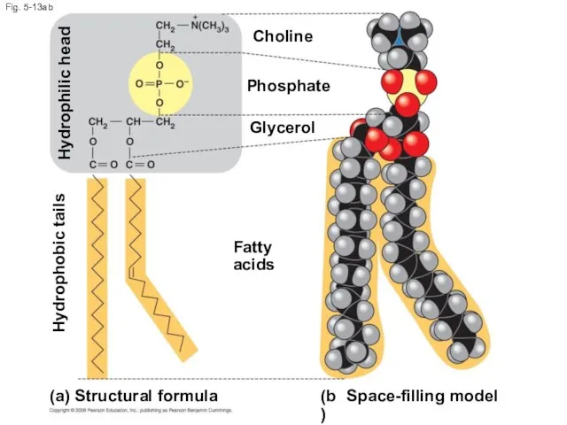
Fig. 5-13ab
(b)
Space-filling model
(a)
Structural formula
Fatty acids
Choline
Phosphate
Glycerol
Hydrophobic tails
Hydrophilic head
Fig. 5-13ab
(b)
Space-filling model
(a)
Structural formula
Fatty acids
Choline
Phosphate
Glycerol
Hydrophobic tails
Hydrophilic head
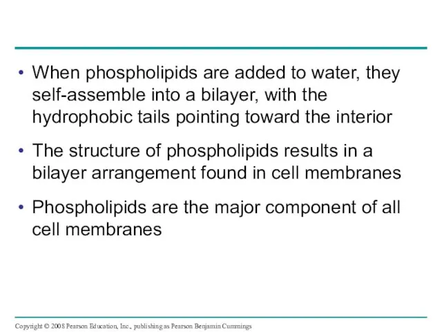
When phospholipids are added to water, they self-assemble into a bilayer,
When phospholipids are added to water, they self-assemble into a bilayer,
Fig. 5-14
Hydrophilic
head
Hydrophobic
tail
WATER
WATER
Fig. 5-14
Hydrophilic
head
Hydrophobic
tail
WATER
WATER
Steroids
Steroids are lipids characterized by a carbon skeleton consisting of four
Steroids
Steroids are lipids characterized by a carbon skeleton consisting of four
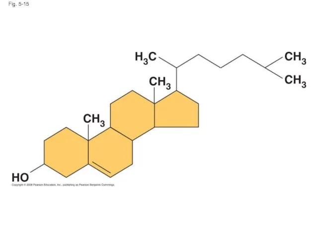
Fig. 5-15
Fig. 5-15
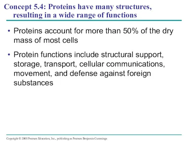
Concept 5.4: Proteins have many structures, resulting in a wide range
Concept 5.4: Proteins have many structures, resulting in a wide range
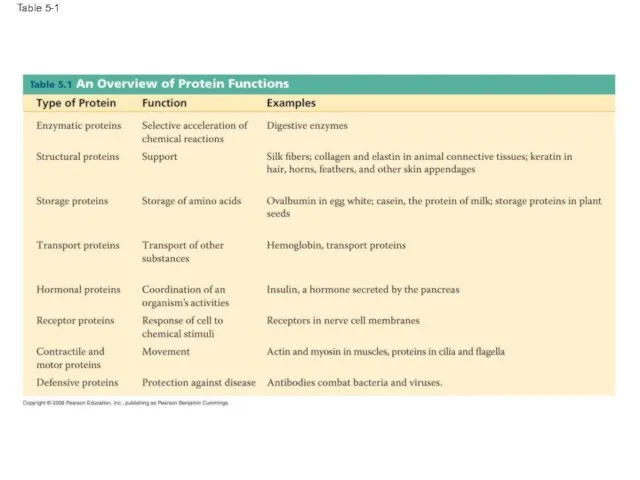
Table 5-1
Table 5-1
Animation: Structural Proteins
Animation: Storage Proteins
Animation: Transport Proteins
Animation: Receptor Proteins
Animation: Contractile Proteins
Animation:
Animation: Structural Proteins
Animation: Storage Proteins
Animation: Transport Proteins
Animation: Receptor Proteins
Animation: Contractile Proteins
Animation:
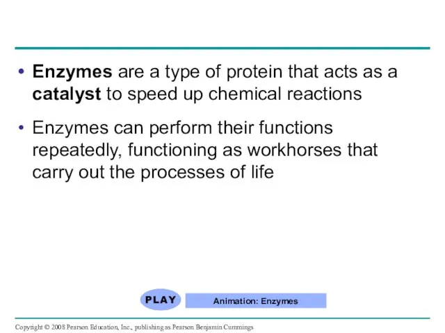
Enzymes are a type of protein that acts as a catalyst
Enzymes are a type of protein that acts as a catalyst
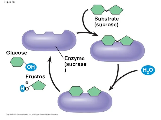
Fig. 5-16
Enzyme
(sucrase)
Substrate
(sucrose)
Fructose
Glucose
OH
H
O
H2O
Fig. 5-16
Enzyme
(sucrase)
Substrate
(sucrose)
Fructose
Glucose
OH
H
O
H2O
Polypeptides
Polypeptides are polymers built from the same set of 20 amino
Polypeptides
Polypeptides are polymers built from the same set of 20 amino
Amino Acid Monomers
Amino acids are organic molecules with carboxyl and amino
Amino Acid Monomers
Amino acids are organic molecules with carboxyl and amino
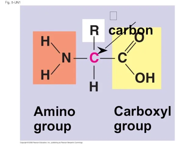
Fig. 5-UN1
Amino
group
Carboxyl
group
carbon
Fig. 5-UN1
Amino
group
Carboxyl
group
carbon
Fig. 5-17
Nonpolar
Glycine
(Gly or G)
Alanine
(Ala or A)
Valine
(Val or V)
Leucine
(Leu or L)
Isoleucine
(Ile or
Fig. 5-17
Nonpolar
Glycine
(Gly or G)
Alanine
(Ala or A)
Valine
(Val or V)
Leucine
(Leu or L)
Isoleucine
(Ile or
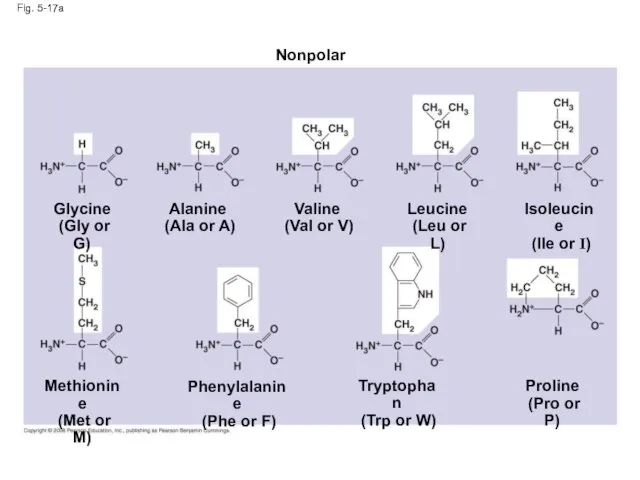
Fig. 5-17a
Nonpolar
Glycine
(Gly or G)
Alanine
(Ala or A)
Valine
(Val or V)
Leucine
Fig. 5-17a
Nonpolar
Glycine
(Gly or G)
Alanine
(Ala or A)
Valine
(Val or V)
Leucine
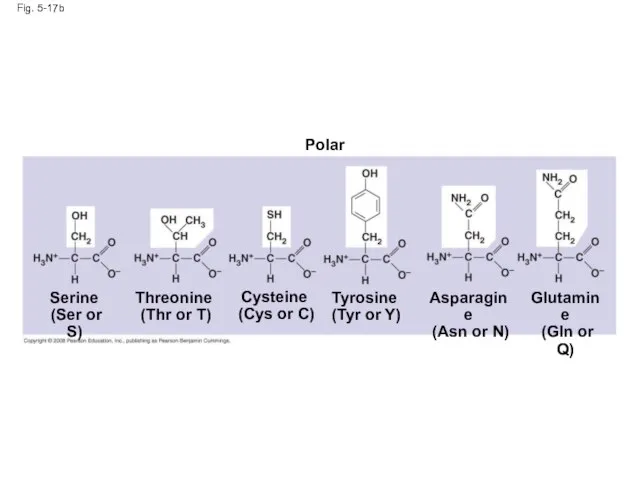
Fig. 5-17b
Polar
Asparagine
(Asn or N)
Glutamine
(Gln or Q)
Serine
(Ser or S)
Threonine
Fig. 5-17b
Polar
Asparagine
(Asn or N)
Glutamine
(Gln or Q)
Serine
(Ser or S)
Threonine
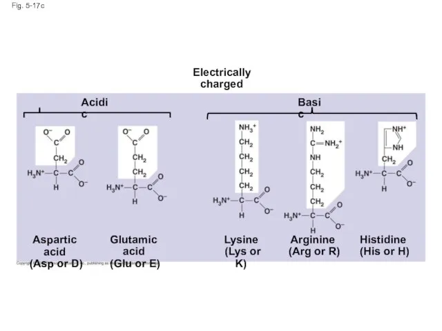
Fig. 5-17c
Acidic
Arginine
(Arg or R)
Histidine
(His or H)
Aspartic acid
(Asp or
Fig. 5-17c
Acidic
Arginine
(Arg or R)
Histidine
(His or H)
Aspartic acid
(Asp or
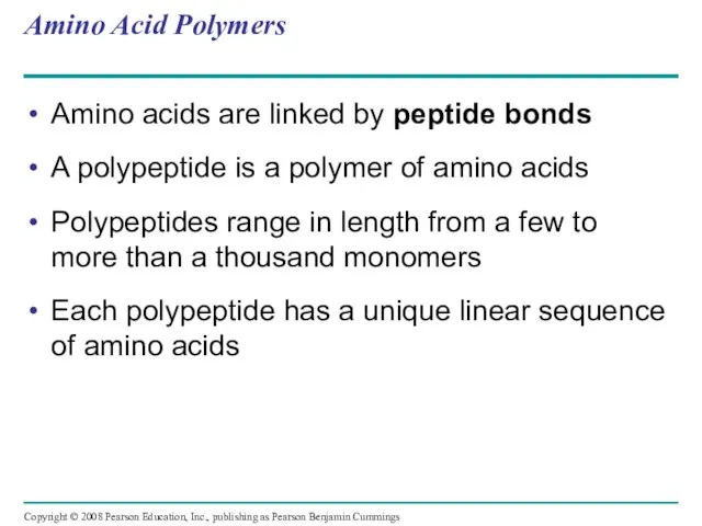
Amino Acid Polymers
Amino acids are linked by peptide bonds
A polypeptide is
Amino Acid Polymers
Amino acids are linked by peptide bonds
A polypeptide is
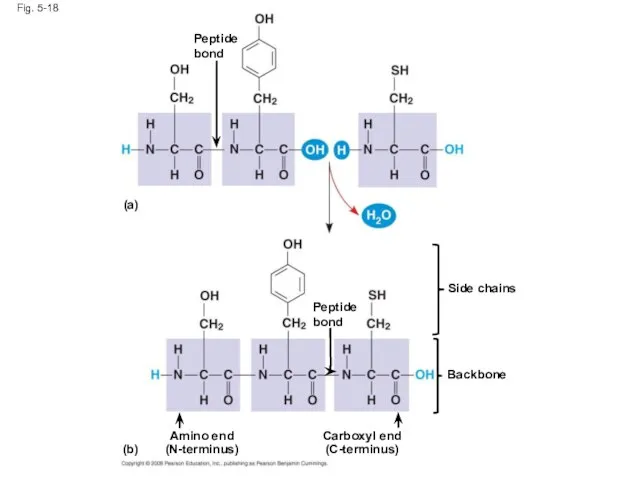
Peptide
bond
Fig. 5-18
Amino end
(N-terminus)
Peptide
bond
Side chains
Backbone
Carboxyl end
(C-terminus)
(a)
(b)
Peptide
bond
Fig. 5-18
Amino end
(N-terminus)
Peptide
bond
Side chains
Backbone
Carboxyl end
(C-terminus)
(a)
(b)
Protein Structure and Function
A functional protein consists of one or more
Protein Structure and Function
A functional protein consists of one or more
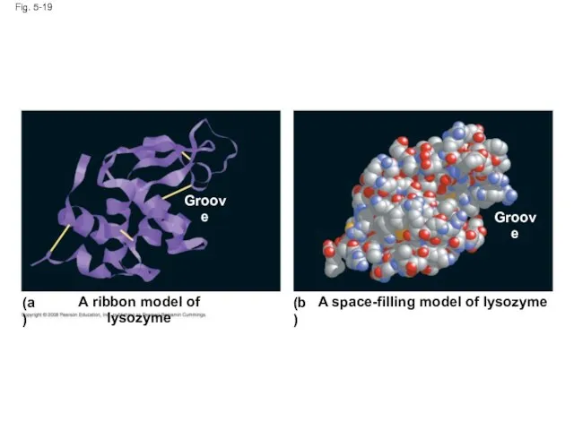
Fig. 5-19
A ribbon model of lysozyme
(a)
(b)
A space-filling model of lysozyme
Groove
Groove
Fig. 5-19
A ribbon model of lysozyme
(a)
(b)
A space-filling model of lysozyme
Groove
Groove
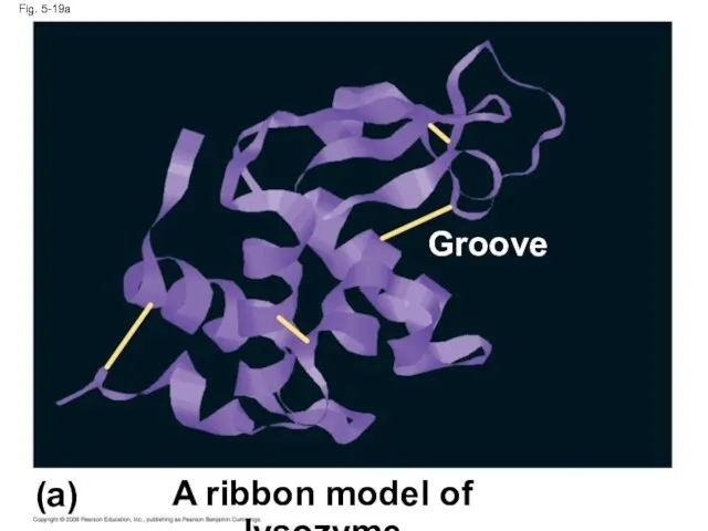
Fig. 5-19a
A ribbon model of lysozyme
(a)
Groove
Fig. 5-19a
A ribbon model of lysozyme
(a)
Groove
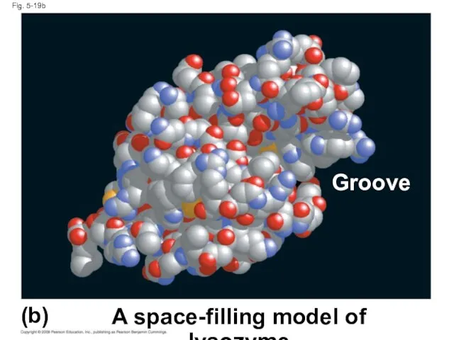
Fig. 5-19b
(b)
A space-filling model of lysozyme
Groove
Fig. 5-19b
(b)
A space-filling model of lysozyme
Groove
The sequence of amino acids determines a protein’s three-dimensional structure
A protein’s
The sequence of amino acids determines a protein’s three-dimensional structure
A protein’s
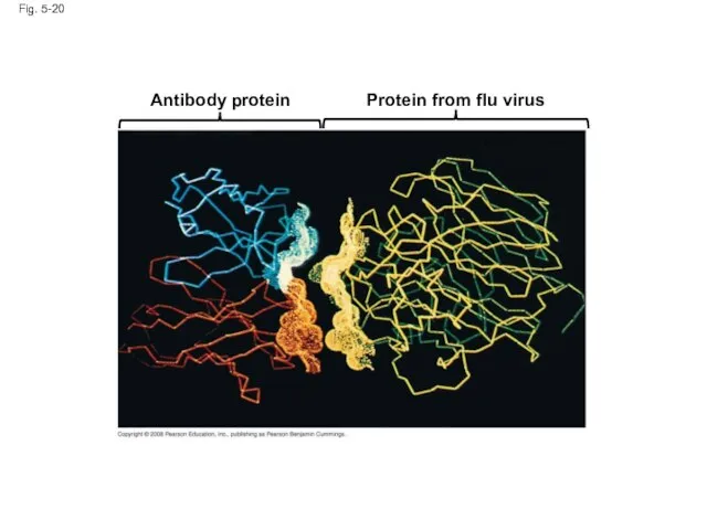
Fig. 5-20
Antibody protein
Protein from flu virus
Fig. 5-20
Antibody protein
Protein from flu virus
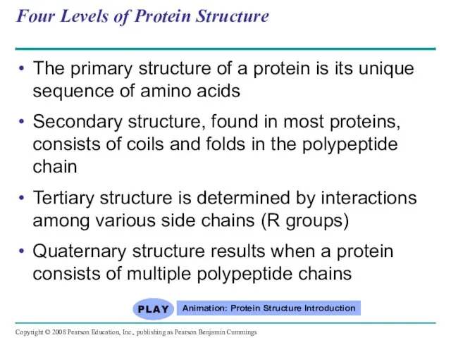
Four Levels of Protein Structure
The primary structure of a protein is
Four Levels of Protein Structure
The primary structure of a protein is
Primary structure, the sequence of amino acids in a protein, is
Primary structure, the sequence of amino acids in a protein, is
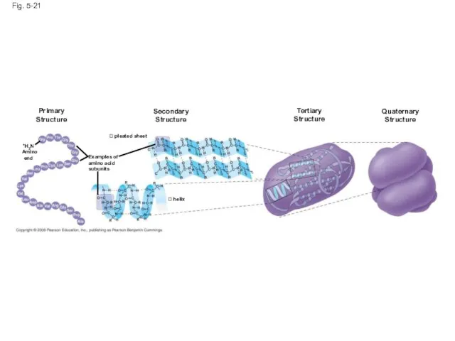
Fig. 5-21
Primary
Structure
Secondary
Structure
Tertiary
Structure
pleated sheet
Examples of
amino acid
subunits
+H3N
Amino end
helix
Quaternary
Structure
Fig. 5-21
Primary
Structure
Secondary
Structure
Tertiary
Structure
pleated sheet
Examples of
amino acid
subunits
+H3N
Amino end
helix
Quaternary
Structure
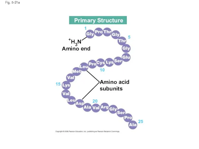
Fig. 5-21a
Amino acid
subunits
+H3N
Amino end
25
20
15
10
5
1
Primary Structure
Fig. 5-21a
Amino acid
subunits
+H3N
Amino end
25
20
15
10
5
1
Primary Structure
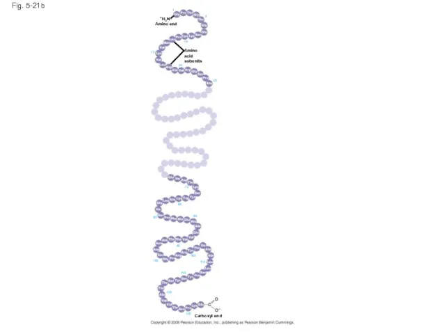
Fig. 5-21b
Amino acid
subunits
+H3N
Amino end
Carboxyl end
125
120
115
110
105
100
95
90
85
80
75
20
25
15
10
5
1
Fig. 5-21b
Amino acid
subunits
+H3N
Amino end
Carboxyl end
125
120
115
110
105
100
95
90
85
80
75
20
25
15
10
5
1
The coils and folds of secondary structure result from hydrogen bonds
The coils and folds of secondary structure result from hydrogen bonds
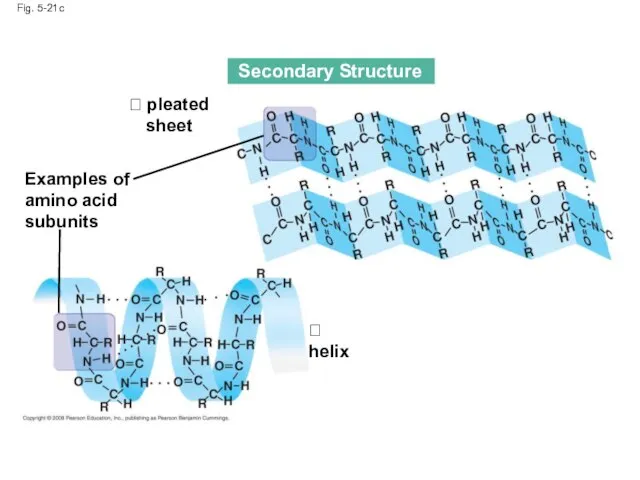
Fig. 5-21c
Secondary Structure
pleated sheet
Examples of
amino acid
subunits
helix
Fig. 5-21c
Secondary Structure
pleated sheet
Examples of
amino acid
subunits
helix
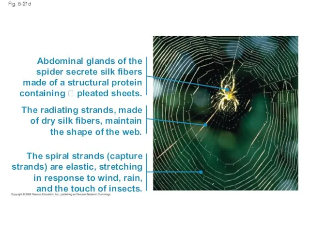
Fig. 5-21d
Abdominal glands of the
spider secrete silk fibers
made of a structural
Fig. 5-21d
Abdominal glands of the
spider secrete silk fibers
made of a structural
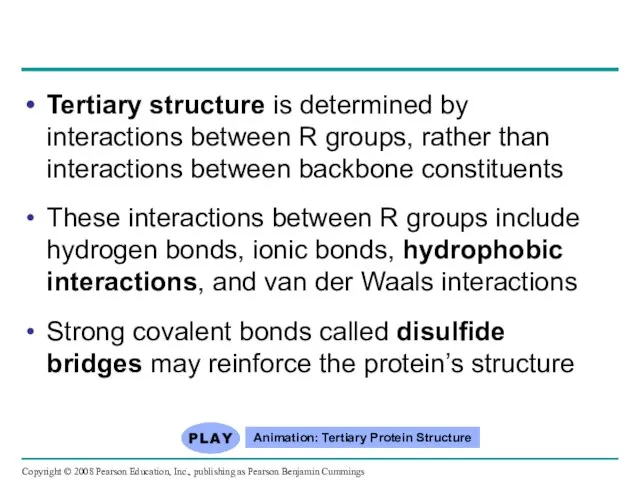
Tertiary structure is determined by interactions between R groups, rather than
Tertiary structure is determined by interactions between R groups, rather than
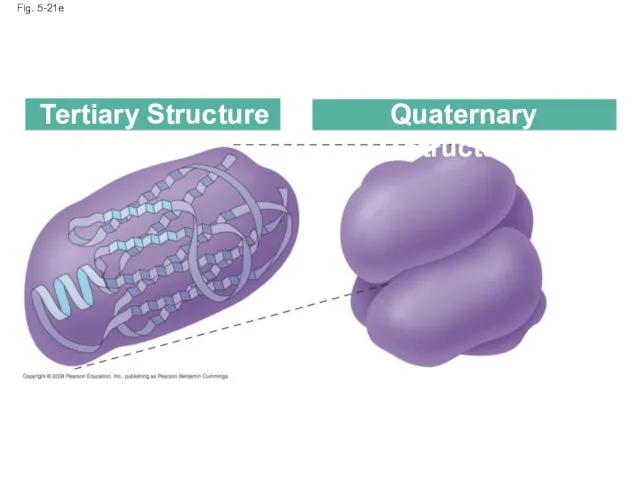
Fig. 5-21e
Tertiary Structure
Quaternary Structure
Fig. 5-21e
Tertiary Structure
Quaternary Structure
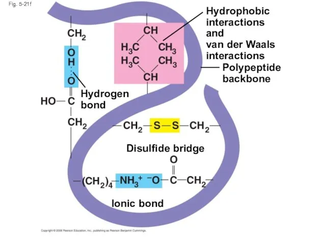
Fig. 5-21f
Polypeptide
backbone
Hydrophobic
interactions and
van der Waals
interactions
Disulfide bridge
Ionic bond
Hydrogen
bond
Fig. 5-21f
Polypeptide
backbone
Hydrophobic
interactions and
van der Waals
interactions
Disulfide bridge
Ionic bond
Hydrogen
bond
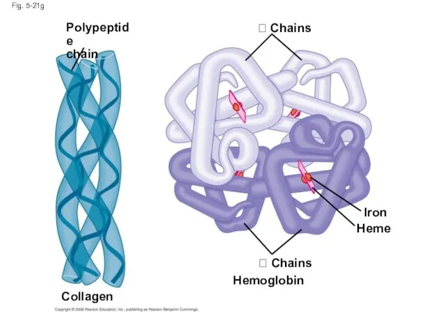
Fig. 5-21g
Polypeptide
chain
Chains
Heme
Iron
Chains
Collagen
Hemoglobin
Fig. 5-21g
Polypeptide
chain
Chains
Heme
Iron
Chains
Collagen
Hemoglobin
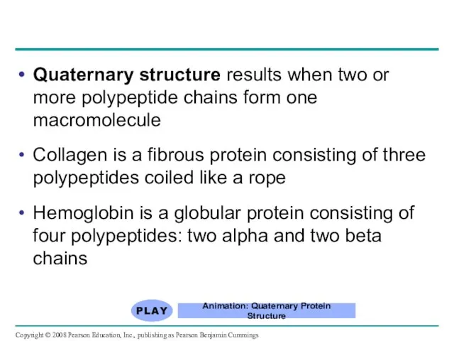
Quaternary structure results when two or more polypeptide chains form one
Quaternary structure results when two or more polypeptide chains form one
Sickle-Cell Disease: A Change in
Primary Structure
A slight change in primary
Sickle-Cell Disease: A Change in
Primary Structure
A slight change in primary
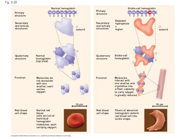
Fig. 5-22
Primary
structure
Secondary
and tertiary
structures
Quaternary
structure
Normal
hemoglobin
(top view)
Primary
structure
Secondary
and tertiary
structures
Quaternary
structure
Function
Function
subunit
Molecules do
not associate
with one
another; each
carries oxygen.
Red
Fig. 5-22
Primary
structure
Secondary
and tertiary
structures
Quaternary
structure
Normal
hemoglobin
(top view)
Primary
structure
Secondary
and tertiary
structures
Quaternary
structure
Function
Function
subunit
Molecules do
not associate
with one
another; each
carries oxygen.
Red
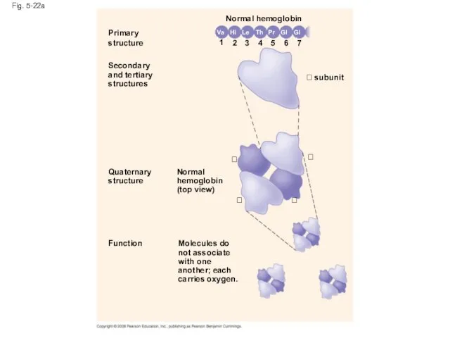
Fig. 5-22a
Primary
structure
Secondary
and tertiary
structures
Function
Quaternary
structure
Molecules do
not associate
with one
another; each
carries oxygen.
Normal
hemoglobin
(top view)
subunit
Normal hemoglobin
7
6
5
4
3
2
1
Glu
Val
His
Leu
Thr
Pro
Glu
Fig. 5-22a
Primary
structure
Secondary
and tertiary
structures
Function
Quaternary
structure
Molecules do
not associate
with one
another; each
carries oxygen.
Normal
hemoglobin
(top view)
subunit
Normal hemoglobin
7
6
5
4
3
2
1
Glu
Val
His
Leu
Thr
Pro
Glu
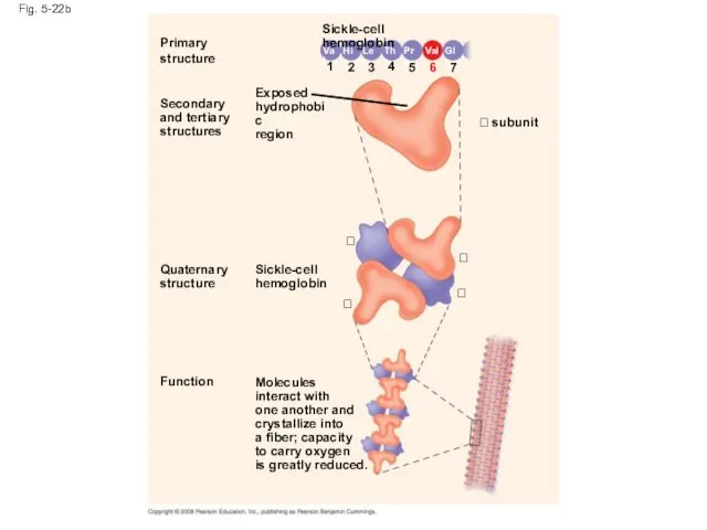
Fig. 5-22b
Primary
structure
Secondary
and tertiary
structures
Function
Quaternary
structure
Molecules
interact with
one another and
crystallize into
a fiber;
Fig. 5-22b
Primary
structure
Secondary
and tertiary
structures
Function
Quaternary
structure
Molecules
interact with
one another and
crystallize into
a fiber;
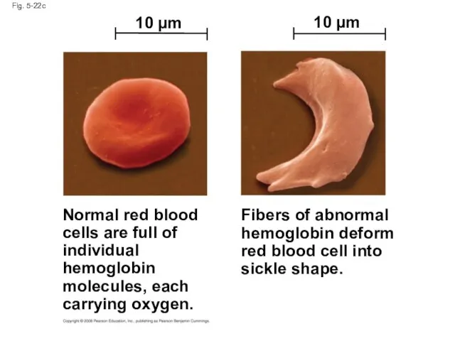
Fig. 5-22c
Normal red blood
cells are full of
individual
hemoglobin
molecules, each
carrying oxygen.
Fibers of
Fig. 5-22c
Normal red blood
cells are full of
individual
hemoglobin
molecules, each
carrying oxygen.
Fibers of
What Determines Protein Structure?
In addition to primary structure, physical and chemical
What Determines Protein Structure?
In addition to primary structure, physical and chemical
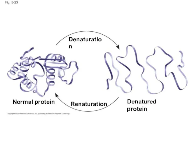
Fig. 5-23
Normal protein
Denatured protein
Denaturation
Renaturation
Fig. 5-23
Normal protein
Denatured protein
Denaturation
Renaturation
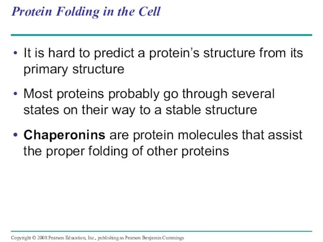
Protein Folding in the Cell
It is hard to predict a protein’s
Protein Folding in the Cell
It is hard to predict a protein’s
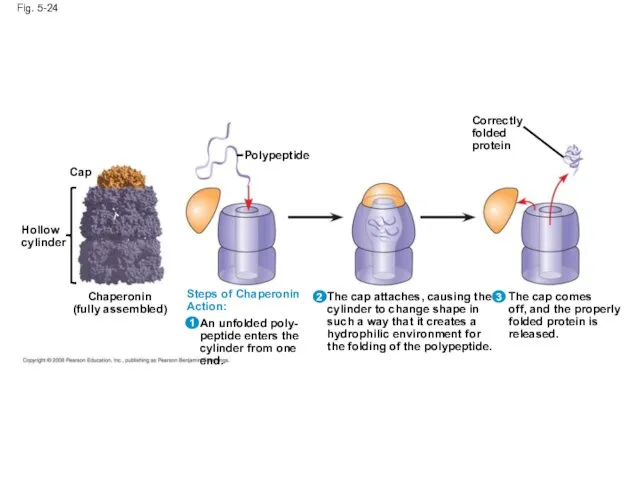
Fig. 5-24
Hollow
cylinder
Cap
Chaperonin
(fully assembled)
Polypeptide
Steps of Chaperonin
Action:
An unfolded poly-
peptide enters the
cylinder from one
Fig. 5-24
Hollow
cylinder
Cap
Chaperonin
(fully assembled)
Polypeptide
Steps of Chaperonin
Action:
An unfolded poly-
peptide enters the
cylinder from one
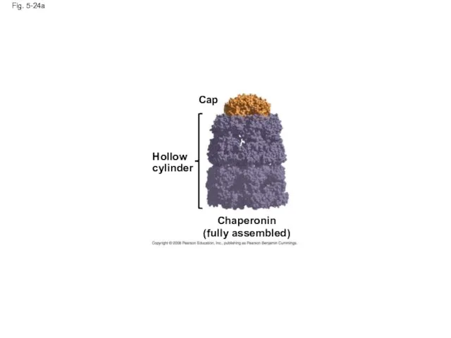
Fig. 5-24a
Hollow
cylinder
Chaperonin
(fully assembled)
Cap
Fig. 5-24a
Hollow
cylinder
Chaperonin
(fully assembled)
Cap
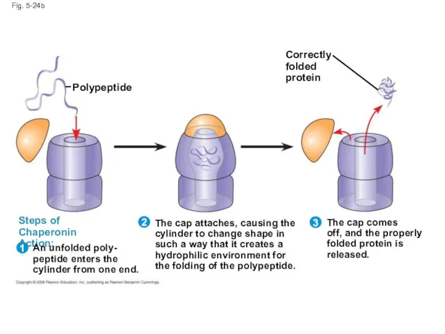
Fig. 5-24b
Correctly
folded
protein
Polypeptide
Steps of Chaperonin
Action:
1
2
An unfolded poly-
peptide enters the
cylinder from one end.
The
Fig. 5-24b
Correctly
folded
protein
Polypeptide
Steps of Chaperonin
Action:
1
2
An unfolded poly-
peptide enters the
cylinder from one end.
The
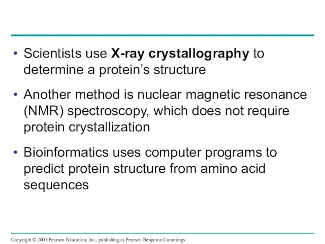
Scientists use X-ray crystallography to determine a protein’s structure
Another method is
Scientists use X-ray crystallography to determine a protein’s structure
Another method is
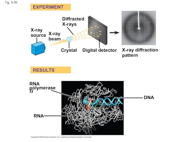
Fig. 5-25
EXPERIMENT
RESULTS
X-ray
source
X-ray
beam
Diffracted
X-rays
Crystal
Digital detector
X-ray diffraction
pattern
RNA
polymerase II
RNA
DNA
Fig. 5-25
EXPERIMENT
RESULTS
X-ray
source
X-ray
beam
Diffracted
X-rays
Crystal
Digital detector
X-ray diffraction
pattern
RNA
polymerase II
RNA
DNA
Fig. 5-25a
Diffracted
X-rays
EXPERIMENT
X-ray
source
X-ray
beam
Crystal
Digital detector
X-ray diffraction
pattern
Fig. 5-25a
Diffracted
X-rays
EXPERIMENT
X-ray
source
X-ray
beam
Crystal
Digital detector
X-ray diffraction
pattern
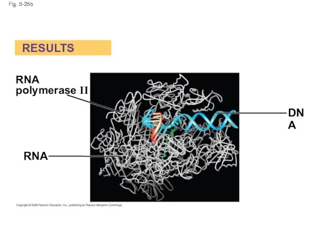
Fig. 5-25b
RESULTS
RNA
RNA
polymerase II
DNA
Fig. 5-25b
RESULTS
RNA
RNA
polymerase II
DNA
Concept 5.5: Nucleic acids store and transmit hereditary information
The amino acid
Concept 5.5: Nucleic acids store and transmit hereditary information
The amino acid
The Roles of Nucleic Acids
There are two types of nucleic acids:
Deoxyribonucleic
The Roles of Nucleic Acids
There are two types of nucleic acids:
Deoxyribonucleic
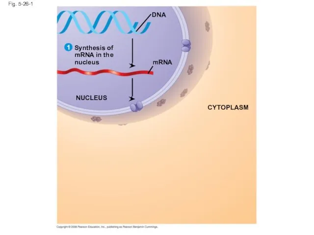
Fig. 5-26-1
mRNA
Synthesis of
mRNA in the
nucleus
DNA
NUCLEUS
CYTOPLASM
1
Fig. 5-26-1
mRNA
Synthesis of
mRNA in the
nucleus
DNA
NUCLEUS
CYTOPLASM
1
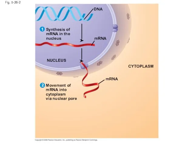
Fig. 5-26-2
mRNA
Synthesis of
mRNA in the
nucleus
DNA
NUCLEUS
mRNA
CYTOPLASM
Movement of
mRNA into cytoplasm
via nuclear pore
1
2
Fig. 5-26-2
mRNA
Synthesis of
mRNA in the
nucleus
DNA
NUCLEUS
mRNA
CYTOPLASM
Movement of
mRNA into cytoplasm
via nuclear pore
1
2
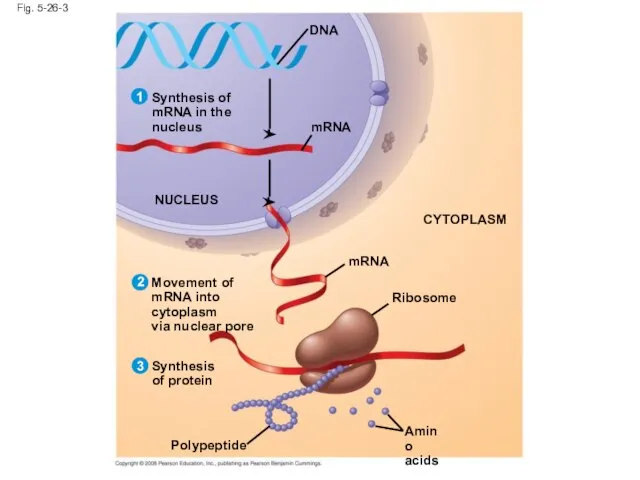
Fig. 5-26-3
mRNA
Synthesis of
mRNA in the
nucleus
DNA
NUCLEUS
mRNA
CYTOPLASM
Movement of
mRNA into cytoplasm
via nuclear pore
Ribosome
Amino
acids
Polypeptide
Synthesis
of protein
1
2
3
Fig. 5-26-3
mRNA
Synthesis of
mRNA in the
nucleus
DNA
NUCLEUS
mRNA
CYTOPLASM
Movement of
mRNA into cytoplasm
via nuclear pore
Ribosome
Amino
acids
Polypeptide
Synthesis
of protein
1
2
3
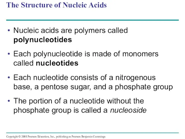
The Structure of Nucleic Acids
Nucleic acids are polymers called polynucleotides
Each polynucleotide
The Structure of Nucleic Acids
Nucleic acids are polymers called polynucleotides
Each polynucleotide
Fig. 5-27
5 end
Nucleoside
Nitrogenous
base
Phosphate
group
Sugar
(pentose)
(b) Nucleotide
(a) Polynucleotide, or nucleic acid
3 end
3C
3C
5C
5C
Nitrogenous bases
Pyrimidines
Cytosine (C)
Thymine
Fig. 5-27
5 end
Nucleoside
Nitrogenous
base
Phosphate
group
Sugar
(pentose)
(b) Nucleotide
(a) Polynucleotide, or nucleic acid
3 end
3C
3C
5C
5C
Nitrogenous bases
Pyrimidines
Cytosine (C)
Thymine
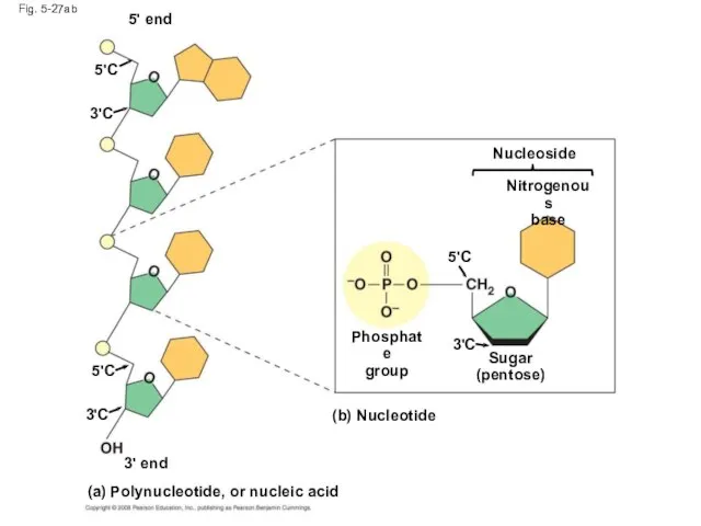
Fig. 5-27ab
5' end
5'C
3'C
5'C
3'C
3' end
(a) Polynucleotide, or nucleic acid
(b) Nucleotide
Nucleoside
Nitrogenous
base
3'C
5'C
Phosphate
group
Sugar
(pentose)
Fig. 5-27ab
5' end
5'C
3'C
5'C
3'C
3' end
(a) Polynucleotide, or nucleic acid
(b) Nucleotide
Nucleoside
Nitrogenous
base
3'C
5'C
Phosphate
group
Sugar
(pentose)
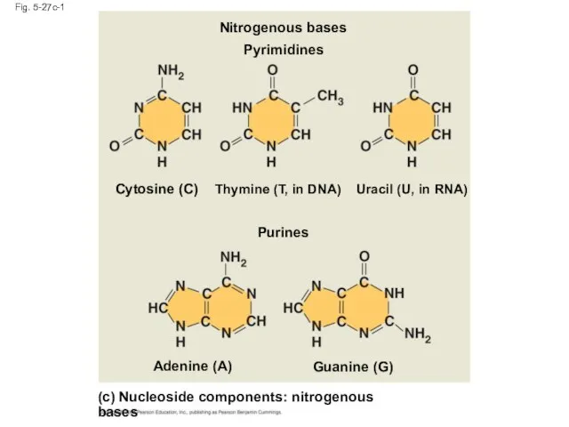
Fig. 5-27c-1
(c) Nucleoside components: nitrogenous bases
Purines
Guanine (G)
Adenine (A)
Cytosine (C)
Thymine (T, in
Fig. 5-27c-1
(c) Nucleoside components: nitrogenous bases
Purines
Guanine (G)
Adenine (A)
Cytosine (C)
Thymine (T, in
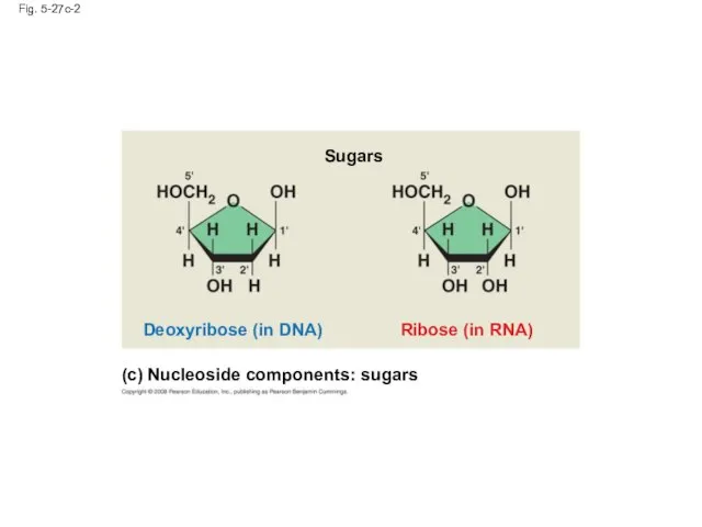
Fig. 5-27c-2
Ribose (in RNA)
Deoxyribose (in DNA)
Sugars
(c) Nucleoside components: sugars
Fig. 5-27c-2
Ribose (in RNA)
Deoxyribose (in DNA)
Sugars
(c) Nucleoside components: sugars
Nucleotide Monomers
Nucleoside = nitrogenous base + sugar
There are two families of
Nucleotide Monomers
Nucleoside = nitrogenous base + sugar
There are two families of
Nucleotide Polymers
Nucleotide polymers are linked together to build a polynucleotide
Adjacent nucleotides
Nucleotide Polymers
Nucleotide polymers are linked together to build a polynucleotide
Adjacent nucleotides
The DNA Double Helix
A DNA molecule has two polynucleotides spiraling around
The DNA Double Helix
A DNA molecule has two polynucleotides spiraling around
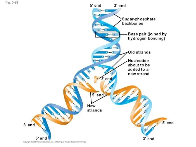
Fig. 5-28
Sugar-phosphate
backbones
3' end
3' end
3' end
3' end
5' end
5' end
5' end
5' end
Base pair
Fig. 5-28
Sugar-phosphate
backbones
3' end
3' end
3' end
3' end
5' end
5' end
5' end
5' end
Base pair
DNA and Proteins as Tape Measures of Evolution
The linear sequences of
DNA and Proteins as Tape Measures of Evolution
The linear sequences of
The Theme of Emergent Properties in the Chemistry of Life: A
The Theme of Emergent Properties in the Chemistry of Life: A
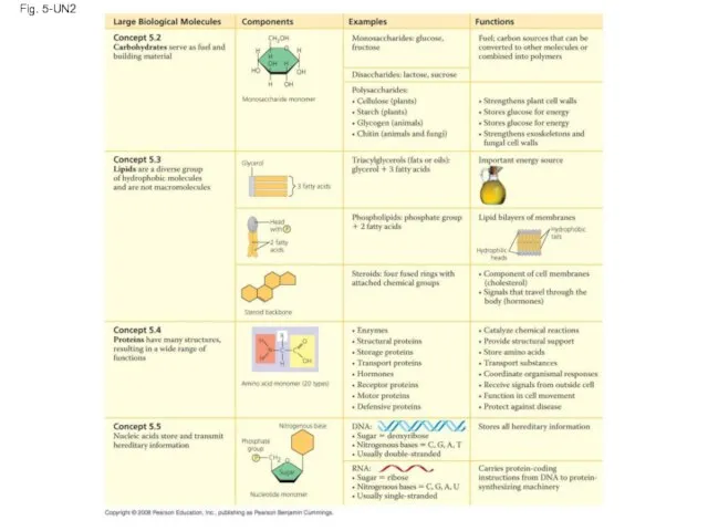
Fig. 5-UN2
Fig. 5-UN2
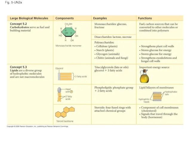
Fig. 5-UN2a
Fig. 5-UN2a
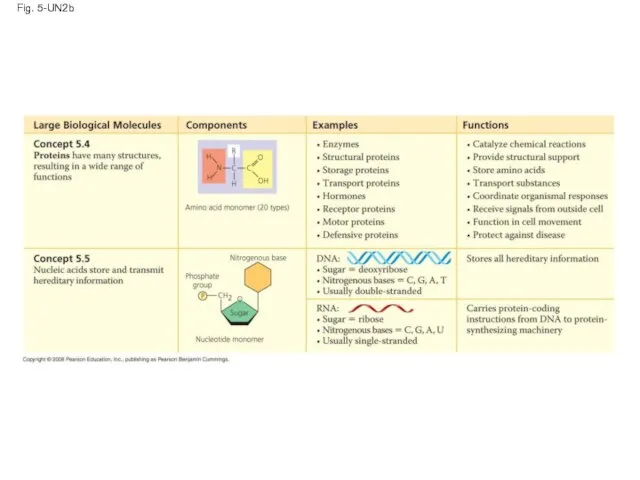
Fig. 5-UN2b
Fig. 5-UN2b
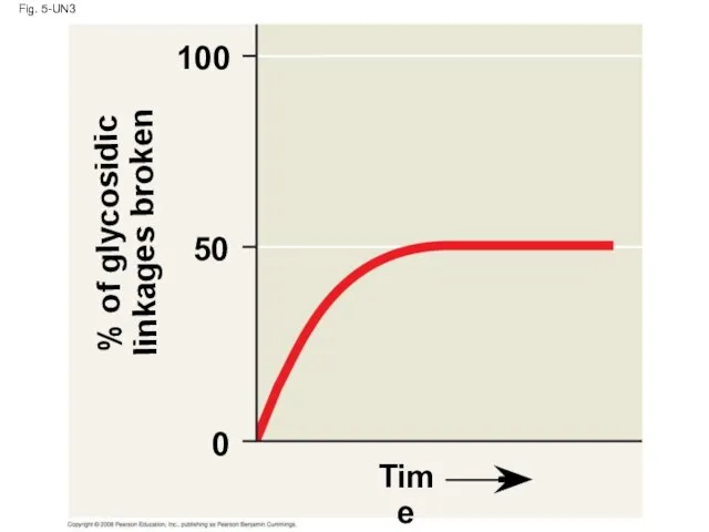
Fig. 5-UN3
% of glycosidic
linkages broken
100
50
0
Time
Fig. 5-UN3
% of glycosidic
linkages broken
100
50
0
Time
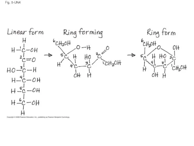
Fig. 5-UN4
Fig. 5-UN4
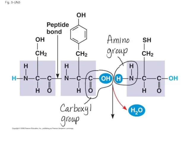
Fig. 5-UN5
Fig. 5-UN5
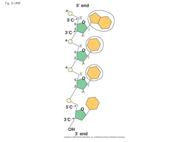
Fig. 5-UN6
Fig. 5-UN6
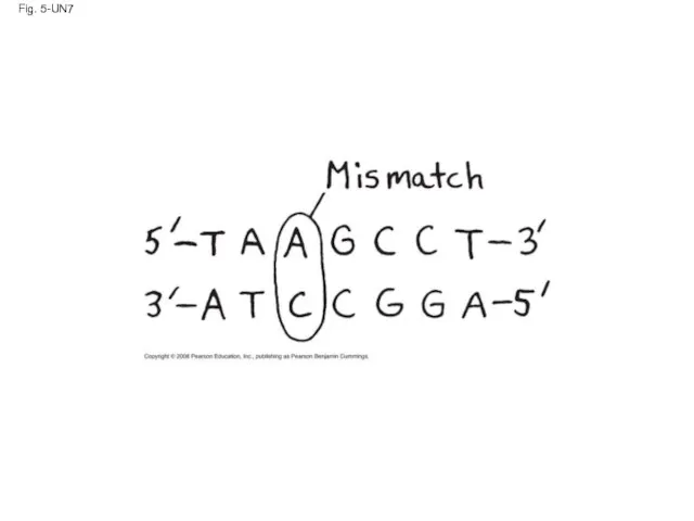
Fig. 5-UN7
Fig. 5-UN7
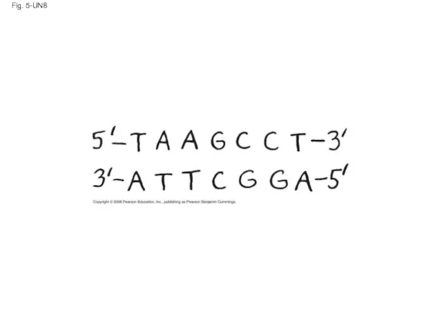
Fig. 5-UN8
Fig. 5-UN8
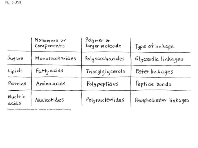
Fig. 5-UN9
Fig. 5-UN9
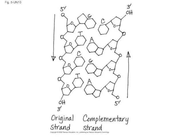
Fig. 5-UN10
Fig. 5-UN10
You should now be able to:
List and describe the four major
You should now be able to:
List and describe the four major
 Тип Плоские черви Презентация для 7 класса
 Презентация на тему Отряд Жуки или Жесткокрылые
  ТЕМА: «Класс рыб» Автор Самойленко Э.А., учитель биологии.
  Сон
Сон Этапы формирования и развития представлений о клетке
 Автор: учитель биологии высшей категории МОУ «Засосенская СОШ имени Героя Советского Союза Н.Л.Яценко» Ковшов Анатолий Владимир
 Обмен белков
 Профілактика інфекційних захворювань Бартош Наталії 11-А клас
  Тип Хордовые класс Млекопитающие Спесивцева О.А.
 Опорно-двигательная система, филогенез
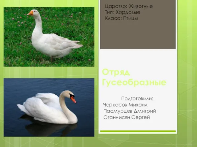 Отряд гусеобразные
 Урок литературного чтения в 4 классе Автор: учитель начальных классов МБОУ СОШ № 6 Федченко Валентина Николаевна
  Зерновые культуры
 Прикладная анатомия гортани
 Секвестрэктомия у пустынного канюка
 Презентация на тему "Вода – растворитель" - скачать презентации по Биологии
 Синичкин день
 Органические вещества, входящие в состав клетки
 Санитарная микробиология
 Презентация на тему В Арктике. Растительный и животный мир Арктики.
 Самые удивительные растения мира Растительный мир очень красив и многообразен. Сегодня существует около 360.000 различных видов
  Будова й значення дихальної системи
 Нуклеиновые кислоты
 Физиология желчного пузыря
 Разнообразие животных
 Мейоз. Механизм мейоза
 Программированный опрос по биологии
 Какие бывают животные