- breast pathology presentation

Содержание
- 2. Pathology of the breast normal anatomy physiologic changes developmental abnormalities inflammations fibrocystic changes tumors benign malignant
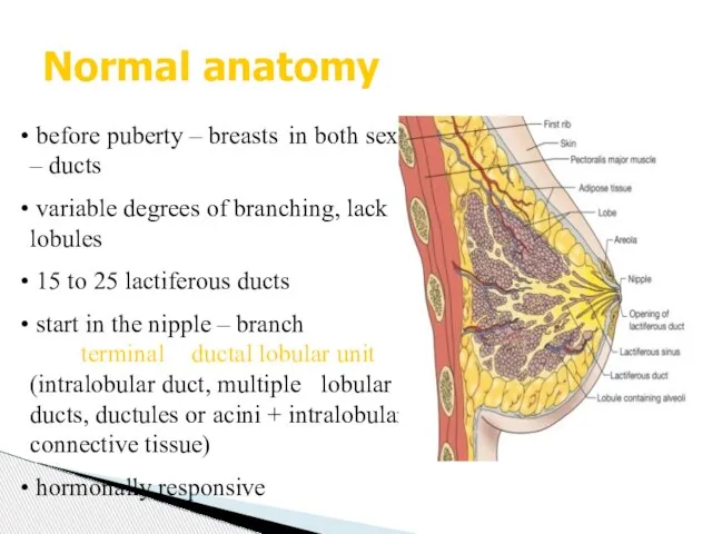
- 3. Normal anatomy before puberty – breasts in both sexes – ducts variable degrees of branching, lack
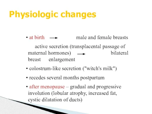
- 4. Physiologic changes at birth male and female breasts active secretion (transplacental passage of maternal hormones) bilateral
- 5. Macromastia diffuse enlargement of both breasts adolescence or pregnancy exaggerated response to hormonal stimulation Pubertal (Virginal)
- 6. Developmental abnormalities Aplasia and hypoplasia uncommon – associated with overdevelopment of the contralateral breast acquired (irradiation
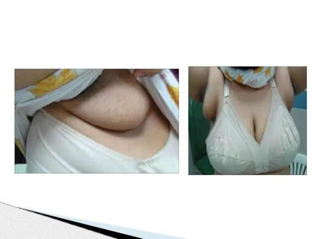
- 8. Ectopic breast supernumerary breast (from ectopic breast tissue – along the milk lines (midaxillae – normal
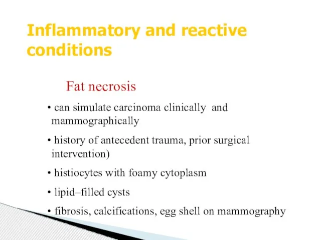
- 10. Inflammatory and reactive conditions Fat necrosis can simulate carcinoma clinically and mammographically history of antecedent trauma,
- 11. Inflammatory and reactive conditions Hemorrhagic necrosis with coagulopathy Warfarin treatment – shortly after initiation edema, hemorrhage,
- 12. Puerperal mastitis early stages (2nd and 3rd W) of lactation – 5% stasis of milk in
- 14. Benign proliferative lesions pathologic spectrum of seemingly related clinically benign breast abnormalities palpably irregular and painful
- 15. Benign proliferative lesions Adenosis elongation of the terminal ductules caricature of the lobule sclerosing adenosis apocrine
- 16. Benign tumors Fibroadenoma proliferation of epithelial and stromal elements most common breast tumor in adolescent and
- 17. Tubular adenoma far less common than fibroadenomas young women, discrete, freely movable masses uniform sized ducts
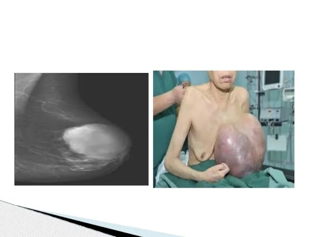
- 19. Cystosarcoma phyllodes (phyllodes tumor) initial description - over 150 years ago - fleshy tumor, leaf-like pattern
- 22. Proliferative changes ductal and lobular hyperplasia atypical ductal and lobular hyperplasia higher risk for the cancer
- 23. Breast carcinoma most frequent malignant tumor in females (followed by cervix and colon) highest incidence –
- 24. Breast carcinoma - classification IN SITU INVASIVE DUCTAL LOBULAR Ductal in situ (intraductal) Lobular in situ
- 25. Carcinoma in situ preinvasive - does not form a palpable tumor not detected clinically (only X-ray
- 26. Invasive carcinoma Invasive ductal carcinoma largest group (65 to 80 % of mammary carcinomas) mid to
- 27. other types: tubular, mucinous, medullary, inflammatory – together about 10 % of breast ca metastases: regional
- 28. Paget‘s disease of the nipple result of intraepithelial spread of intraductal carcinoma large pale-staining cells within
- 29. Paget’s disease
- 30. PAGET’S DISEASE OF NIPPLE Rare manifestation of breast CA. U/l erythematous eruption, Pruritus. Malignant cells/PAGET CELLS
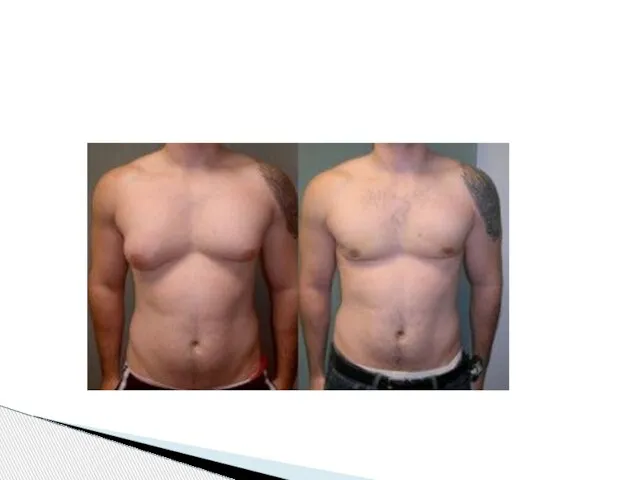
- 31. Pathology of the male breast Gynecomastia most common clinical and pathologic abnormality of the male breast
- 35. Скачать презентацию
Pathology of the breast
normal anatomy
physiologic changes
developmental abnormalities
Pathology of the breast
normal anatomy
physiologic changes
developmental abnormalities
Normal anatomy
before puberty – breasts in both sexes – ducts
Normal anatomy
before puberty – breasts in both sexes – ducts
Physiologic changes
at birth male and female breasts
active secretion (transplacental
Physiologic changes
at birth male and female breasts
active secretion (transplacental
Macromastia
diffuse enlargement of both breasts
adolescence or pregnancy
Macromastia
diffuse enlargement of both breasts
adolescence or pregnancy
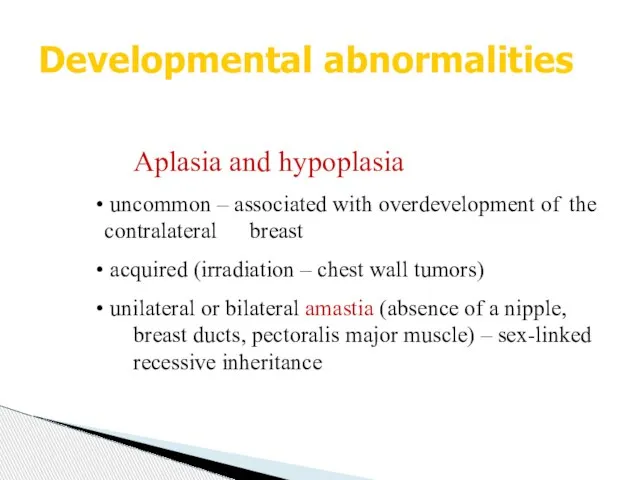
Developmental abnormalities
Aplasia and hypoplasia
uncommon – associated with overdevelopment of
Developmental abnormalities
Aplasia and hypoplasia
uncommon – associated with overdevelopment of

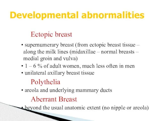
Ectopic breast
supernumerary breast (from ectopic breast tissue – along the
Ectopic breast
supernumerary breast (from ectopic breast tissue – along the
Inflammatory and reactive conditions
Fat necrosis
can simulate carcinoma clinically and
Inflammatory and reactive conditions
Fat necrosis
can simulate carcinoma clinically and
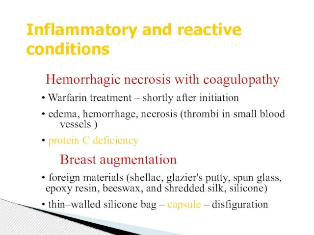
Inflammatory and reactive conditions
Hemorrhagic necrosis with coagulopathy
Warfarin treatment – shortly
Inflammatory and reactive conditions
Hemorrhagic necrosis with coagulopathy
Warfarin treatment – shortly
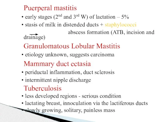
Puerperal mastitis
early stages (2nd and 3rd W) of lactation
Puerperal mastitis
early stages (2nd and 3rd W) of lactation

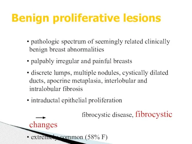
Benign proliferative lesions
pathologic spectrum of seemingly related clinically benign breast
Benign proliferative lesions
pathologic spectrum of seemingly related clinically benign breast
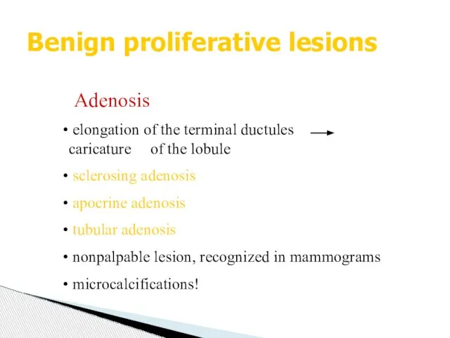
Benign proliferative lesions
Adenosis
elongation of the terminal ductules caricature of
Benign proliferative lesions
Adenosis
elongation of the terminal ductules caricature of
Benign tumors
Fibroadenoma
proliferation of epithelial and stromal elements
most common breast
Benign tumors
Fibroadenoma
proliferation of epithelial and stromal elements
most common breast
Tubular adenoma
far less common than fibroadenomas
young women, discrete, freely
Tubular adenoma
far less common than fibroadenomas
young women, discrete, freely

Cystosarcoma phyllodes
(phyllodes tumor)
initial description - over 150 years ago -
Cystosarcoma phyllodes
(phyllodes tumor)
initial description - over 150 years ago -

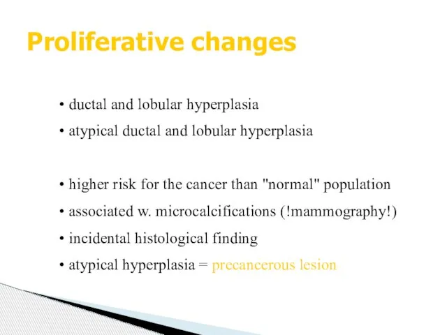
Proliferative changes
ductal and lobular hyperplasia
atypical ductal and lobular hyperplasia
Proliferative changes
ductal and lobular hyperplasia
atypical ductal and lobular hyperplasia
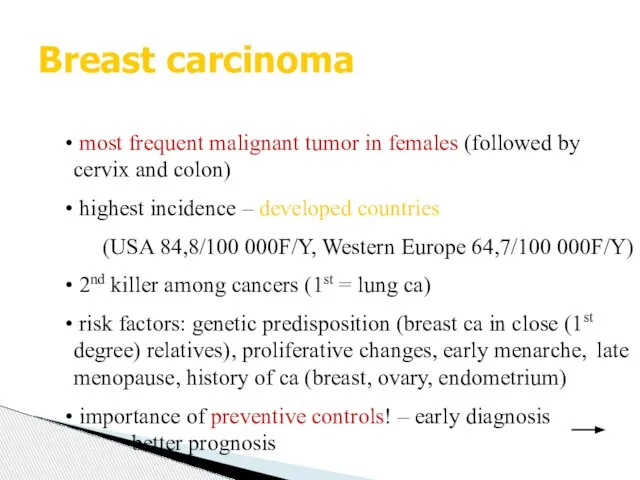
Breast carcinoma
most frequent malignant tumor in females (followed by cervix
Breast carcinoma
most frequent malignant tumor in females (followed by cervix
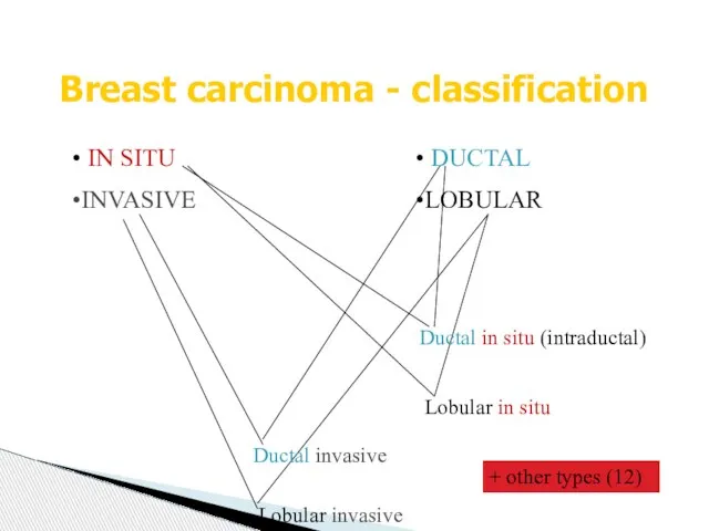
Breast carcinoma - classification
IN SITU
INVASIVE
DUCTAL
LOBULAR
Ductal in situ (intraductal)
Lobular
Breast carcinoma - classification
IN SITU
INVASIVE
DUCTAL
LOBULAR
Ductal in situ (intraductal)
Lobular
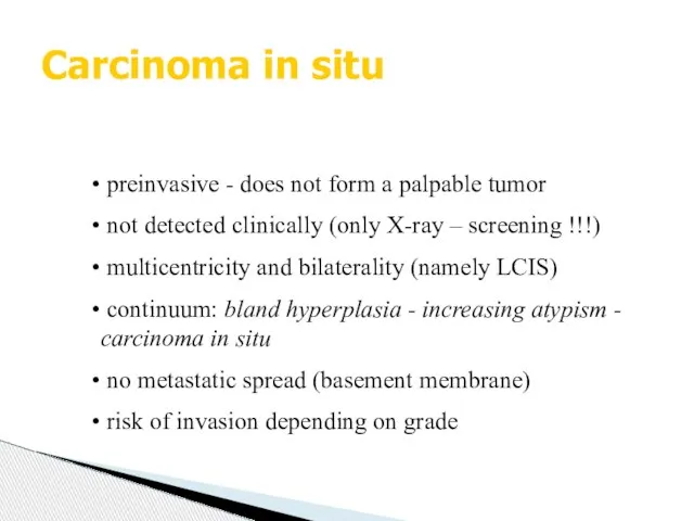
Carcinoma in situ
preinvasive - does not form a palpable tumor
Carcinoma in situ
preinvasive - does not form a palpable tumor
Invasive carcinoma
Invasive ductal carcinoma
largest group (65 to 80 % of
Invasive carcinoma
Invasive ductal carcinoma
largest group (65 to 80 % of

other types: tubular, mucinous, medullary, inflammatory – together about 10
other types: tubular, mucinous, medullary, inflammatory – together about 10
Paget‘s disease of the nipple
result of intraepithelial spread of intraductal
Paget‘s disease of the nipple
result of intraepithelial spread of intraductal
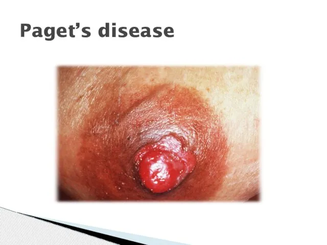
Paget’s disease
Paget’s disease
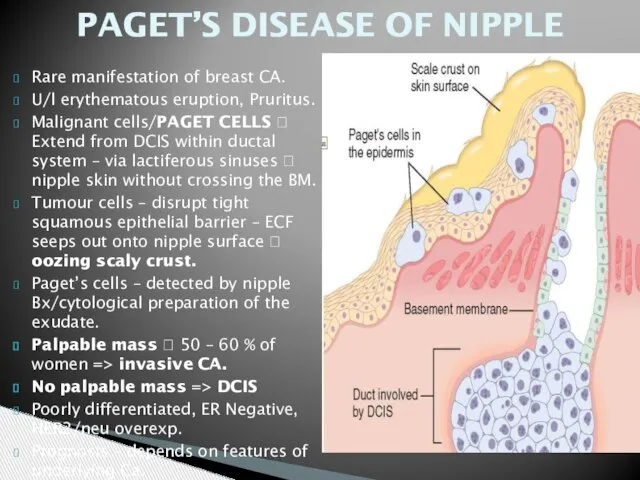
PAGET’S DISEASE OF NIPPLE
Rare manifestation of breast CA.
U/l erythematous eruption, Pruritus.
Malignant
PAGET’S DISEASE OF NIPPLE
Rare manifestation of breast CA.
U/l erythematous eruption, Pruritus.
Malignant
Pathology of the male breast
Gynecomastia
most common clinical and pathologic abnormality
Pathology of the male breast
Gynecomastia
most common clinical and pathologic abnormality

 Противомикробные средства
 Желудочно-кишечные кровотечения
 Доғалы протездің құрылымы, қолдану көрсеткіштері, қарсы көрсеткіштері. Артықшылықтары мен кемшіліктері
 Псевдоперитонеальды синдром
 Воспалительный процесс
 Патология восприятия и мышления
 Презентация по медицине Фенилкетонурия ФКУ финилпировиноградная олигофрения болезнь Фёллинга
  Мейірбикелік үрдіс
 Пузырные дерматозы
 Гонартроз. Артроз колінного суглобу (гонартроз, деформуючий артроз)
 Кардиотонические, антиангинальные и антиаритмические средства
 Триггеры. Чтобы решить задачу, нужно сделать так, чтобы сработал триггер
 Введение в патологическую анатомию
 Производные аминоалкилбензолов. (Тема 3)
 Острые инфекции у детей
 СПИД - синдром приобретенного иммунодефицита
 Клинические формы ихтиоза
 Тератология
 Аудиометрия. Звуковые методы исследования в медицине: перкуссия, аускультация. Фонокардиография
 ZhVP
ZhVP Диагностическое выскабливание слизистой матки
 Ауру сезімі және терапиялық стоматология клиникасында жансыздандыру
 Pneumonia
 Перитонит. Классификация, диагностика, лечение
 Основы_сбалансированного_питания
 Туберкулезді спецификалық алдын алу
 Микробиологическая диагностика заболеваний, передающихся половым путем
 Философия совершенного гостеприимства