- Diseases of kidney

Содержание
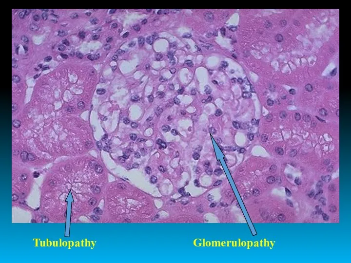
- 3. - Glomerulopathy - Tubulopathy - Interstitial diseases - Tumors - Congenital anomalies Diseases of Kidney
- 5. Renal and extrarenal symptoms
- 6. Glomerulonephritis is an infectious and allergic disease or disease of unknown nature, which is based on
- 7. Pathogenesis
- 8. 1) NEPHROTIC 2) NEPHRITIC 3) SLOWLY PROGRESSIVE UREMIA MAIN CLINICAL SYNDROMES
- 9. NEPHRITIC VS. NEPHROTIC SYNDROMES
- 10. Palpebral edema Anasarca
- 11. 1. Primary, secondary, hereditary. 2. Acute, subacute, chronic. 3. Intracapillary, extracapillary. 4. Exudative, proliferative, mixed. 7.
- 12. According to the etiological factors: Primary glomerulonephritis: Acute diffuse proliferative glomerulonephritis; Rapidly progressive glomerulonephritis; Membranous glomerulonephritis;
- 13. MORPHOLOGICAL CLASSIFICATION OF GLOMERULONEPHRITIS Diffuse intracapillary glomerulonephritis (acute glomerulonephritis). Extracapillary glomerulonephritis with crescents (rapidly progressive glomerulonephritis).
- 14. Glomerulonephritis Intracapillary Extracapillary
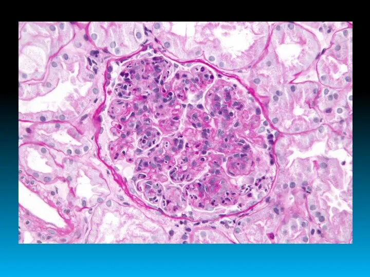
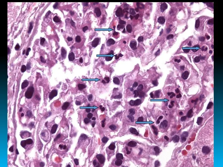
- 15. ACUTE GLOMERULONEPHRITIS
- 16. WITH NEPHRITIC SYNDROME Edema, shortness of breath, headache, nausea, vomiting, weakness; Arterial hypertension; Hematuria; Diuresis ↓;
- 17. “MOTTLED” KIDNEYS
- 20. Immune complexes on basal membrane and/or mesangial cells + Deposits of IgG, IgM and C3 along
- 21. SUBACUTE RAPIDLY PROGRESSIVE GLOMERULONEPHRITIS MAIN FEATURE IS RAPID DEVELOPMENT AND PROGRESSION OF CHRONIC RENAL FAILURE
- 22. CHRONIC GLOMERULONEPHRITIS LATENT WITH HYPERTENSIVE SYNDROME WITH HEMATURIA WITH NEPHROTIC SYNDROME MIXED
- 23. “LARGE WHITE KIDNEY” Accumulation of lipids in proximal tubules GLOMERULONEPHRITIS WITH MINIMAL CHANGES (LIPOID NEPHROSIS)
- 24. GLOMERULONEPHRITIS WITH MINIMAL CHANGES (LIPOID NEPHROSIS)
- 25. MEMBRANOUS NEPHROPATHY
- 26. “SPIKES AND DOMES” ON BASAL MEMBRANE
- 27. MESANGIAL PROLIFERATIVE GLOMERULONEPHRITIS
- 28. FOCAL SEGMENTAL GLOMERULOSCLEROSIS
- 29. MEMBRANOPROLIFERATIVE GLOMERULONEPHRITIS Light micrograph in membranoproliferative glomerulonephritis showing a lobular appearance of the glomerular tuft with
- 30. FIBROPLASTIC GLOMERULONEPHRITIS It is an outcome of any above mentioned glomerulonephritis Morphologically: - Glomerular sclerosis -
- 31. The most common causes are: Tuberculosis; Chronic diseases of lungs and bronchi; Chronic osteomyelitis and rheumatoid
- 33. CONGO RED STAINING
- 34. Amyloid (А) can be seen as fibrillar masses under the basal membrane (BM). ELECTRON MICROSCOPY
- 35. ACUTE RENAL FAILURE
- 36. NECROTIC NEPHROSIS ACUTE TUBULAR NECROSIS
- 37. NECROTIC NEPHROSIS
- 38. PYELONEPHRITIS
- 39. ACUTE PYELONEPHRITIS
- 40. COMPLICATIONS OF ACUTE PYELONEPHRITIS Necrosis of the renal papillae (papillonecrosis) – necrotic papillae can be rejected
- 41. PYONEPHROSIS
- 42. CHRONIC PYELONEPHRITIS
- 43. “THYROID KIDNEY”
- 44. CHRONIC RENAL FAILURE
- 45. CHRONIC RENAL FAILURE
- 47. Скачать презентацию
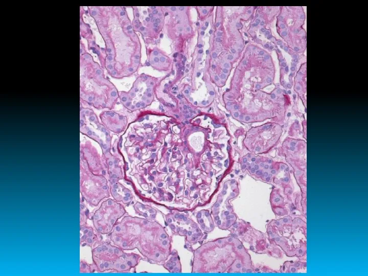
- Glomerulopathy
- Tubulopathy
- Interstitial diseases
- Tumors
- Congenital anomalies
Diseases of Kidney
- Glomerulopathy
- Tubulopathy
- Interstitial diseases
- Tumors
- Congenital anomalies
Diseases of Kidney
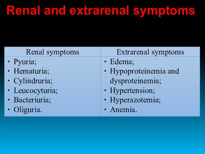
Renal and extrarenal symptoms
Renal and extrarenal symptoms
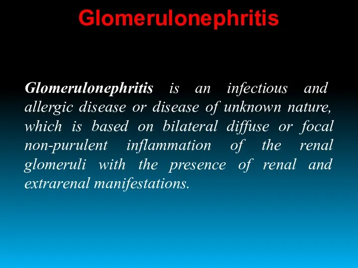
Glomerulonephritis is an infectious and allergic disease or disease of unknown
Glomerulonephritis is an infectious and allergic disease or disease of unknown
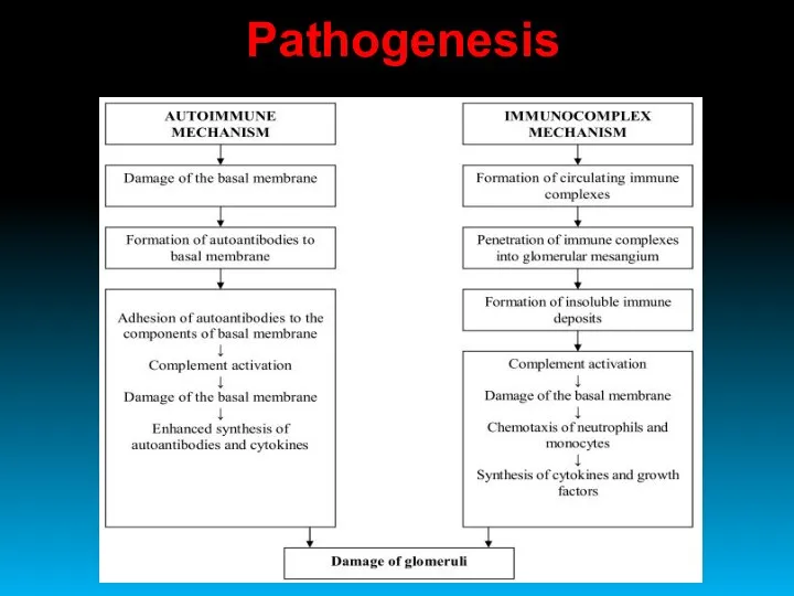
Pathogenesis
Pathogenesis
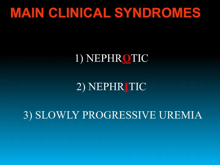
1) NEPHROTIC
2) NEPHRITIC
3) SLOWLY PROGRESSIVE UREMIA
MAIN CLINICAL SYNDROMES
1) NEPHROTIC
2) NEPHRITIC
3) SLOWLY PROGRESSIVE UREMIA
MAIN CLINICAL SYNDROMES
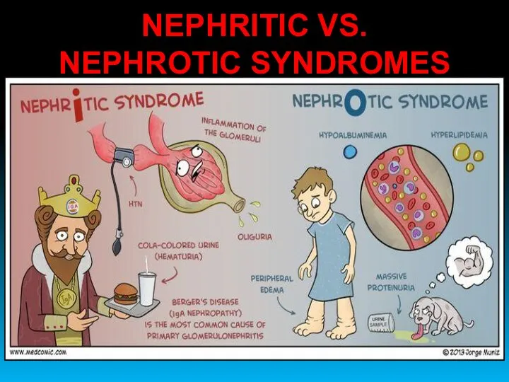
NEPHRITIC VS.
NEPHROTIC SYNDROMES
NEPHRITIC VS.
NEPHROTIC SYNDROMES
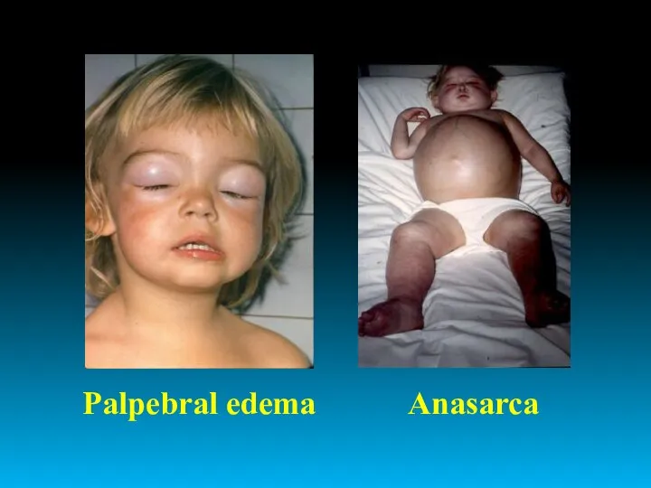
Palpebral edema
Anasarca
Palpebral edema
Anasarca
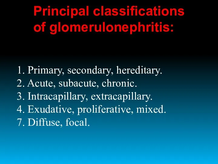
1. Primary, secondary, hereditary.
2. Acute, subacute, chronic.
3. Intracapillary, extracapillary.
4. Exudative, proliferative, mixed.
7. Diffuse, focal.
Principal classifications
of glomerulonephritis:
1. Primary, secondary, hereditary.
2. Acute, subacute, chronic.
3. Intracapillary, extracapillary.
4. Exudative, proliferative, mixed.
7. Diffuse, focal.
Principal classifications
of glomerulonephritis:
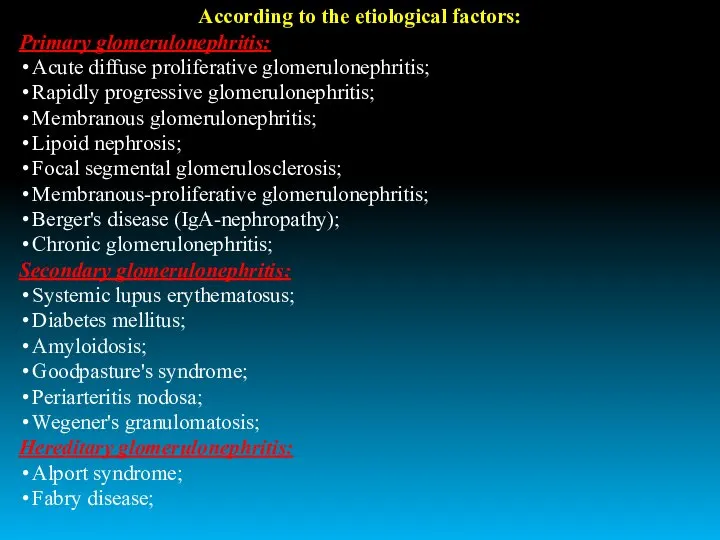
According to the etiological factors:
Primary glomerulonephritis:
Acute diffuse proliferative glomerulonephritis;
Rapidly progressive glomerulonephritis;
Membranous
According to the etiological factors:
Primary glomerulonephritis:
Acute diffuse proliferative glomerulonephritis;
Rapidly progressive glomerulonephritis;
Membranous
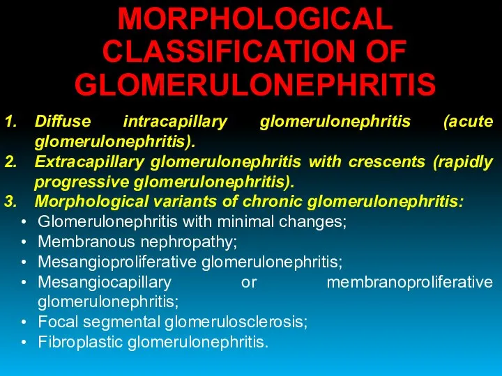
MORPHOLOGICAL CLASSIFICATION OF GLOMERULONEPHRITIS
Diffuse intracapillary glomerulonephritis (acute glomerulonephritis).
Extracapillary glomerulonephritis with crescents
MORPHOLOGICAL CLASSIFICATION OF GLOMERULONEPHRITIS
Diffuse intracapillary glomerulonephritis (acute glomerulonephritis).
Extracapillary glomerulonephritis with crescents
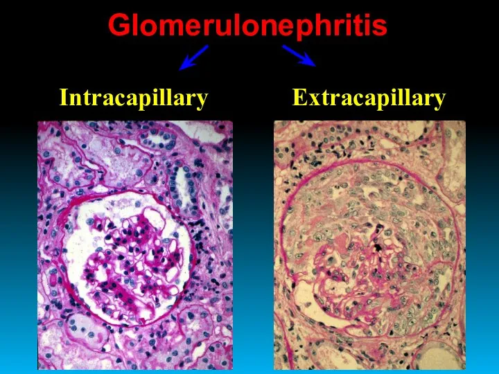
Glomerulonephritis
Intracapillary
Extracapillary
Glomerulonephritis
Intracapillary
Extracapillary
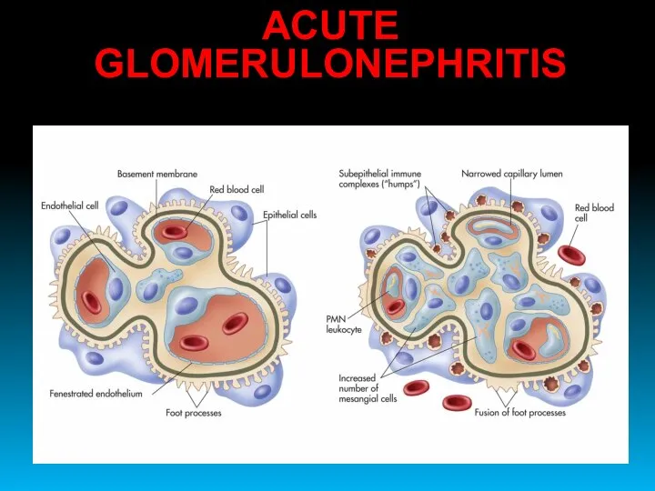
ACUTE GLOMERULONEPHRITIS
ACUTE GLOMERULONEPHRITIS
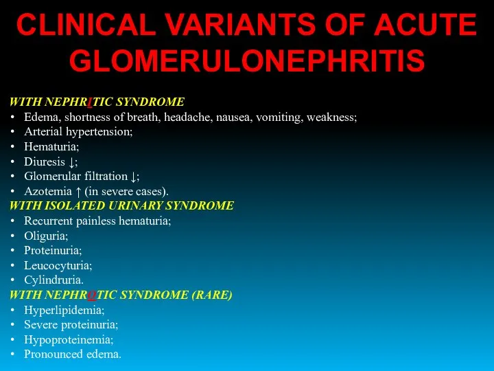
WITH NEPHRITIC SYNDROME
Edema, shortness of breath, headache, nausea, vomiting, weakness;
Arterial hypertension;
Hematuria;
Diuresis
WITH NEPHRITIC SYNDROME
Edema, shortness of breath, headache, nausea, vomiting, weakness;
Arterial hypertension;
Hematuria;
Diuresis
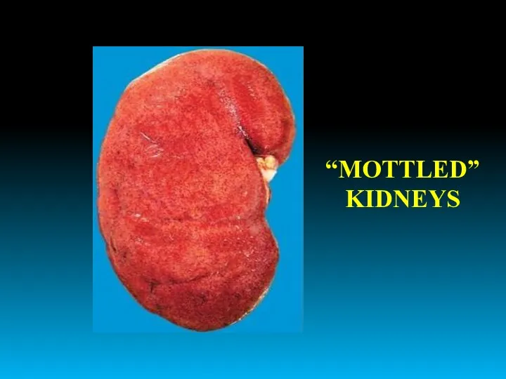
“MOTTLED”
KIDNEYS
“MOTTLED”
KIDNEYS
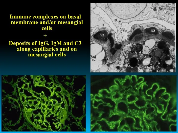
Immune complexes on basal membrane and/or mesangial cells
+
Deposits of IgG, IgM
Immune complexes on basal membrane and/or mesangial cells
+
Deposits of IgG, IgM
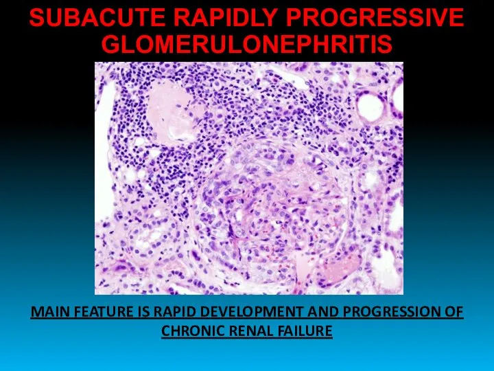
SUBACUTE RAPIDLY PROGRESSIVE GLOMERULONEPHRITIS
MAIN FEATURE IS RAPID DEVELOPMENT AND PROGRESSION OF
SUBACUTE RAPIDLY PROGRESSIVE GLOMERULONEPHRITIS
MAIN FEATURE IS RAPID DEVELOPMENT AND PROGRESSION OF
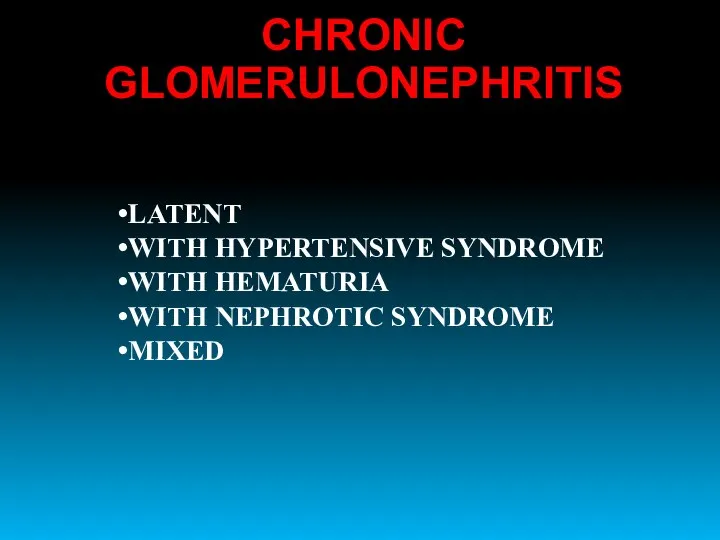
CHRONIC GLOMERULONEPHRITIS
LATENT
WITH HYPERTENSIVE SYNDROME
WITH HEMATURIA
WITH NEPHROTIC SYNDROME
MIXED
CHRONIC GLOMERULONEPHRITIS
LATENT
WITH HYPERTENSIVE SYNDROME
WITH HEMATURIA
WITH NEPHROTIC SYNDROME
MIXED
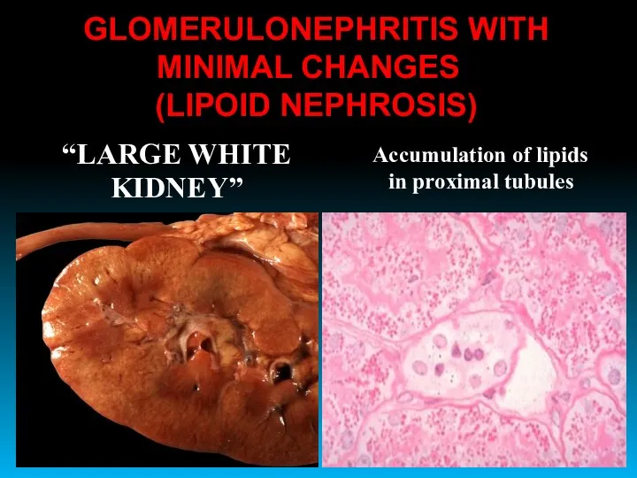
“LARGE WHITE
KIDNEY”
Accumulation of lipids in proximal tubules
GLOMERULONEPHRITIS WITH MINIMAL CHANGES
(LIPOID NEPHROSIS)
“LARGE WHITE
KIDNEY”
Accumulation of lipids in proximal tubules
GLOMERULONEPHRITIS WITH MINIMAL CHANGES
(LIPOID NEPHROSIS)
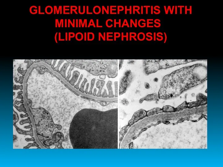
GLOMERULONEPHRITIS WITH MINIMAL CHANGES
(LIPOID NEPHROSIS)
GLOMERULONEPHRITIS WITH MINIMAL CHANGES
(LIPOID NEPHROSIS)
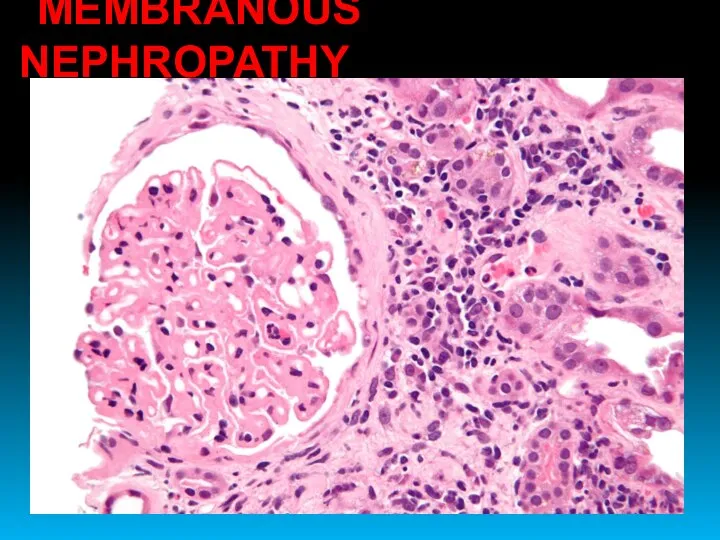
MEMBRANOUS NEPHROPATHY
MEMBRANOUS NEPHROPATHY
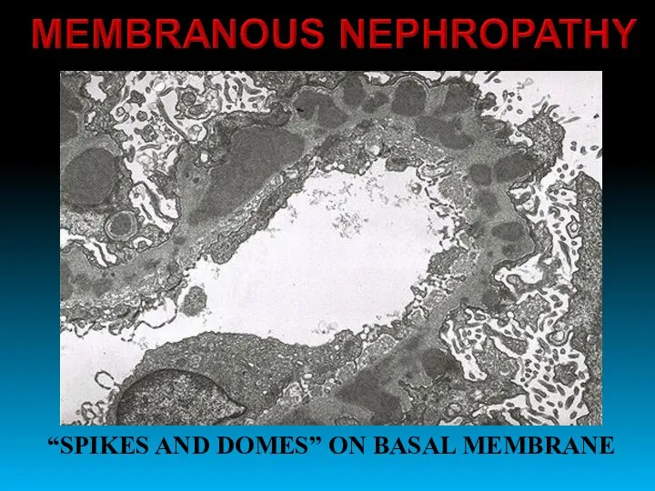
“SPIKES AND DOMES” ON BASAL MEMBRANE
“SPIKES AND DOMES” ON BASAL MEMBRANE
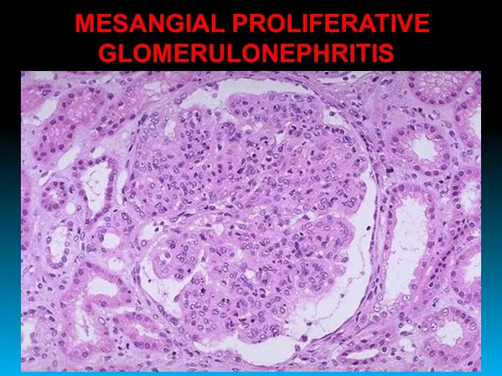
MESANGIAL PROLIFERATIVE GLOMERULONEPHRITIS
MESANGIAL PROLIFERATIVE GLOMERULONEPHRITIS
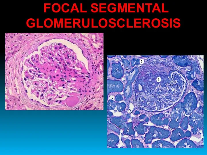
FOCAL SEGMENTAL GLOMERULOSCLEROSIS
FOCAL SEGMENTAL GLOMERULOSCLEROSIS
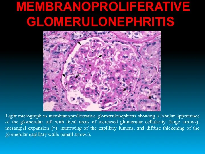
MEMBRANOPROLIFERATIVE GLOMERULONEPHRITIS
Light micrograph in membranoproliferative glomerulonephritis showing a lobular appearance of
MEMBRANOPROLIFERATIVE GLOMERULONEPHRITIS
Light micrograph in membranoproliferative glomerulonephritis showing a lobular appearance of
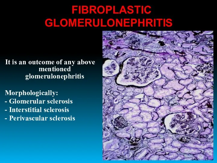
FIBROPLASTIC GLOMERULONEPHRITIS
It is an outcome of any above mentioned glomerulonephritis
Morphologically:
- Glomerular
FIBROPLASTIC GLOMERULONEPHRITIS
It is an outcome of any above mentioned glomerulonephritis
Morphologically:
- Glomerular
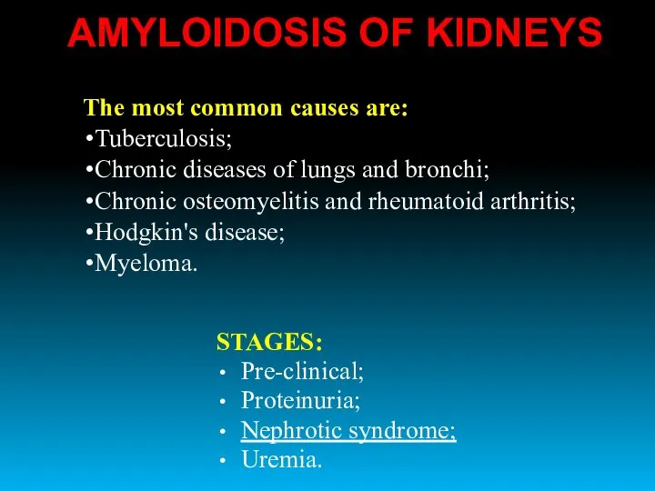
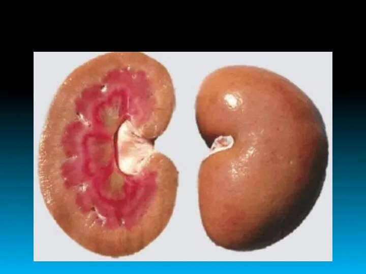
The most common causes are:
Tuberculosis;
Chronic diseases of lungs and bronchi;
Chronic osteomyelitis
The most common causes are:
Tuberculosis;
Chronic diseases of lungs and bronchi;
Chronic osteomyelitis
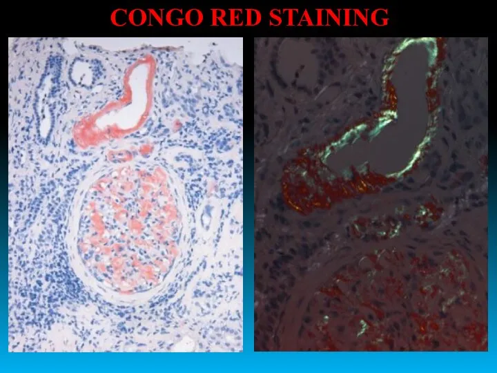
CONGO RED STAINING
CONGO RED STAINING
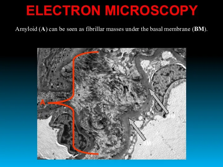
Amyloid (А) can be seen as fibrillar masses under the basal
Amyloid (А) can be seen as fibrillar masses under the basal
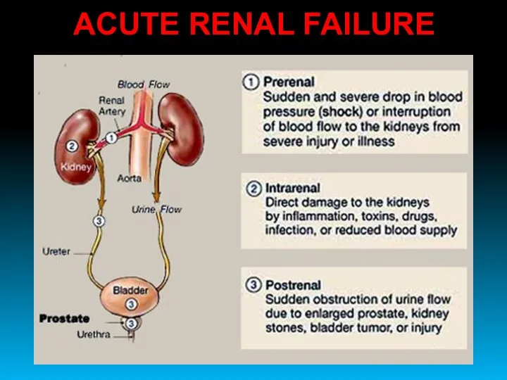
ACUTE RENAL FAILURE
ACUTE RENAL FAILURE
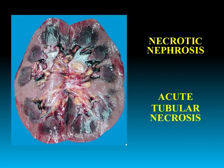
NECROTIC NEPHROSIS
ACUTE
TUBULAR NECROSIS
NECROTIC NEPHROSIS
ACUTE
TUBULAR NECROSIS
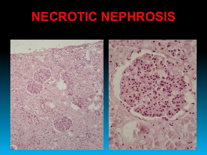
NECROTIC NEPHROSIS
NECROTIC NEPHROSIS
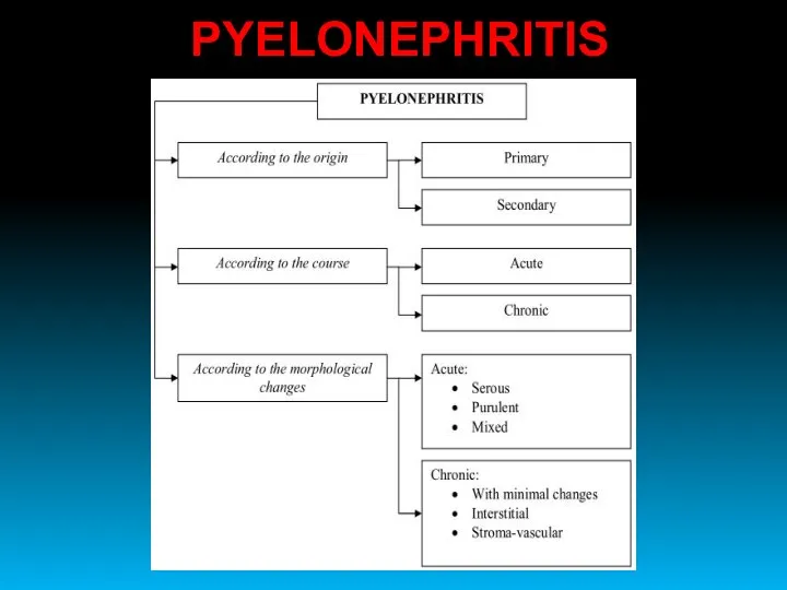
PYELONEPHRITIS
PYELONEPHRITIS
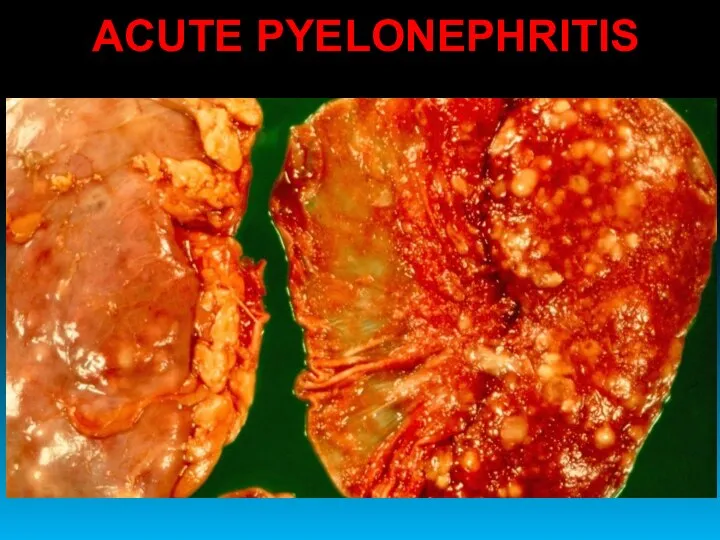
ACUTE PYELONEPHRITIS
ACUTE PYELONEPHRITIS
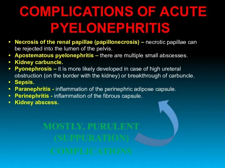
COMPLICATIONS OF ACUTE PYELONEPHRITIS
Necrosis of the renal papillae (papillonecrosis) – necrotic
COMPLICATIONS OF ACUTE PYELONEPHRITIS
Necrosis of the renal papillae (papillonecrosis) – necrotic
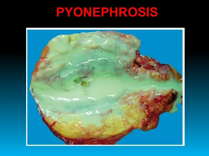
PYONEPHROSIS
PYONEPHROSIS
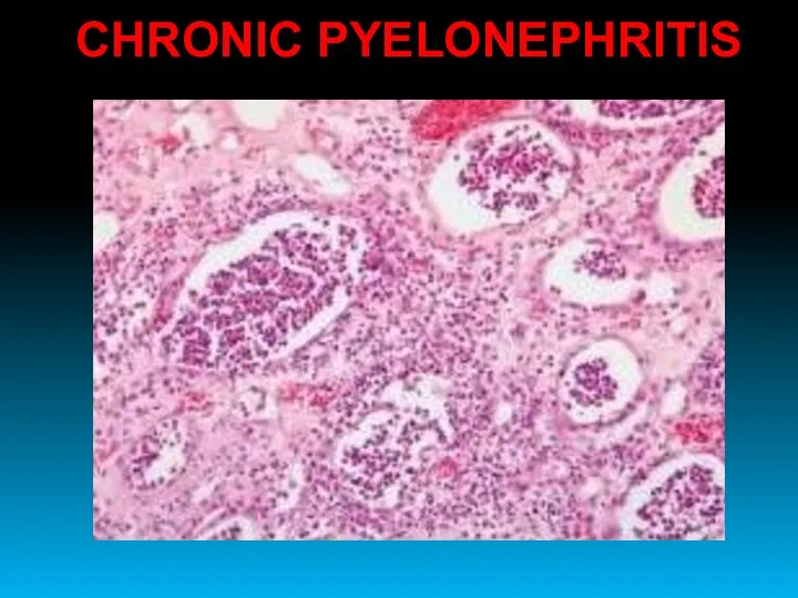
CHRONIC PYELONEPHRITIS
CHRONIC PYELONEPHRITIS
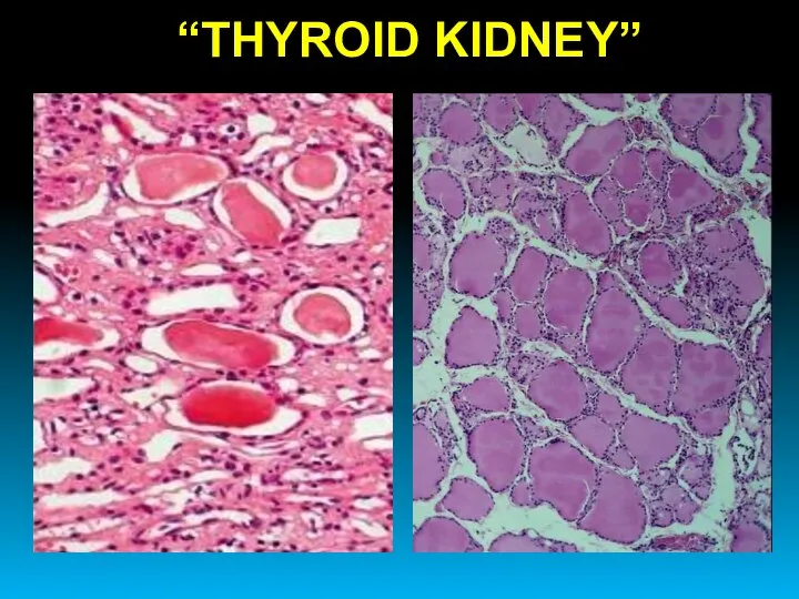
“THYROID KIDNEY”
“THYROID KIDNEY”
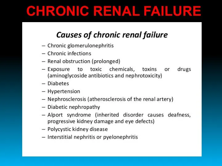
CHRONIC RENAL FAILURE
CHRONIC RENAL FAILURE
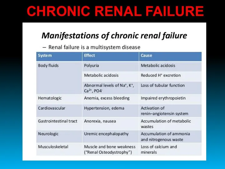
CHRONIC RENAL FAILURE
CHRONIC RENAL FAILURE
 Қан жасау және лимфоидты тіндер ісіктерінің патоморфологиясы, классификациясы. Тромбопения және тромбоцитопатиялардың
 ЖИТС-пен ауыратын науқастардағы комплаенс
 Советы для ведения личного дневника
 Мониторинг распространенности различных форм хронического тонзиллита у детей
 Влияние погодных факторов на здоровье. Профилактика гелиометеотропных реакций (кейс-задание)
 Вирусные заболевания кожи
 Түсті металлургия саласындағы жұмысшылардың кәсіби аурушаңдығы және алдын - алу жолдары
 Lektsia_Besplodny_brak
 Психология
 Паллиативная помощь в онкологии
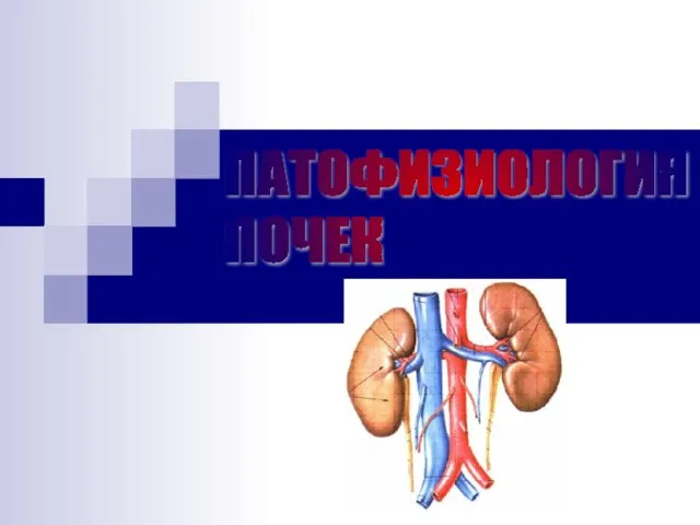 Патофизиология почек
 Заболевания бронхолегочной системы
 График работы врачей ОАПП №1 18.09.2021
 Лучевая диагностика туберкулёза органов дыхания
 Психология, как наука и ее структура
 Последствия ранней половой жизни
 Қантсыз диабет
 Пневмония. Эпидемиология пневмоний
 Инфекционные болезни. Воздушно-капельные инфекции, бактериальной этиологии
 Фармацевтические аэрозоли. Определение, технологическая схема производства
 Ортобиотическая система комплексной реабилитации и социальной адаптации детей с тяжёлыми нарушениями речи
 Рак гайморовой пазухи, клиника, диагностика, лечение
 Первая помощь при кровотечении
 Бактериальные инфекции: Кандидоз слизистой оболочки полости рта
 Congenital heart diseases
 Некроз. Смерть, признаки смерти. Посмертные изменения
 Лимфатическая система. Влияние окружающей среды на здоровье и иммунитет человека
 Оптимальные методы визуализации и дифференциальная диагностика заболеваний печени и желчевыводящих путей