- Injections. Punctures

Содержание
- 2. INJECTIONS An injection is an infusion method of putting fluid into the body, usually with a
- 3. METHODS OF INJECTION There are several methods of injection or infusion used in animals, including intradermal,
- 4. INTRAMUSCULAR INJECTIONS Choose muscle tissue of lesser value to consumers for intramuscular injections. In cattle, for
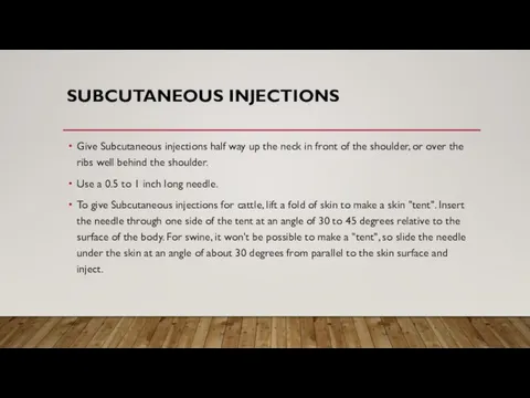
- 6. SUBCUTANEOUS INJECTIONS Give Subcutaneous injections half way up the neck in front of the shoulder, or
- 8. INTRAVENOUS THERAPY Intravenous therapy is the infusion of liquid substances directly into a vein. Intravenous simply
- 10. CONSEQUENCES OF POOR INJECTION TECHNIQUES Treatment failure, if product absorption is delayed or blocked. Drug residues
- 11. RECORDS OF TREATMENT All treatments given to food animals should be permanently recorded to ensure withdrawal
- 12. CARDIAC PUNCTURE Cardiac puncture is a suitable technique to obtain a single, large, good quality sample
- 13. ARTHROCENTESIS Arthrocentesis is performed in cases of suspected inflammation or infection of a peripheral joint. Cytologic
- 14. TECHNIQUE Sedation and local anesthesia - Many animals require mild sedation for the procedure. Ill and
- 15. Procedure - The skin over the joint(s) is clipped and prepared using aseptic technique. The joint
- 16. Arthrocentesis of the carpus is best performed with the carpus partially flexed. The needle is usually
- 18. Скачать презентацию

INJECTIONS
An injection is an infusion method of putting fluid into the
INJECTIONS
An injection is an infusion method of putting fluid into the

METHODS OF INJECTION
There are several methods of injection or infusion used
METHODS OF INJECTION
There are several methods of injection or infusion used

INTRAMUSCULAR INJECTIONS
Choose muscle tissue of lesser value to consumers for
INTRAMUSCULAR INJECTIONS
Choose muscle tissue of lesser value to consumers for

SUBCUTANEOUS INJECTIONS
Give Subcutaneous injections half way up the neck in front
SUBCUTANEOUS INJECTIONS
Give Subcutaneous injections half way up the neck in front


INTRAVENOUS THERAPY
Intravenous therapy is the infusion of liquid substances directly into
INTRAVENOUS THERAPY
Intravenous therapy is the infusion of liquid substances directly into


CONSEQUENCES OF POOR INJECTION TECHNIQUES
Treatment failure, if product absorption is delayed
CONSEQUENCES OF POOR INJECTION TECHNIQUES
Treatment failure, if product absorption is delayed

RECORDS OF TREATMENT
All treatments given to food animals should be permanently
RECORDS OF TREATMENT
All treatments given to food animals should be permanently

CARDIAC PUNCTURE
Cardiac puncture is a suitable technique to obtain a single,
CARDIAC PUNCTURE
Cardiac puncture is a suitable technique to obtain a single,
ARTHROCENTESIS
Arthrocentesis is performed in cases of suspected inflammation or infection of
ARTHROCENTESIS
Arthrocentesis is performed in cases of suspected inflammation or infection of
TECHNIQUE
Sedation and local anesthesia - Many animals require mild sedation for the
TECHNIQUE
Sedation and local anesthesia - Many animals require mild sedation for the

Procedure - The skin over the joint(s) is clipped and prepared using
Procedure - The skin over the joint(s) is clipped and prepared using

Arthrocentesis of the carpus is best performed with the carpus partially
Arthrocentesis of the carpus is best performed with the carpus partially
 Цветок ромашки
 Урок естествознания на тему: Лекарства
 Реабилитационная медико-психологическая программа для больных с первично диагностированными злокачественными новообразованиями
 Влияние вредных факторов на плод
 Дәріге тәуелділіктің әлеуметтік мәселелері
 Скриниг-диагностика ВПС
 Анализаторы. Кодирование информации
 Эритремия
 Avoiding sports injures. Grade 7
 Тамақтандыру түрлері мен құрамы
 Комплексний підхід до оцінки імунного статусу людини. Імунограма
 Маркетинговые технологии в аптечной организации
 Стратегия Всемирной Организации Здравоохранения в области охраны здоровья населения
 Подготовка публичного выступления
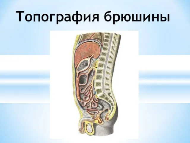 Топография брюшины
 Гемолитическая болезнь новорождённых
 Неотложная помощь при пароксизмальной фибрилляции предсердий
 Правила гигиены питания
 Асоциальное поведение и конфликты
 Неотложные состояния в урологии
 И.И. Мечников (1845-1916)
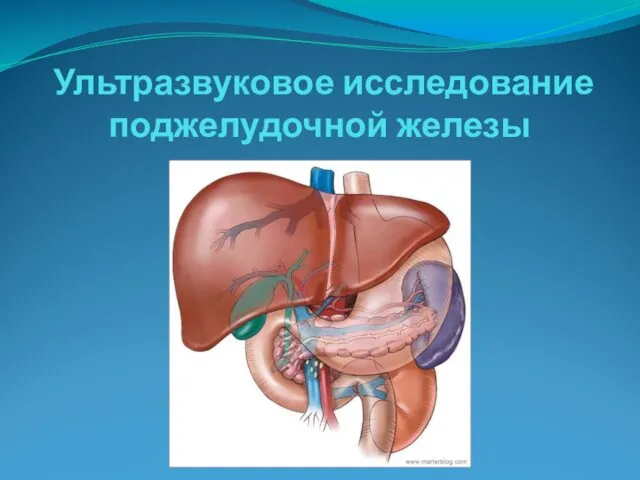 Ультразвуковое исследование поджелудочной железы
 Общая и частная психопатология
 Организация ухода за больными кардиологического профиля
 Тоны сердца, верхушечный толчок. Фонокардиография ее клиническое значение
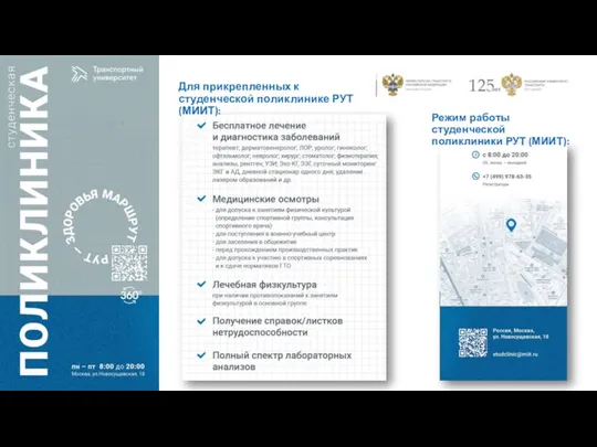 Студенческая поликлиника РУТ
 Ранения магистральных сосудов. Кровотечения и кровопотери
 Гинекологиялық науқастарды диспансерлеу. Әйелдердің репродуктивті жүйесі