- The BLUE-Protocol and the Diagnosis of Pneumonia

Содержание
- 2. The BLUE-protocol is an ultrasound approach of the lung, and of the venous network in appropriate
- 3. WHICH DEVICE, WHICH PROBE?
- 4. Note: Lung sliding has been described as a shimmering appearance of the pleura, or like tiny
- 5. DESCRIBE THE MAIN PATHOLOGICAL PROFILE (PROFILE B) This aspect of multiple B lines and accompanied by
- 6. Pleural effusion. Left and middle: minute pleural effusion at the PLAPS-point. Below the pleural line, a
- 7. BASIC IDENTIFICATION . The A-line indicates gas below the pleural line ULCs (or B-lines) associated with
- 8. Merlin's space defines the area located between the pleural line, the shadow of the ribs and
- 9. DESCRIBE PROFILE A (NORMAL PULMONARY SURFACE image obtained in the normal subject when the probe is
- 10. DESCRIBE PROFILE B' (MAIN PATHOLOGICAL PROFILE) This video shows an interstitial syndrome close to the B
- 11. DESCRIBE PROFILE A' (PNEUMOTHORAX) This video shows a totally abolished pleural slip (with the stratosphere sign
- 12. Lung point This video shows a characteristic appearance of frontal lung point, an all-or-nothing law where
- 13. DESCRIBE PROFILE C (PNEUMOPATHY The detection of alveolar disorders (regardless of number and size) on the
- 14. DESCRIBE A/B PROFILE (PNEUMOPATHY) An anterior asymmetry, with a profile A on one side and profile
- 15. DESCRIBE A/PLAPS PROFILE (PNEUMOPATHY) It is defined by the association of a profile A (thus anterior)
- 16. DESCRIBE THE NUDE PROFILE /NON PLAP/COPD/ASTHMA Everything is normal in the nude profile, the appearance of
- 18. Скачать презентацию
The BLUE-protocol is an ultrasound approach of the lung, and of
The BLUE-protocol is an ultrasound approach of the lung, and of
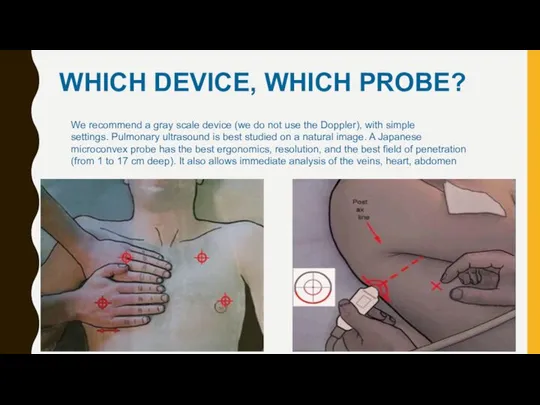
WHICH DEVICE, WHICH PROBE?
WHICH DEVICE, WHICH PROBE?

Note: Lung sliding has been described as a shimmering appearance of the
Note: Lung sliding has been described as a shimmering appearance of the
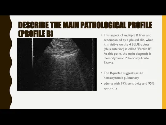
DESCRIBE THE MAIN PATHOLOGICAL PROFILE (PROFILE B)
This aspect of multiple B
DESCRIBE THE MAIN PATHOLOGICAL PROFILE (PROFILE B)
This aspect of multiple B
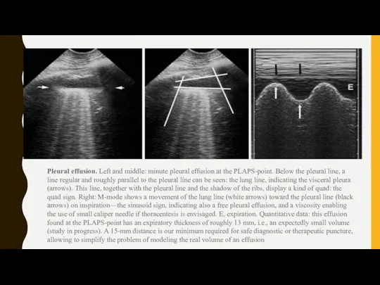
Pleural effusion. Left and middle: minute pleural effusion at the PLAPS-point. Below
Pleural effusion. Left and middle: minute pleural effusion at the PLAPS-point. Below
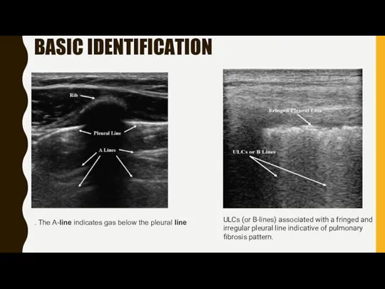
BASIC IDENTIFICATION
. The A-line indicates gas below the pleural line
ULCs (or B-lines) associated
BASIC IDENTIFICATION
. The A-line indicates gas below the pleural line
ULCs (or B-lines) associated

Merlin's space defines the area located between the pleural line, the shadow of the ribs
Merlin's space defines the area located between the pleural line, the shadow of the ribs
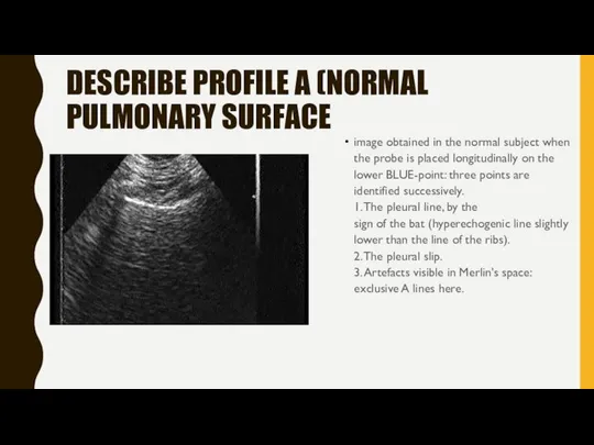
DESCRIBE PROFILE A (NORMAL PULMONARY SURFACE
image obtained in the normal subject
DESCRIBE PROFILE A (NORMAL PULMONARY SURFACE
image obtained in the normal subject

DESCRIBE PROFILE B' (MAIN PATHOLOGICAL PROFILE)
This video shows an interstitial syndrome
DESCRIBE PROFILE B' (MAIN PATHOLOGICAL PROFILE)
This video shows an interstitial syndrome
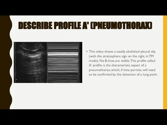
DESCRIBE PROFILE A' (PNEUMOTHORAX)
This video shows a totally abolished pleural slip
DESCRIBE PROFILE A' (PNEUMOTHORAX)
This video shows a totally abolished pleural slip

Lung point
This video shows a characteristic appearance of frontal lung point,
Lung point This video shows a characteristic appearance of frontal lung point,

DESCRIBE PROFILE C (PNEUMOPATHY
The detection of alveolar disorders (regardless of number
DESCRIBE PROFILE C (PNEUMOPATHY
The detection of alveolar disorders (regardless of number

DESCRIBE A/B PROFILE (PNEUMOPATHY)
An anterior asymmetry, with a profile A on
DESCRIBE A/B PROFILE (PNEUMOPATHY)
An anterior asymmetry, with a profile A on

DESCRIBE A/PLAPS PROFILE (PNEUMOPATHY)
It is defined by the association of a
DESCRIBE A/PLAPS PROFILE (PNEUMOPATHY)
It is defined by the association of a
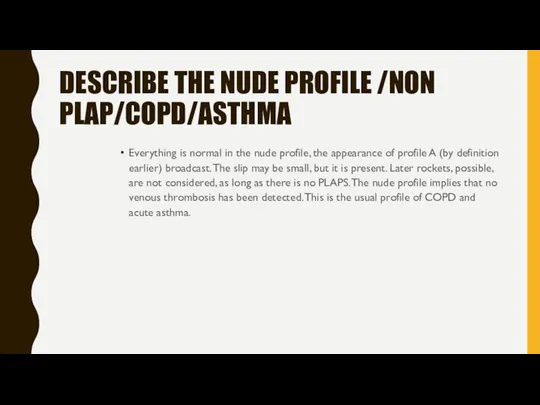
DESCRIBE THE NUDE PROFILE /NON PLAP/COPD/ASTHMA
Everything is normal in the nude
DESCRIBE THE NUDE PROFILE /NON PLAP/COPD/ASTHMA
Everything is normal in the nude
 Алгоритми
 Биметаллические термометры, модель 55, промышленная серия. (S5550; R5502; A5500)
 Valdo 2016 katalogs III daļa -pielabots
 Презентация Час безопасности по БДД август 2022г. Новойл
 Отрасль птицеводства
 Комплекс машин для возделывания картофеля
 Молитва
 Стеклянные товары
 20180417_1f3856b393dcae63533e798ef76677a5
 Ұңғылардың сағасын су астында орналастыру әдісі. Жабдықтарды су астында орналастыру жүйелері. Суасты технологиясының беріктігі
 Conditionals
 Переходные процессы 1-ого порядка с емкостным элементом
 Регистры. Основные понятия и определения
 Плавкие предохранители
 Казка Колобок
 Назначение и сущность архитектуры. Здания и сооружения. Лекция 1
 The climate control
 Zvd engineering - о копмании
 111
111 Домашние животные. Мы в ответе за тех, кого приручили
 Памятники Фатежского района
 Мастер-класс по рисованию пейзажа для детей Утро в городе
 Великий канон cвятого Андрея Критского, читаемый в четверг пятой седмицы Великого поста
 8. Оценивание случайных погрешностей измерения
 География морского транспорта РФ
 20180227_kultura_detstva_v_sssr_1920-1930-e_gody._slayd-shou_k_razrabotke_uroka_stepuninoy_a.s
 История компании Samsung
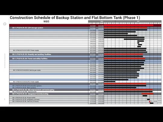 Construction Schedule of Backup Station and Flat Bottom Tank (Phase 1)