- Central Nervous System

Содержание
- 2. Introduction Analogies; telephone switchboard; computer; miracle A fantastically complex and flexible biological organ Cephalization become more
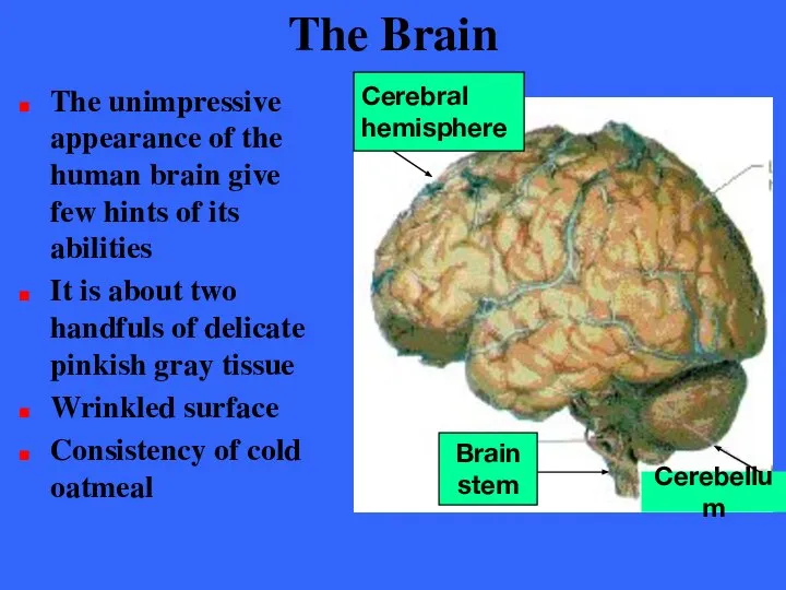
- 3. The Brain The unimpressive appearance of the human brain give few hints of its abilities It
- 4. The Brain Average adult male’s brain weighs about 1600 g (3.5 pounds) Average adult female’s brain
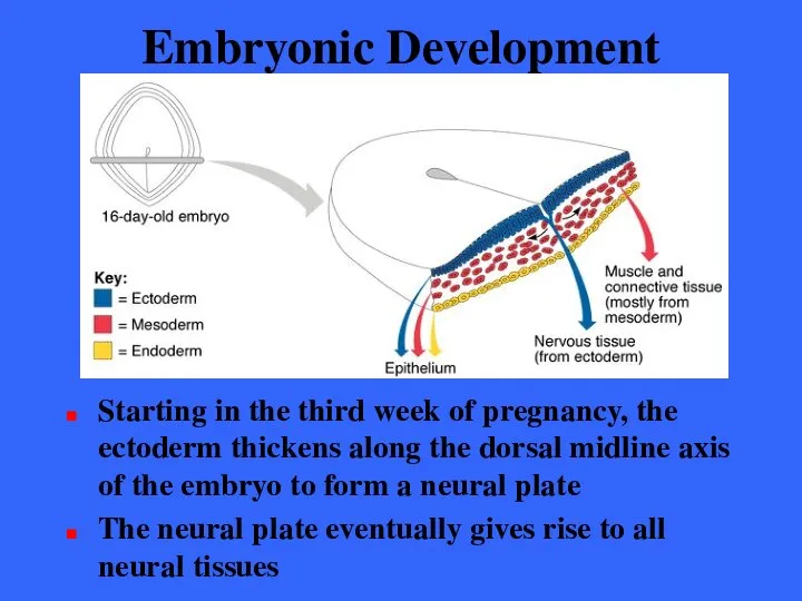
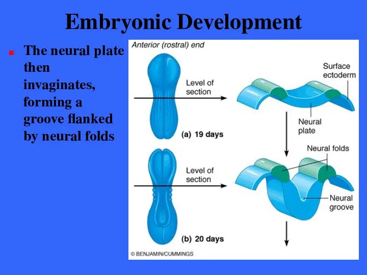
- 5. Embryonic Development Starting in the third week of pregnancy, the ectoderm thickens along the dorsal midline
- 6. Embryonic Development The neural plate then invaginates, forming a groove flanked by neural folds
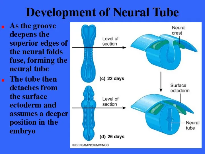
- 7. Development of Neural Tube As the groove deepens the superior edges of the neural folds fuse,
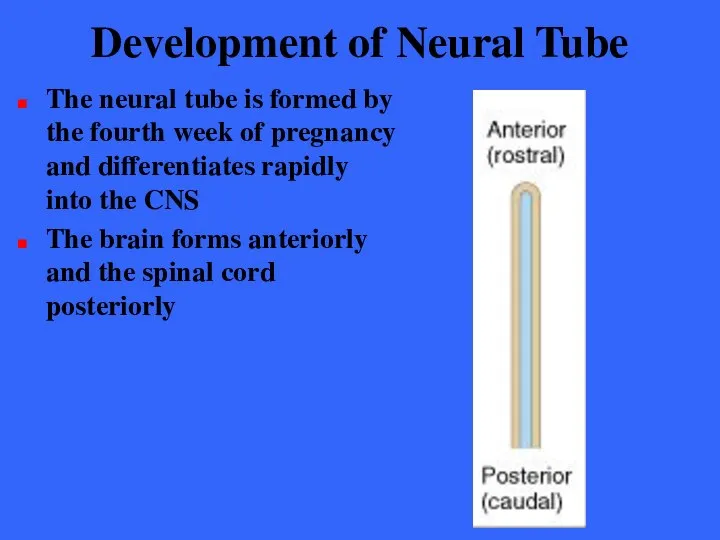
- 8. Development of Neural Tube The neural tube is formed by the fourth week of pregnancy and
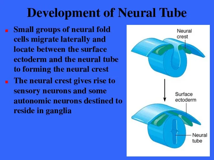
- 9. Development of Neural Tube Small groups of neural fold cells migrate laterally and locate between the
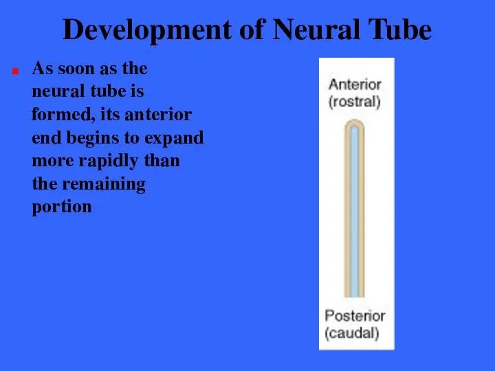
- 10. Development of Neural Tube As soon as the neural tube is formed, its anterior end begins
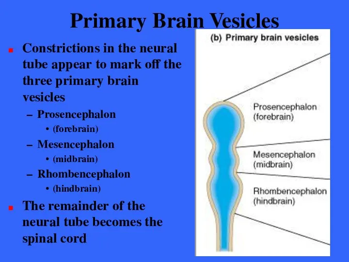
- 11. Primary Brain Vesicles Constrictions in the neural tube appear to mark off the three primary brain
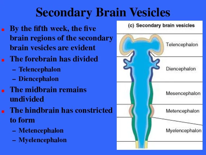
- 12. Secondary Brain Vesicles By the fifth week, the five brain regions of the secondary brain vesicles
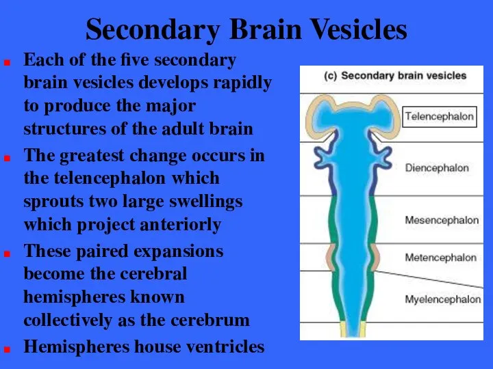
- 13. Secondary Brain Vesicles Each of the five secondary brain vesicles develops rapidly to produce the major
- 14. Secondary Brain Vesicles Various areas of the diencephalon specialize to form Hypothalamus Thalamus Epithalamus
- 15. Secondary Brain Vesicles The mesencephalon develops into Midbrain Brain stem
- 16. Secondary Brain Vesicles Various areas of the Metencephalon specialize to form Brain stem Pons Cerebellum
- 17. Secondary Brain Vesicles Various areas of the Myelencephalon specialize to form Brain stem Medulla oblongata All
- 18. Adult Neural Canal Regions The central canal of the neural tube enlarge in four areas to
- 19. Development of Flexures During this period of rapid brain growth change is also occurring in the
- 20. Effects of Space Restriction A second consequence of restricted space is that the cerebral hemispheres are
- 21. Effects of Space Restriction As a result the hemispheres grow back over and almost completely envelop
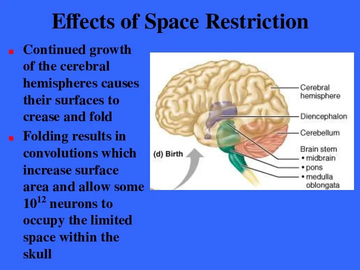
- 22. Effects of Space Restriction Continued growth of the cerebral hemispheres causes their surfaces to crease and
- 23. Effects of Space Restriction The wrinkling of the hemispheres may result from tension on the young
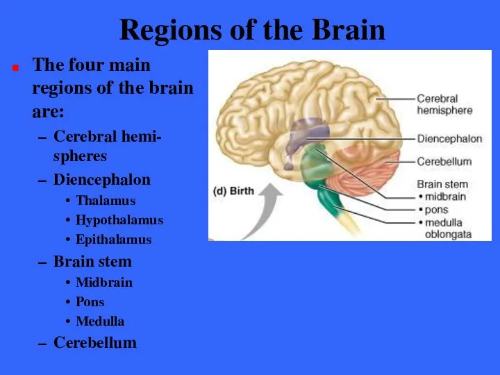
- 24. Regions of the Brain The four main regions of the brain are: Cerebral hemi- spheres Diencephalon
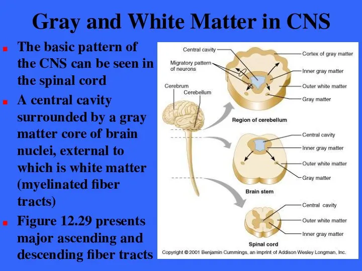
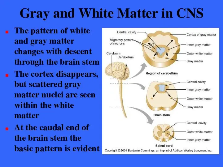
- 25. Gray and White Matter in CNS The basic pattern of the CNS can be seen in
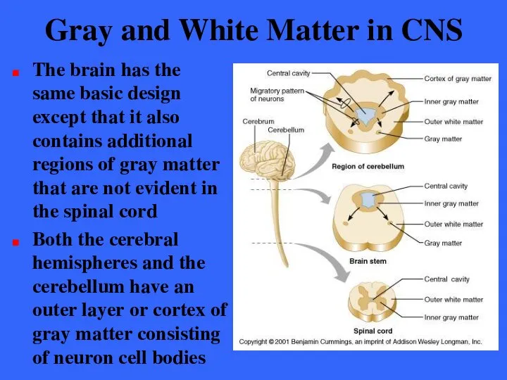
- 26. Gray and White Matter in CNS The brain has the same basic design except that it
- 27. Gray and White Matter in CNS The pattern of white and gray matter changes with descent
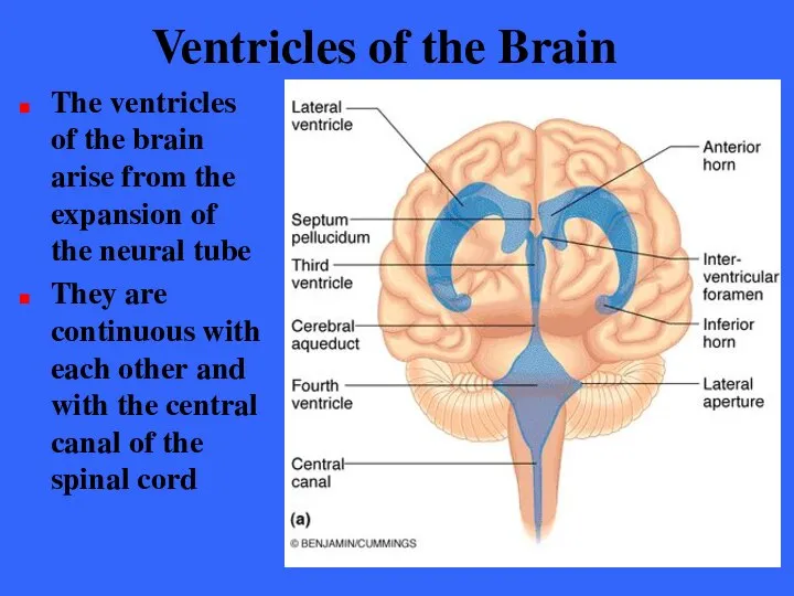
- 28. Ventricles of the Brain The ventricles of the brain arise from the expansion of the neural
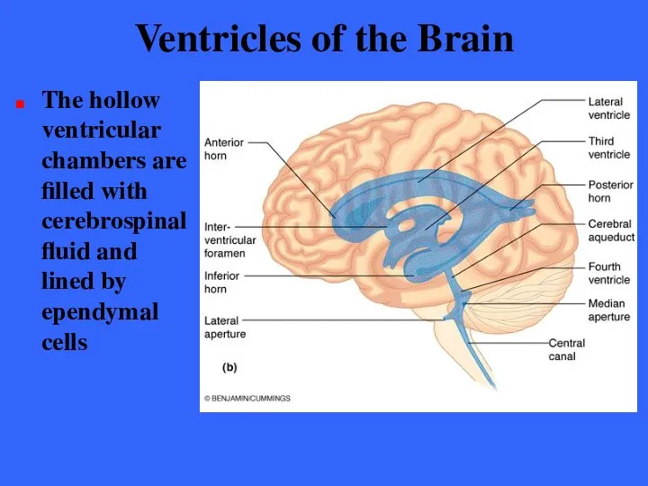
- 29. Ventricles of the Brain The hollow ventricular chambers are filled with cerebrospinal fluid and lined by
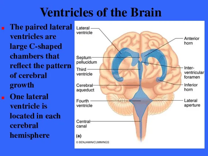
- 30. Ventricles of the Brain The paired lateral ventricles are large C-shaped chambers that reflect the pattern
- 31. Ventricles of the Brain Anteriorly, the lateral ventricles lie close together separated only by a thin
- 32. Ventricles of the Brain Communication occurs through the inter- ventricular foramen (foramen of Moro)
- 33. Ventricles of the Brain The third ventricle is continuous with the fourth ventricle via the canal-like
- 34. Ventricles of the Brain The fourth ventricle which lies dorsal to the pons and posterior to
- 35. Ventricles of the Brain Three openings mark the walls of the fourth ventricle Paired lateral apertures
- 36. The Cerebral Hemispheres The cerebral hemispheres form the superior part of the brain These two structures
- 37. The Cerebral Hemispheres Nearly the entire surface of the cerebral hemispheres is marked by elevated ridges
- 38. The Cerebral Hemispheres Prominent gyri and sulci are similar in all people The median longitudinal fissure
- 39. Lobes of Cerebral Hemispheres Deeper sulci divide each hemisphere into five lobes Frontal lobe Temporal lobe
- 40. Lobes of Cerebral Hemispheres Location of the insula deep within the Lateral sulcus of the hemisphere
- 41. Fissures of Cerebral Hemispheres Sulci divide lobes of the hemispheres Central sulcus Parieto- occipital sulcus Lateral
- 42. Medial Surface of Right Hemisphere Medial surface of the right hemisphere showing the Parieto- occipital sulcus
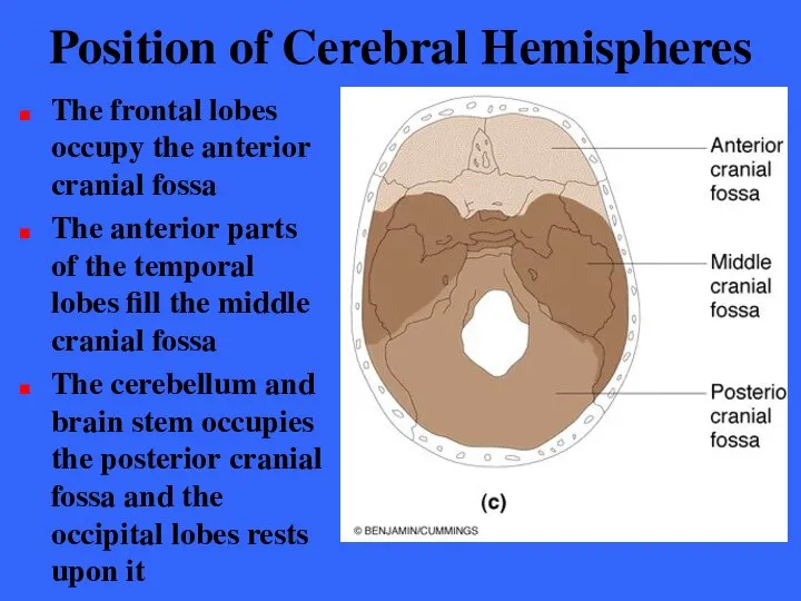
- 43. Position of Cerebral Hemispheres The frontal lobes occupy the anterior cranial fossa The anterior parts of
- 44. Cerebral Cortex The cerebral cortex is the “executive suite” of the nervous system It enables us
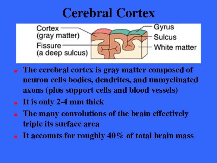
- 45. Cerebral Cortex The cerebral cortex is gray matter composed of neuron cells bodies, dendrites, and unmyelinated
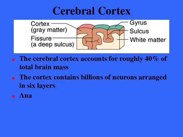
- 46. Cerebral Cortex The cerebral cortex accounts for roughly 40% of total brain mass The cortex contains
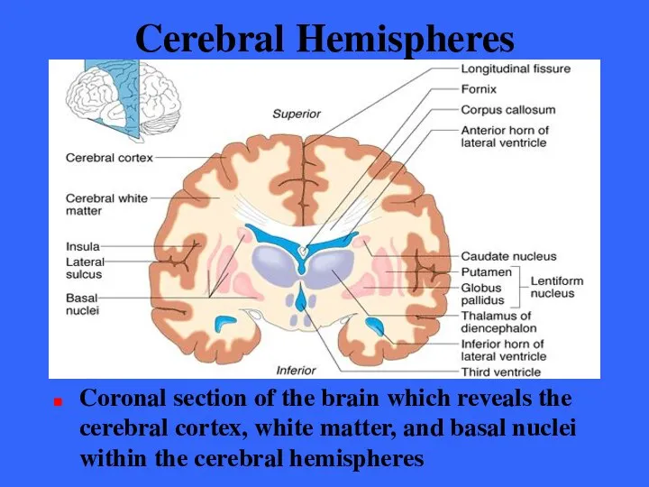
- 47. Cerebral Hemispheres Coronal section of the brain which reveals the cerebral cortex, white matter, and basal
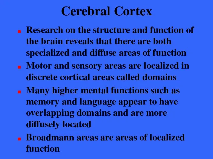
- 48. Cerebral Cortex Research on the structure and function of the brain reveals that there are both
- 49. Cerebral Cortex - Generalizations The cerebral cortex has three types of functional areas Motor areas /
- 50. Cerebral Cortex - Generalizations Although they are largely symmetrical in structure the two hemispheres are not
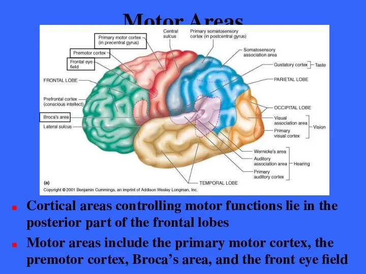
- 51. Motor Areas Cortical areas controlling motor functions lie in the posterior part of the frontal lobes
- 52. Primary Motor Cortex The primary motor cortex is located in the precentral gyrus of the frontal
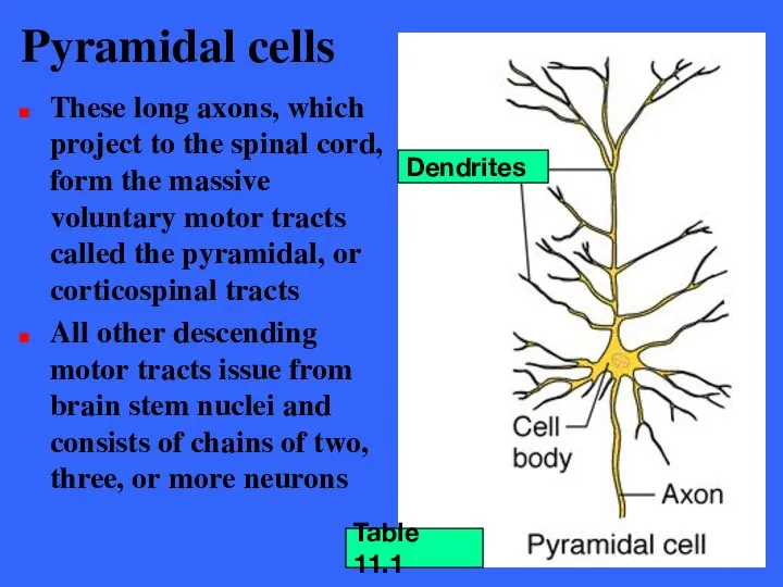
- 53. Pyramidal cells These long axons, which project to the spinal cord, form the massive voluntary motor
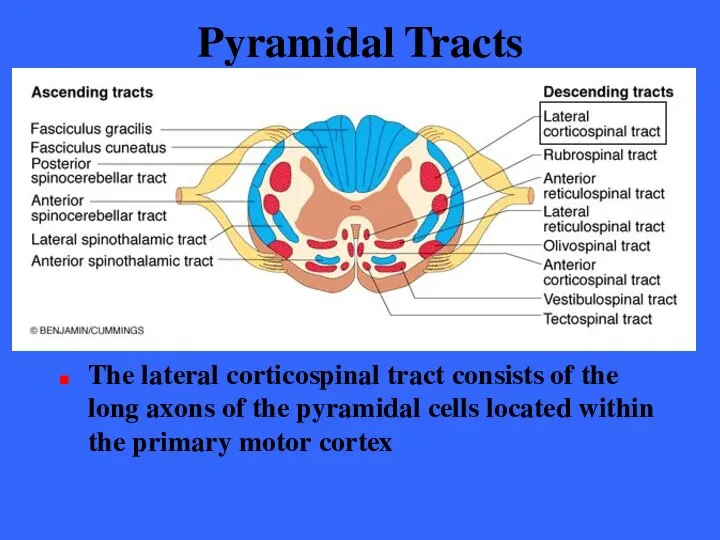
- 54. Pyramidal Tracts The lateral corticospinal tract consists of the long axons of the pyramidal cells located
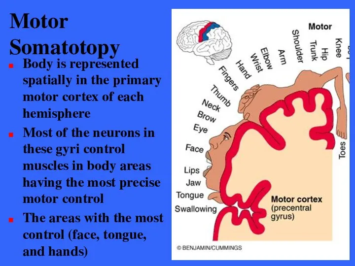
- 55. Motor Somatotopy Body is represented spatially in the primary motor cortex of each hemisphere Most of
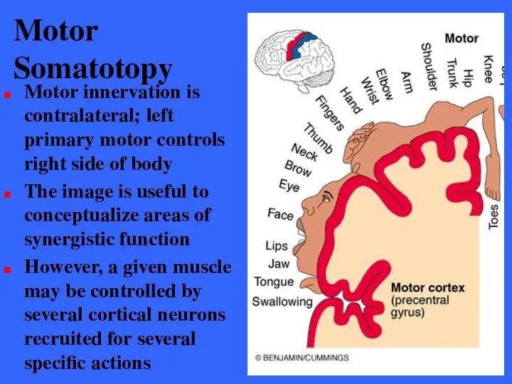
- 56. Motor Somatotopy Motor innervation is contralateral; left primary motor controls right side of body The image
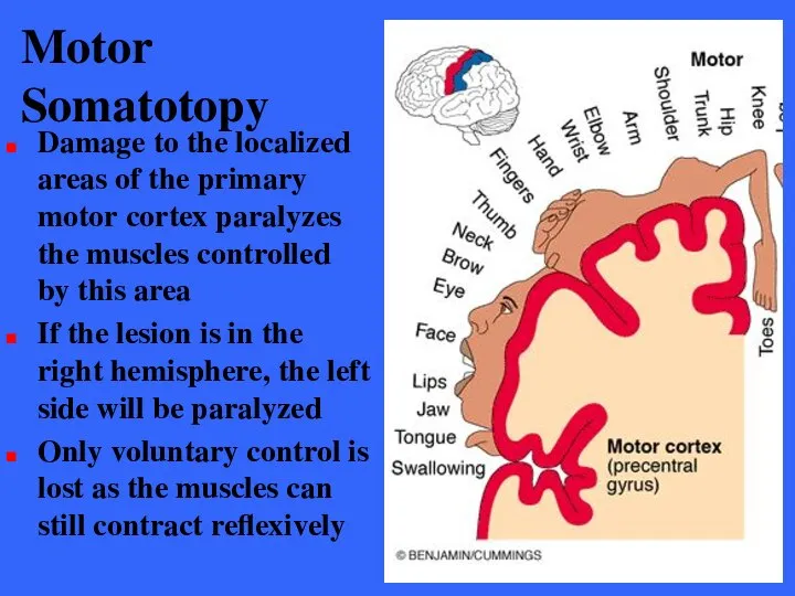
- 57. Motor Somatotopy Damage to the localized areas of the primary motor cortex paralyzes the muscles controlled
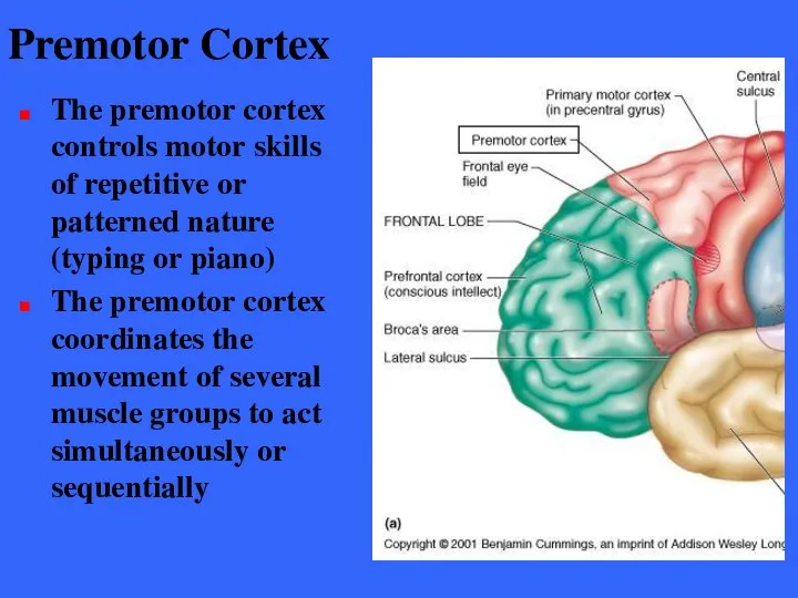
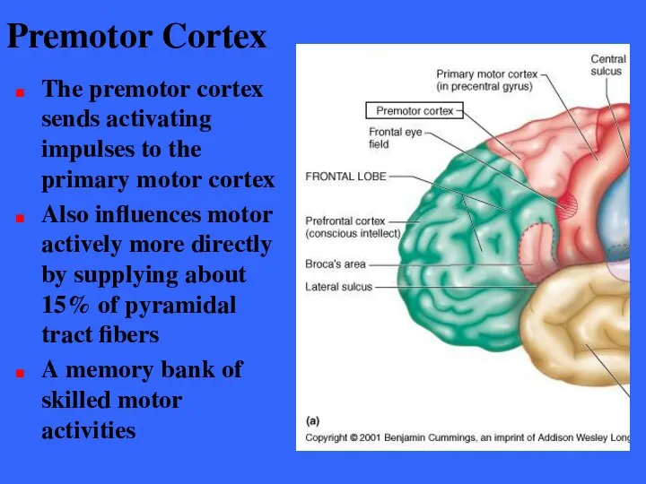
- 58. Premotor Cortex The premotor cortex controls motor skills of repetitive or patterned nature (typing or piano)
- 59. Premotor Cortex The premotor cortex sends activating impulses to the primary motor cortex Also influences motor
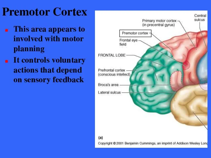
- 60. Premotor Cortex This area appears to involved with motor planning It controls voluntary actions that depend
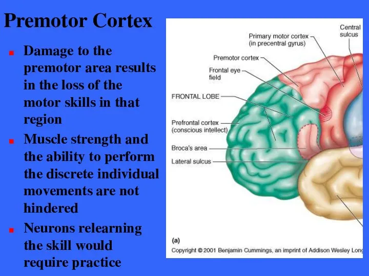
- 61. Premotor Cortex Damage to the premotor area results in the loss of the motor skills in
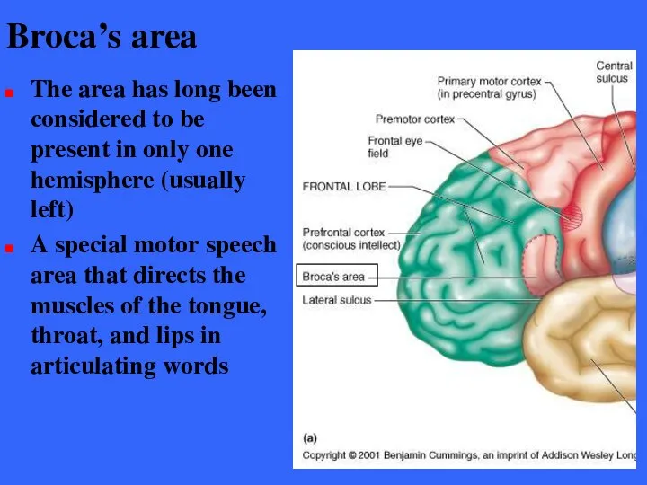
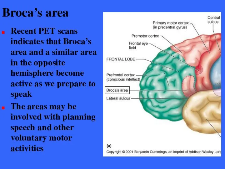
- 62. Broca’s area The area has long been considered to be present in only one hemisphere (usually
- 63. Broca’s area Recent PET scans indicates that Broca’s area and a similar area in the opposite
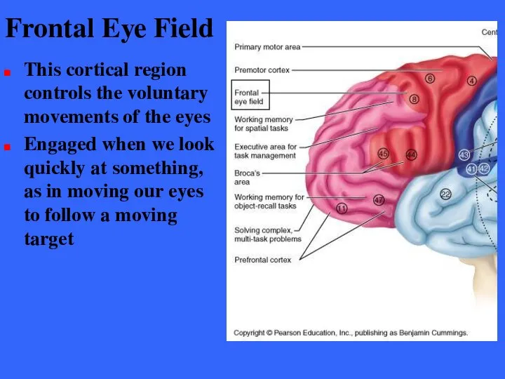
- 64. Frontal Eye Field This cortical region controls the voluntary movements of the eyes Engaged when we
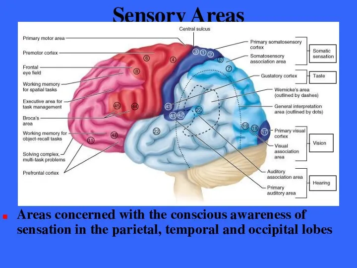
- 65. Sensory Areas Areas concerned with the conscious awareness of sensation in the parietal, temporal and occipital
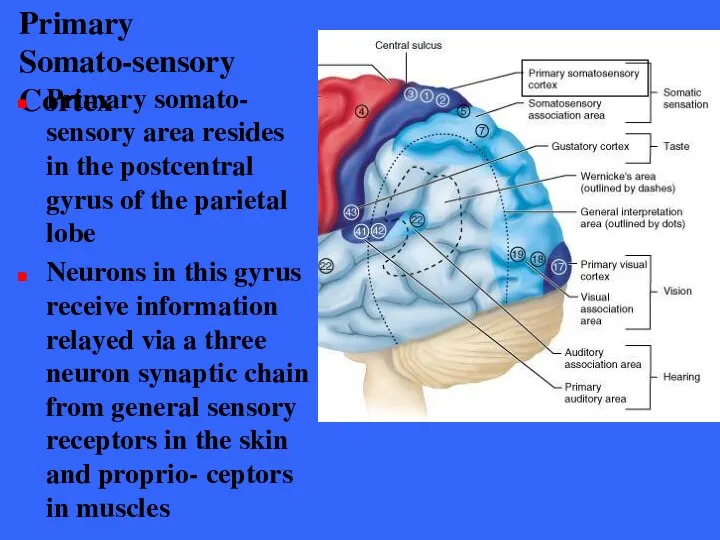
- 66. Primary Somato-sensory Cortex Primary somato- sensory area resides in the postcentral gyrus of the parietal lobe
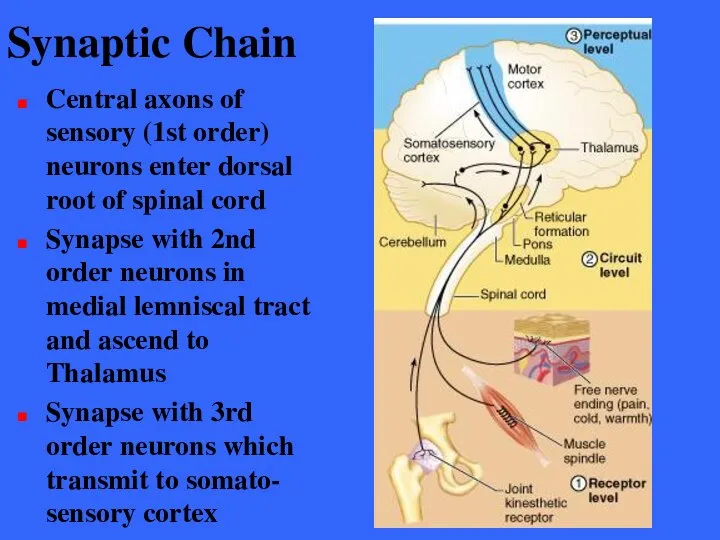
- 67. Synaptic Chain Central axons of sensory (1st order) neurons enter dorsal root of spinal cord Synapse
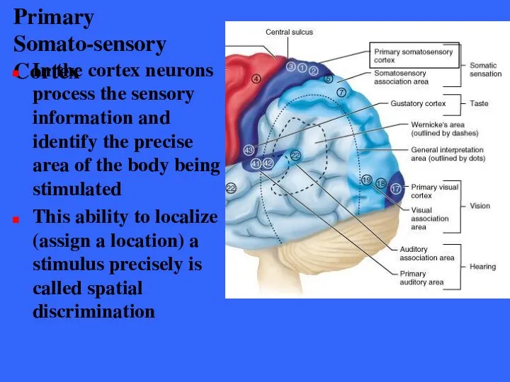
- 68. Primary Somato-sensory Cortex In the cortex neurons process the sensory information and identify the precise area
- 69. Motor and Sensory Somatotopy
- 70. Primary Somato-sensory Cortex The sensory spatial discrimination is contralateral with the right hemisphere receiving inputs from
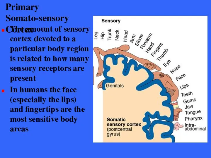
- 71. Primary Somato-sensory Cortex The amount of sensory cortex devoted to a particular body region is related
- 72. Primary Somatosensory Cortex Damage to the primary somatisensory cortex destroys the conscious ability to feel and
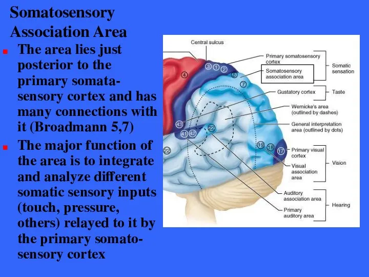
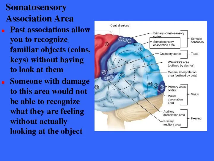
- 73. Somatosensory Association Area The area lies just posterior to the primary somata- sensory cortex and has
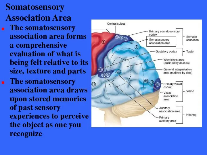
- 74. Somatosensory Association Area The somatosensory association area forms a comprehensive evaluation of what is being felt
- 75. Somatosensory Association Area Past associations allow you to recognize familiar objects (coins, keys) without having to
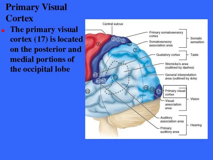
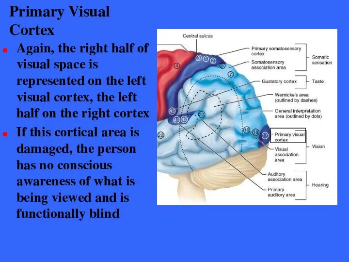
- 76. Primary Visual Cortex The primary visual cortex (17) is located on the posterior and medial portions
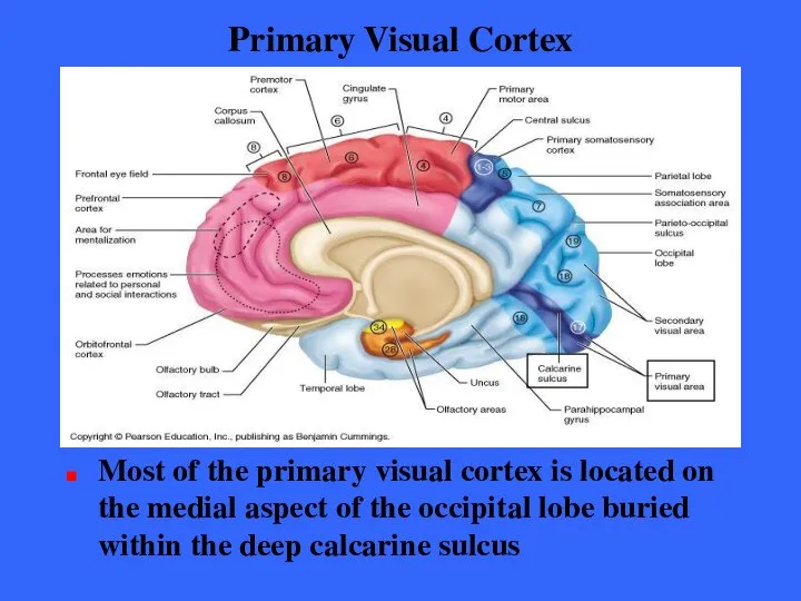
- 77. Primary Visual Cortex Most of the primary visual cortex is located on the medial aspect of
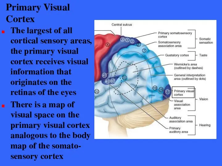
- 78. Primary Visual Cortex The largest of all cortical sensory areas, the primary visual cortex receives visual
- 79. Primary Visual Cortex Again, the right half of visual space is represented on the left visual
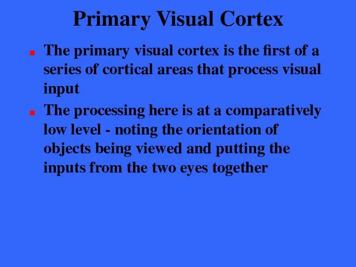
- 80. Primary Visual Cortex The primary visual cortex is the first of a series of cortical areas
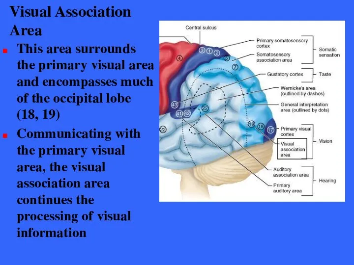
- 81. Visual Association Area This area surrounds the primary visual area and encompasses much of the occipital
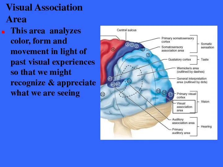
- 82. Visual Association Area This area analyzes color, form and movement in light of past visual experiences
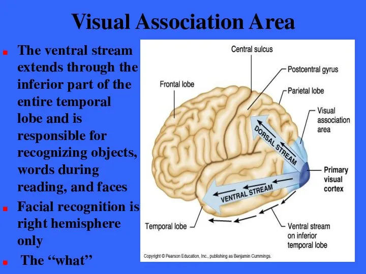
- 83. Visual Association Area Recent neuroimaging has revealed that complex visual processing far forward from the occipital
- 84. Visual Association Area The ventral stream extends through the inferior part of the entire temporal lobe
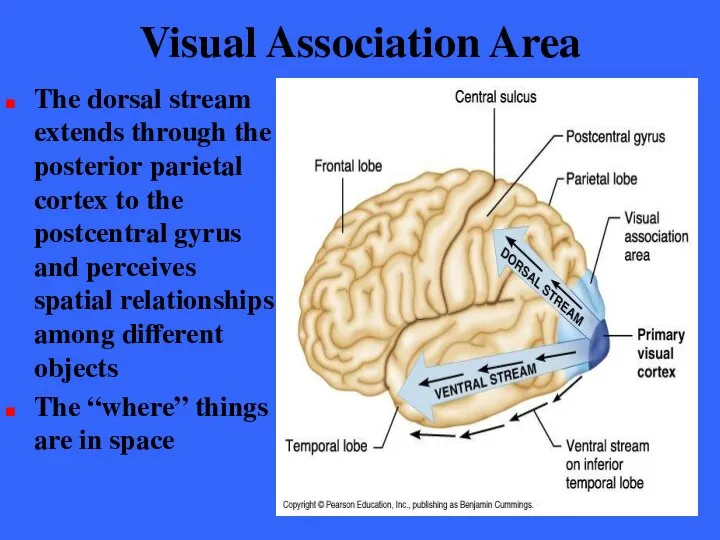
- 85. Visual Association Area The dorsal stream extends through the posterior parietal cortex to the postcentral gyrus
- 86. Visual Association Area The dorsal stream in the parietal lobe is important for spatial perception The
- 87. Visual Areas Damage to the visual cortex results in functional blindness Damage to the visual association
- 88. Primary Auditory Cortex The primary auditory cortex is located on the superior margin of the temporal
- 89. Primary Auditory Cortex Hearing receptors in the cochlear of the inner ear transmit impulses to primary
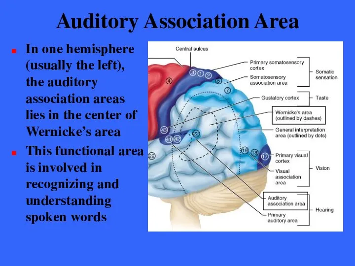
- 90. Auditory Association Area The auditory association area lies just posterior to the primary auditory area Broadmann
- 91. Auditory Association Area In one hemisphere (usually the left), the auditory association areas lies in the
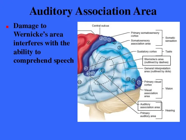
- 92. Auditory Association Area Damage to Wernicke’s area interferes with the ability to comprehend speech
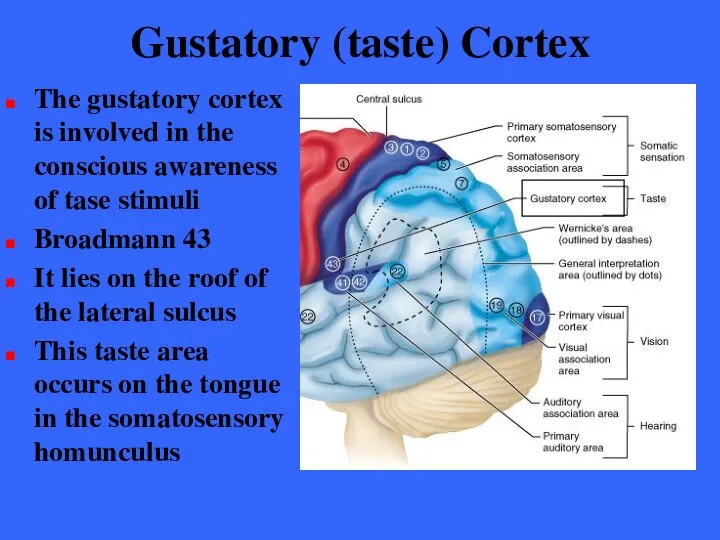
- 93. Gustatory (taste) Cortex The gustatory cortex is involved in the conscious awareness of tase stimuli Broadmann
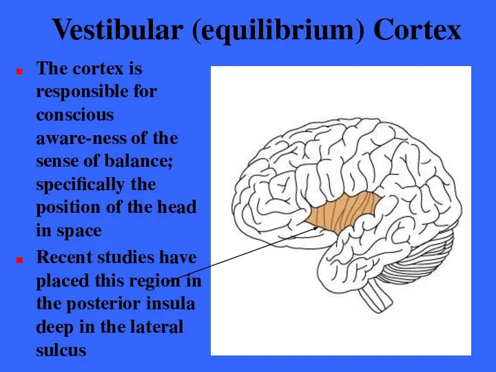
- 94. Vestibular (equilibrium) Cortex The cortex is responsible for conscious aware-ness of the sense of balance; specifically
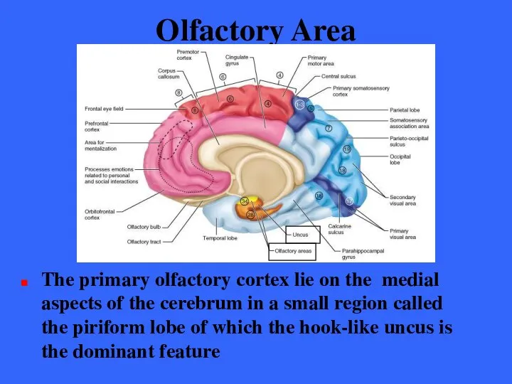
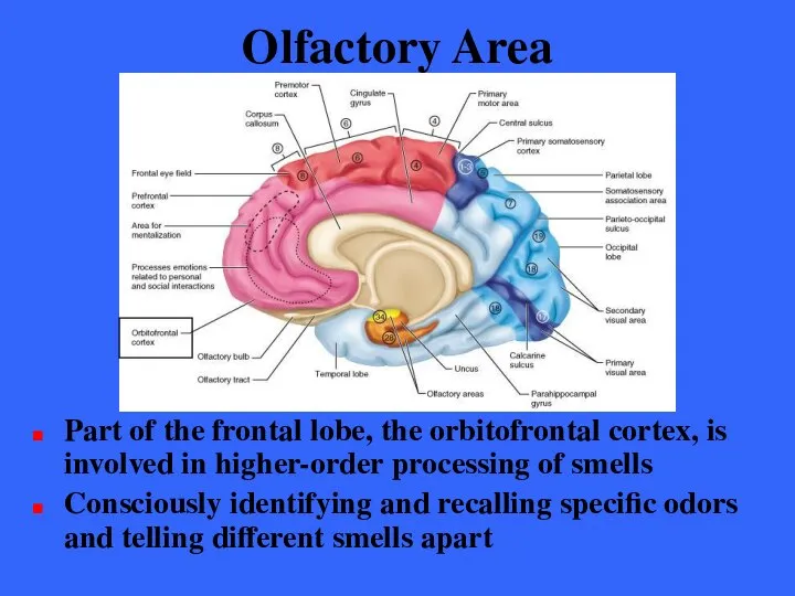
- 95. Olfactory Area The primary olfactory cortex lie on the medial aspects of the cerebrum in a
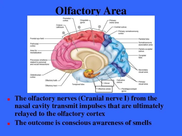
- 96. Olfactory Area The olfactory nerves (Cranial nerve I) from the nasal cavity transmit impulses that are
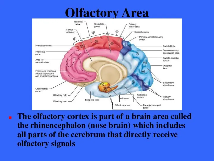
- 97. Olfactory Area The olfactory cortex is part of a brain area called the rhinencephalon (nose brain)
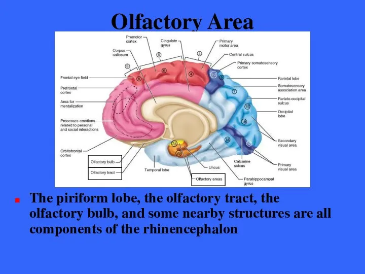
- 98. Olfactory Area The piriform lobe, the olfactory tract, the olfactory bulb, and some nearby structures are
- 99. Olfactory Area The rhinencephalon connects to the brain area that is involved in emotions, the limbic
- 100. Olfactory Area Part of the frontal lobe, the orbitofrontal cortex, is involved in higher-order processing of
- 101. Association Areas Association areas include all cortical areas other than primary sensory and motor areas The
- 102. Association Areas The term association area is fading from use and will probably be replaced by
- 103. Prefrontal Cortex The prefrontal cortex occupies the large region of the frontal lobe anterior to the
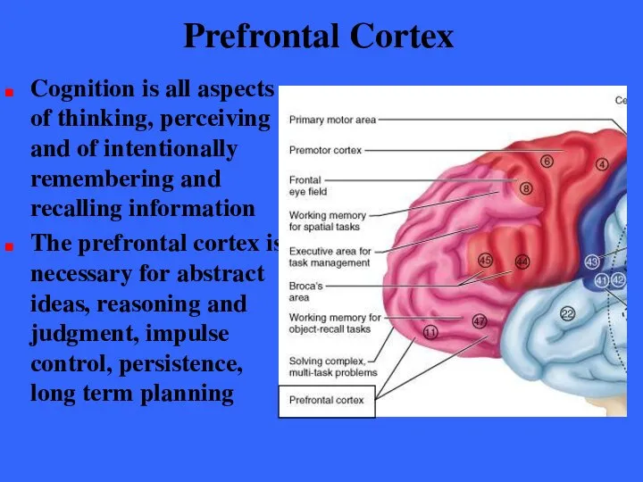
- 104. Prefrontal Cortex Cognition is all aspects of thinking, perceiving and of intentionally remembering and recalling information
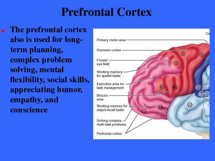
- 105. Prefrontal Cortex The prefrontal cortex also is used for long- term planning, complex problem solving, mental
- 106. Prefrontal Cortex The prefrontal cortex also seems to be related to mood and has close links
- 107. Prefrontal Cortex Functional neuro-imaging techniques have begun to reveal the functions of specific parts of the
- 108. Prefrontal Cortex The working memories of spatial relations are stored in the dorsolateral prefrontal cortex just
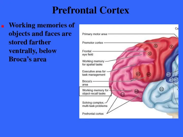
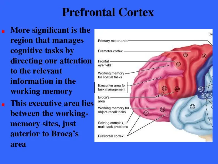
- 109. Prefrontal Cortex Working memories of objects and faces are stored farther ventrally, below Broca’s area
- 110. Prefrontal Cortex More significant is the region that manages cognitive tasks by directing our attention to
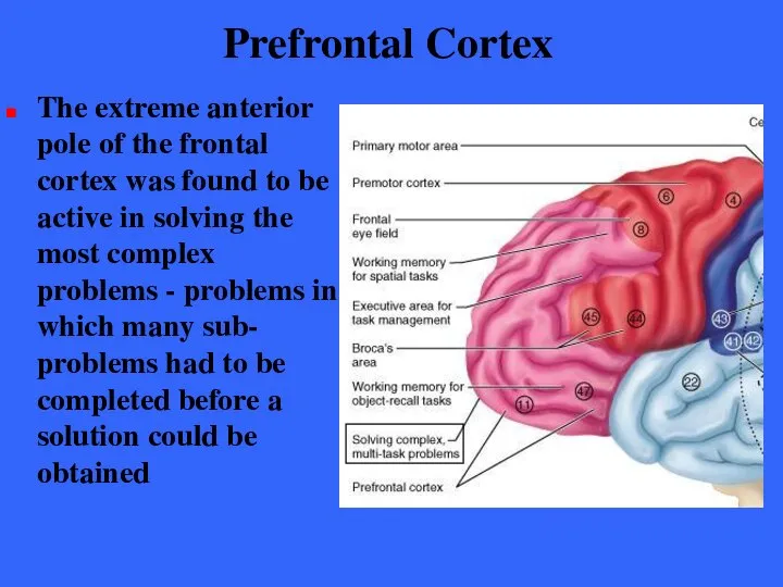
- 111. Prefrontal Cortex The extreme anterior pole of the frontal cortex was found to be active in
- 112. Prefrontal Cortex The new findings suggest support for a general rule of neuroscience that says the
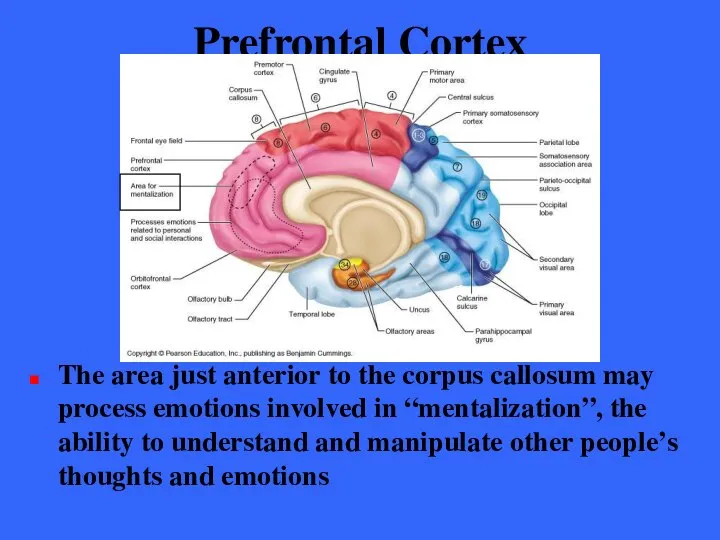
- 113. Prefrontal Cortex The area just anterior to the corpus callosum may process emotions involved in “mentalization”,
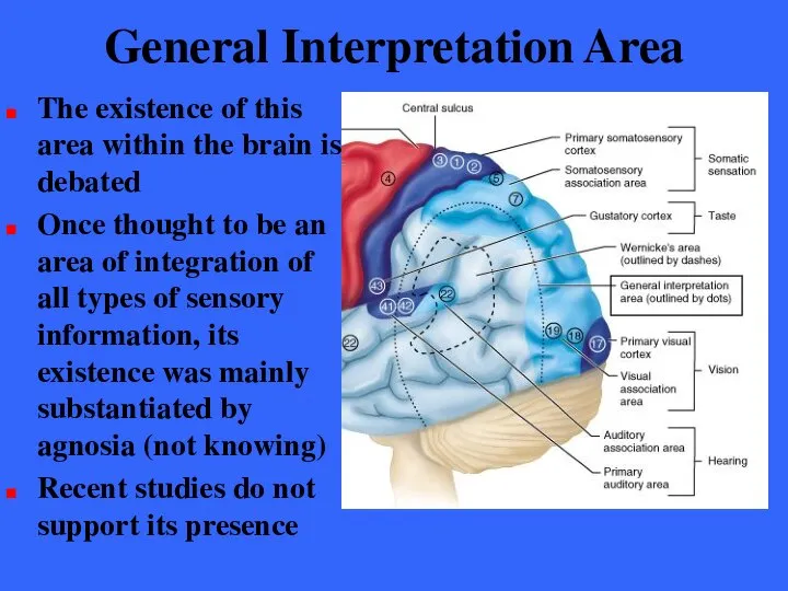
- 114. General Interpretation Area The existence of this area within the brain is debated Once thought to
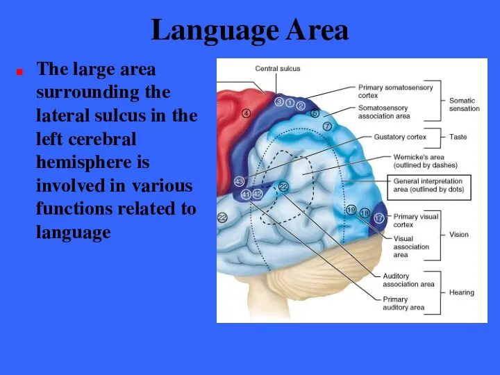
- 115. Language Area The large area surrounding the lateral sulcus in the left cerebral hemisphere is involved
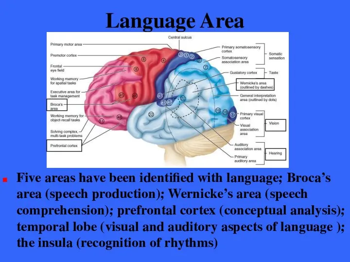
- 116. Language Area Five areas have been identified with language; Broca’s area (speech production); Wernicke’s area (speech
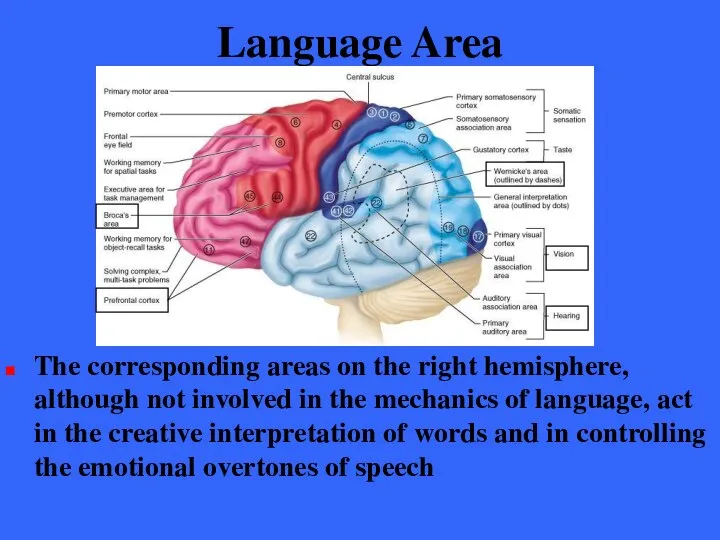
- 117. Language Area The corresponding areas on the right hemisphere, although not involved in the mechanics of
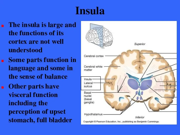
- 118. Insula The insula is large and the functions of its cortex are not well understood Some
- 119. Lateralization of Cortical Function We use both cerebral hemispheres for almost every task and it appears
- 120. Lateralization of Cortical Function In most people (Approx. 90%) the left hemisphere has greater control over
- 121. Lateralization of Cortical Function Most individuals (90%) with left cerebral dominance are right-handed In the remaining
- 122. Lateralization of Cortical Function The two cerebral hemispheres have perfect and almost instantaneous communication with one
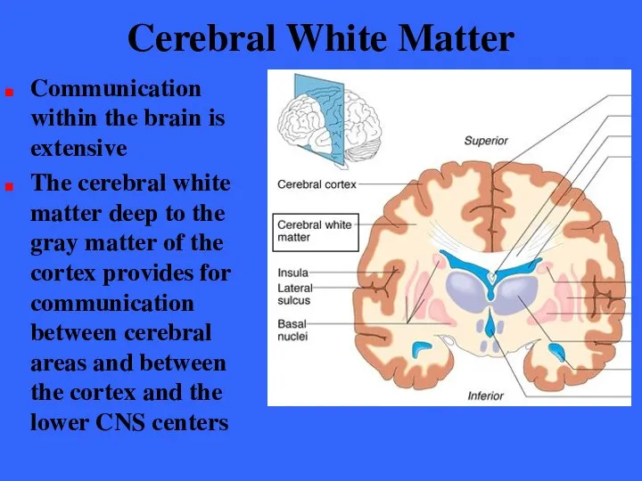
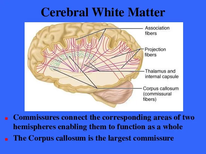
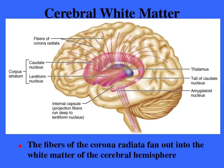
- 123. Cerebral White Matter Communication within the brain is extensive The cerebral white matter deep to the
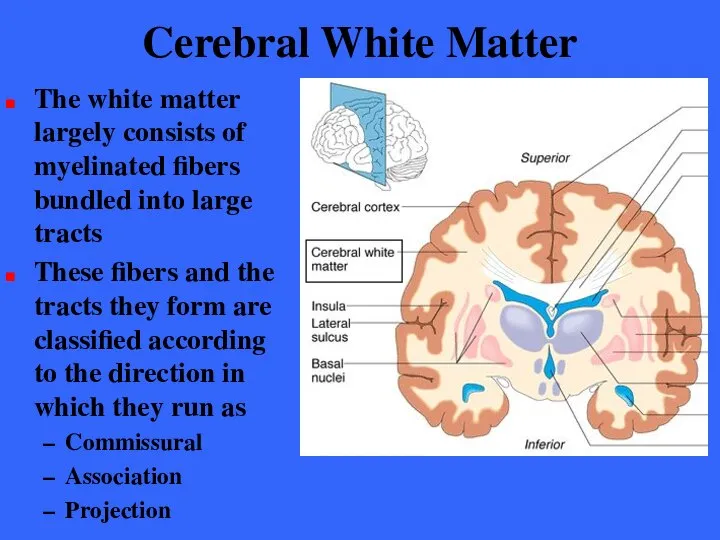
- 124. Cerebral White Matter The white matter largely consists of myelinated fibers bundled into large tracts These
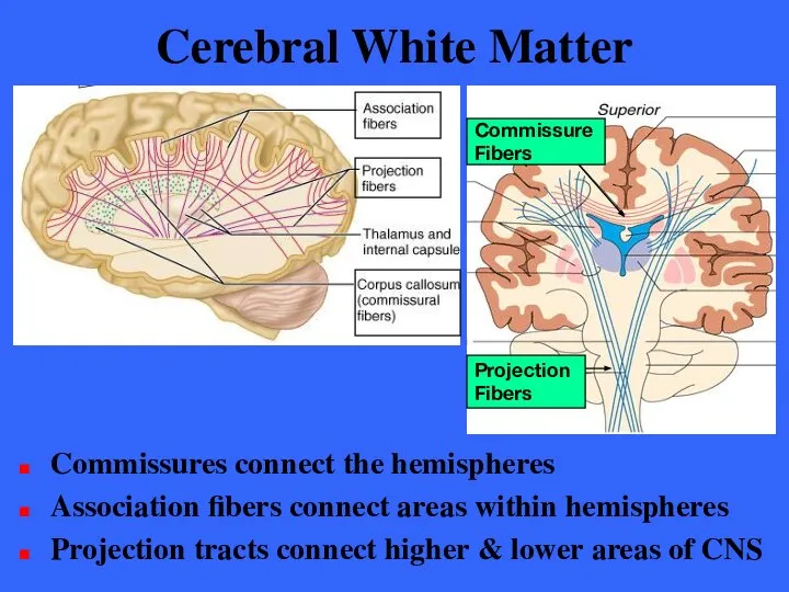
- 125. Cerebral White Matter Commissures connect the hemispheres Association fibers connect areas within hemispheres Projection tracts connect
- 126. Cerebral White Matter Commissures connect the corresponding areas of two hemispheres enabling them to function as
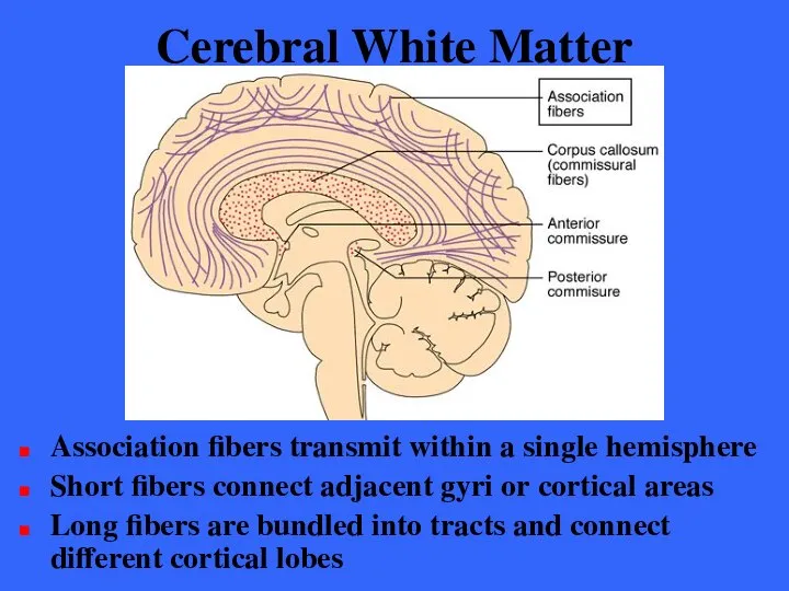
- 127. Cerebral White Matter Association fibers transmit within a single hemisphere Short fibers connect adjacent gyri or
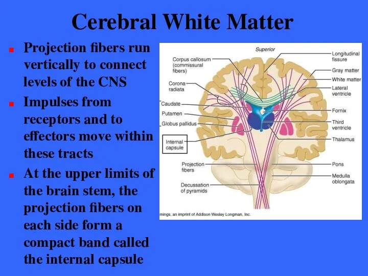
- 128. Cerebral White Matter Projection fibers run vertically to connect levels of the CNS Impulses from receptors
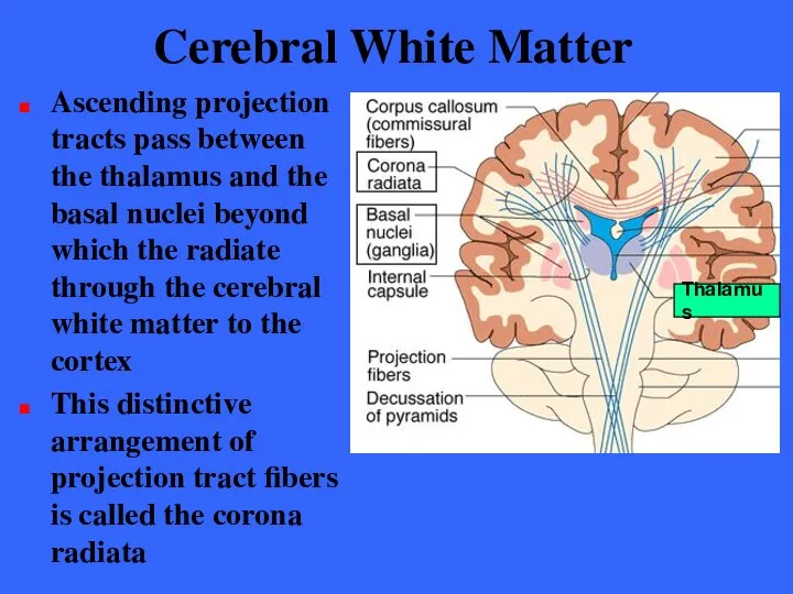
- 129. Cerebral White Matter Ascending projection tracts pass between the thalamus and the basal nuclei beyond which
- 130. Cerebral White Matter The fibers of the corona radiata fan out into the white matter of
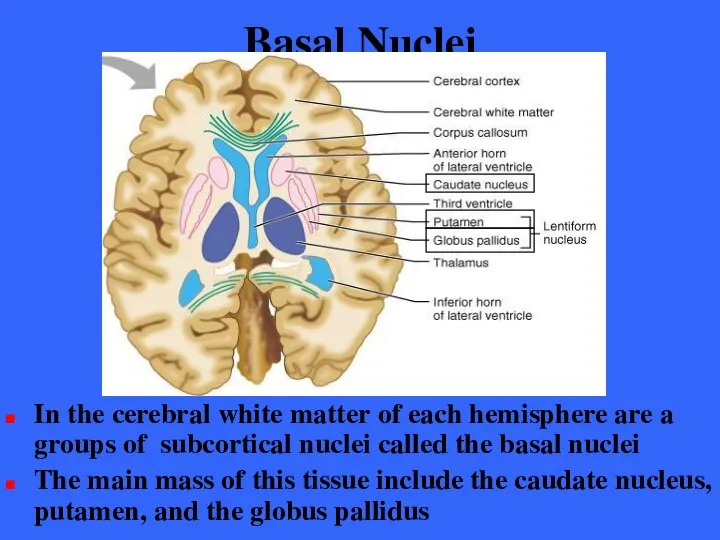
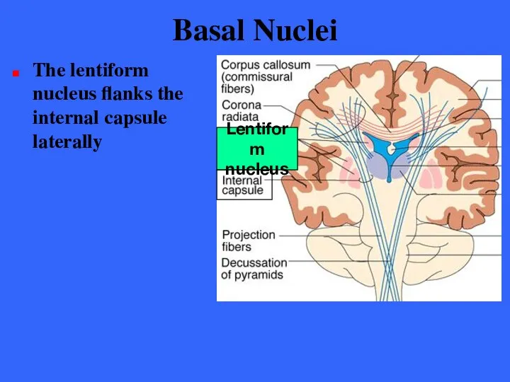
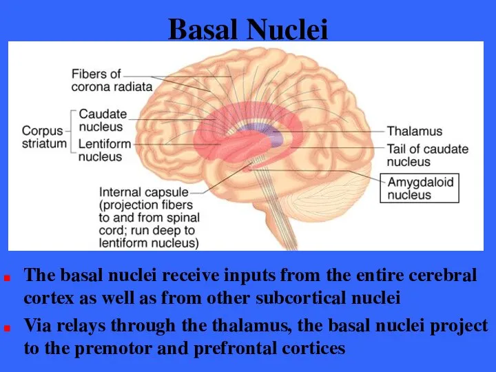
- 131. Basal Nuclei In the cerebral white matter of each hemisphere are a groups of subcortical nuclei
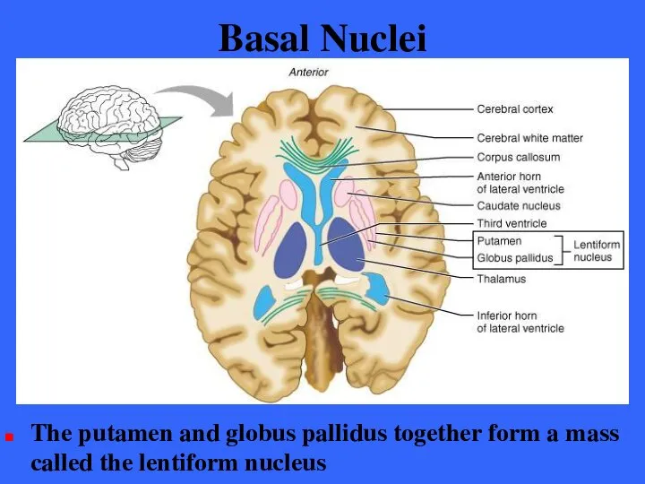
- 132. Basal Nuclei The putamen and globus pallidus together form a mass called the lentiform nucleus
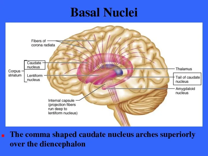
- 133. Basal Nuclei The comma shaped caudate nucleus arches superiorly over the diencephalon
- 134. Basal Nuclei The lentiform nucleus flanks the internal capsule laterally Lentiform nucleus
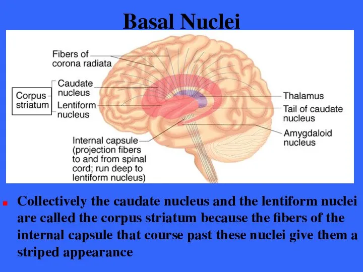
- 135. Basal Nuclei Collectively the caudate nucleus and the lentiform nuclei are called the corpus striatum because
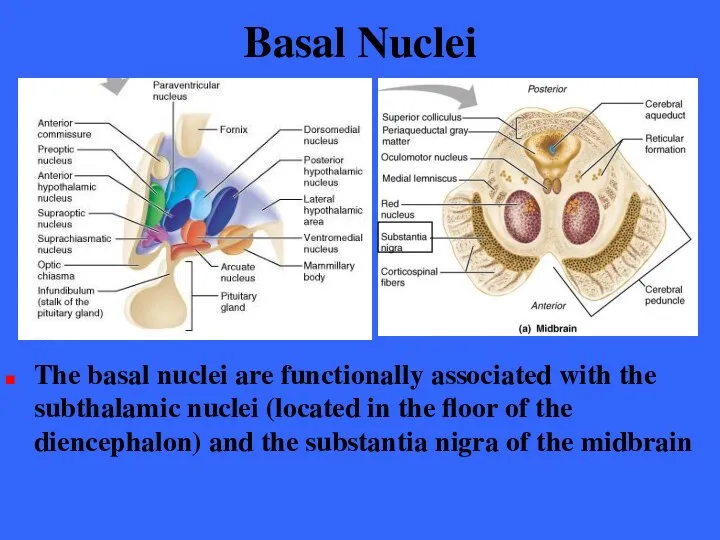
- 136. Basal Nuclei The basal nuclei are functionally associated with the subthalamic nuclei (located in the floor
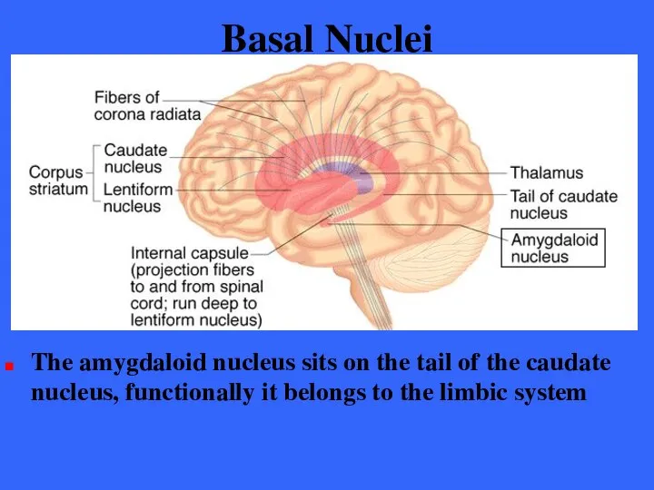
- 137. Basal Nuclei The amygdaloid nucleus sits on the tail of the caudate nucleus, functionally it belongs
- 138. Basal Nuclei Functionally, the basal nuclei can be viewed as complex neural calculators that cooperate with
- 139. Basal Nuclei The basal nuclei receive inputs from the entire cerebral cortex as well as from
- 140. Basal Nuclei Via relays the basal nuclei influence muscle movements directed by the primary motor cortex
- 141. Basal Nuclei The nuclei are involved in monitoring muscle movements that are relatively slow and sustained
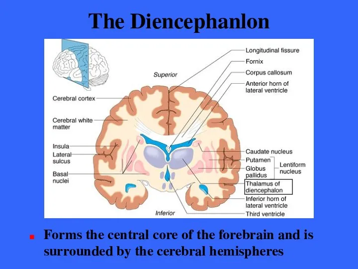
- 142. The Diencephanlon Forms the central core of the forebrain and is surrounded by the cerebral hemispheres
- 143. The Diencephalon The diencephalon consists of three structures Thalamus Hypothalamus Epithalamus These structures effectively enclose the
- 144. The Diencephalon The three structures of the diencephalon Thalamus Hypothalamus Epithalamus These structures are shown with
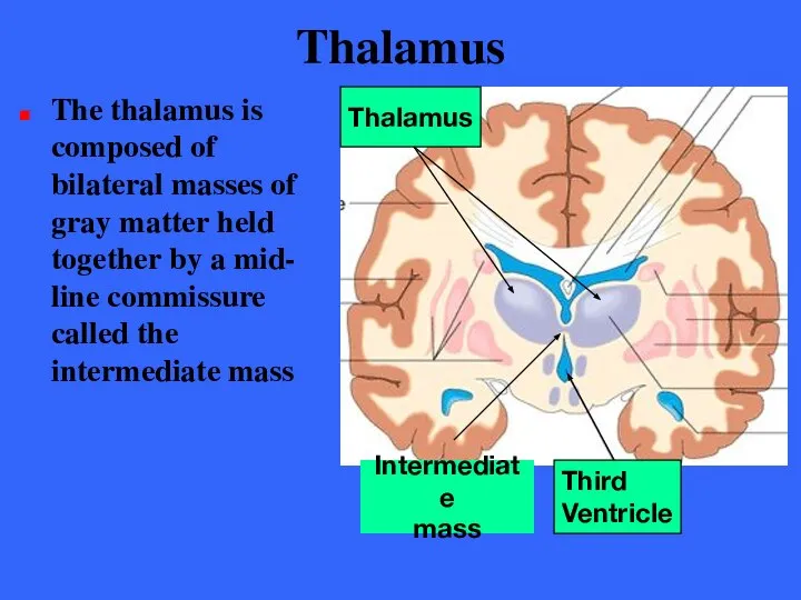
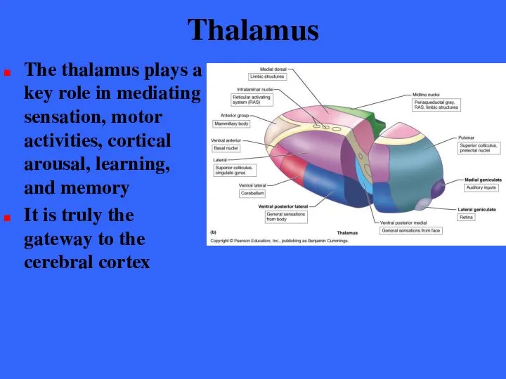
- 145. Thalamus The egg shaped thalamus makes up 80% of the diencephalon and forms the superolateral walls
- 146. Thalamus The thalamus is composed of bilateral masses of gray matter held together by a mid-
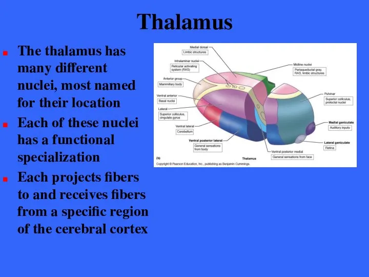
- 147. Thalamus The thalamus has many different nuclei, most named for their location Each of these nuclei
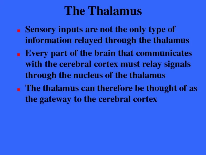
- 148. The Thalamus Sensory inputs are not the only type of information relayed through the thalamus Every
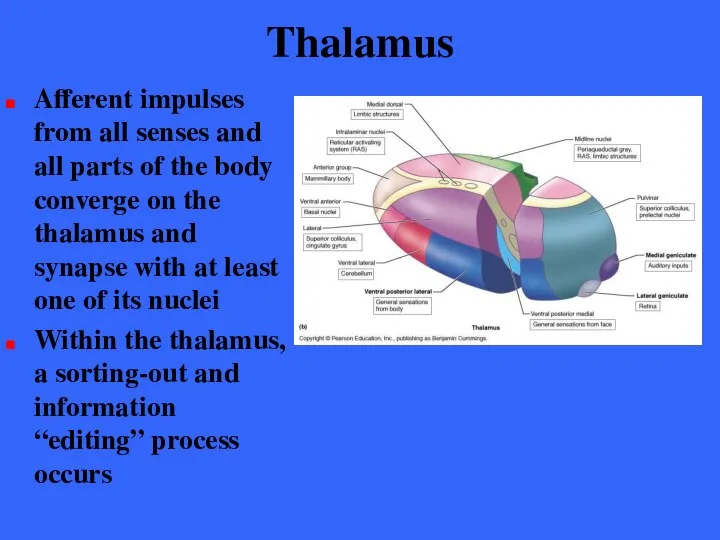
- 149. Thalamus Afferent impulses from all senses and all parts of the body converge on the thalamus
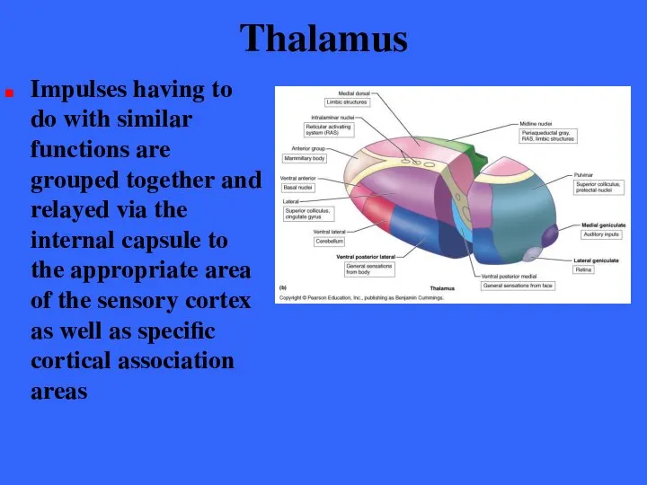
- 150. Thalamus Impulses having to do with similar functions are grouped together and relayed via the internal
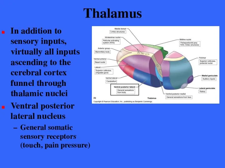
- 151. Thalamus In addition to sensory inputs, virtually all inputs ascending to the cerebral cortex funnel through
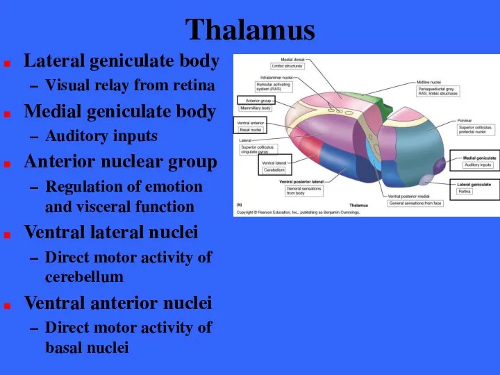
- 152. Thalamus Lateral geniculate body Visual relay from retina Medial geniculate body Auditory inputs Anterior nuclear group
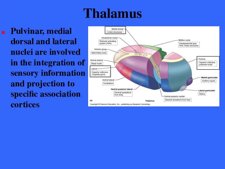
- 153. Thalamus Pulvinar, medial dorsal and lateral nuclei are involved in the integration of sensory information and
- 154. Thalamus The thalamus plays a key role in mediating sensation, motor activities, cortical arousal, learning, and
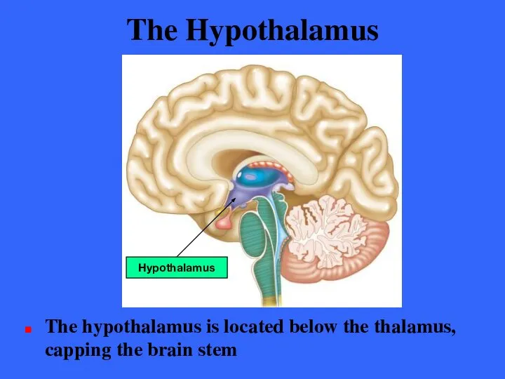
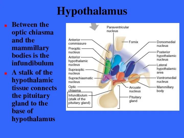
- 155. The Hypothalamus The hypothalamus is located below the thalamus, capping the brain stem Hypothalamus
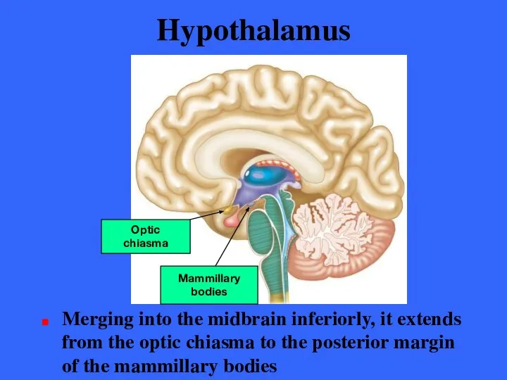
- 156. Hypothalamus Merging into the midbrain inferiorly, it extends from the optic chiasma to the posterior margin
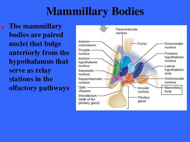
- 157. Mammillary Bodies The mammillary bodies are paired nuclei that bulge anteriorly from the hypothalamus that serve
- 158. Hypothalamus Between the optic chiasma and the mammillary bodies is the infundibulum A stalk of the
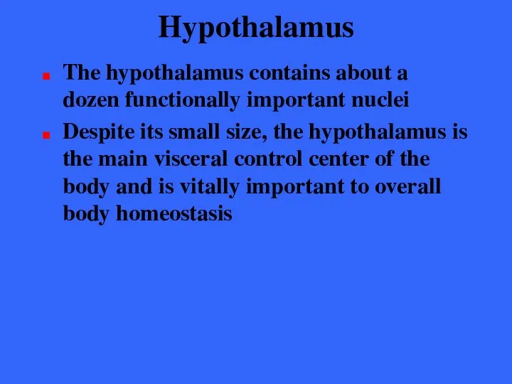
- 159. Hypothalamus The hypothalamus contains about a dozen functionally important nuclei Despite its small size, the hypothalamus
- 160. Autonomic Control Center The hypothalamus regulates involuntary nervous activity by controlling the activity of autonomic centers
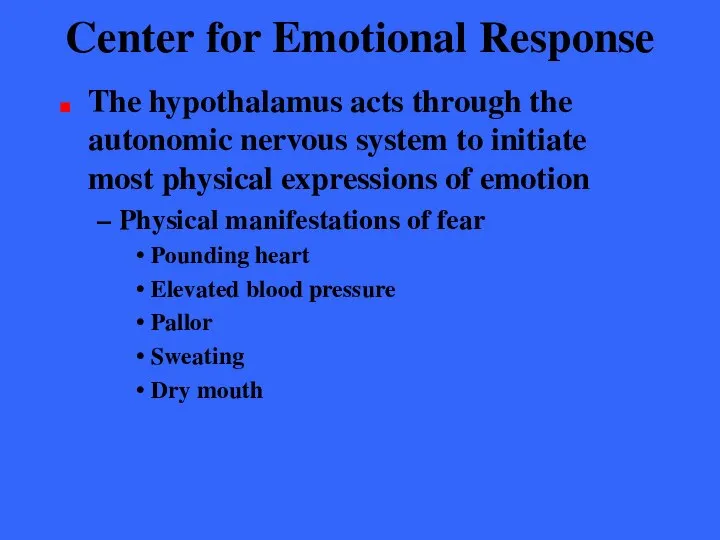
- 161. Center for Emotional Response The hypothalamus has numerous connections with cortical association areas, lower brain stem
- 162. Center for Emotional Response The hypothalamus acts through the autonomic nervous system to initiate most physical
- 163. Body Temperature Regulation The body’s thermostat is in the hypothalamus The hypothalamus receives input from the
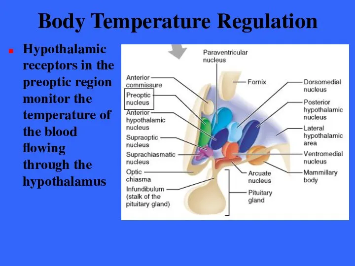
- 164. Body Temperature Regulation Hypothalamic receptors in the preoptic region monitor the temperature of the blood flowing
- 165. Body Temperature Regulation According to signals received by the preoptic nuclei the hypothalamus initiates mechanisms to
- 166. Regulation of Hunger & Thirst In response to changing levels of glucose, amino acids, hormones, and
- 167. Regulation of Water Balance When body fluids become too concentrated, hypothalamic neurons called osmoreceptors are activated
- 168. Regulation of Sleep-Wake Cycles Acting with other brain regions, the hypothalamus helps regulate the complex phenomenon
- 169. Regulation of Sleep-Wake Cycles Hypothalamus through the operation of its suprachiasmatic nucleus (our biological clock) sets
- 170. Control of Endocrine Functioning The hypothalamus acts as the helmsman of the endocrine system By producing
- 171. Formation of Memory The nucleus of the mammillary body receives many inputs from the major memory-processing
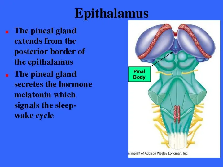
- 172. Epithalamus The epithalamus is the posterior portion of the diencephalon It forms the roof of the
- 173. The Epithalamus The epithalmus consists of one tiny group of nuclei and a small, unpaired knob
- 174. Epithalamus The pineal gland extends from the posterior border of the epithalamus The pineal gland secretes
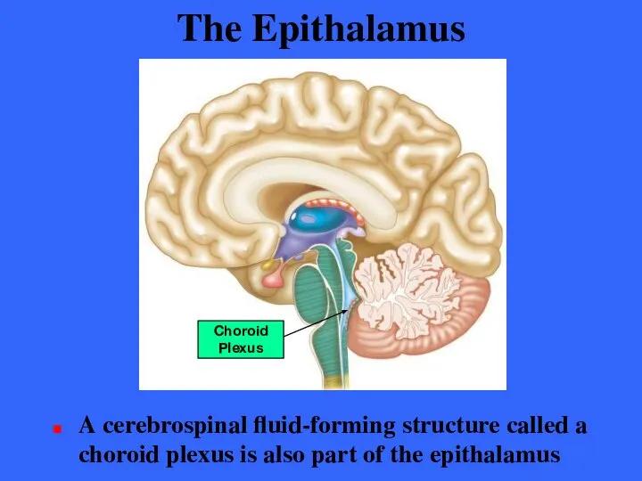
- 175. The Epithalamus A cerebrospinal fluid-forming structure called a choroid plexus is also part of the epithalamus
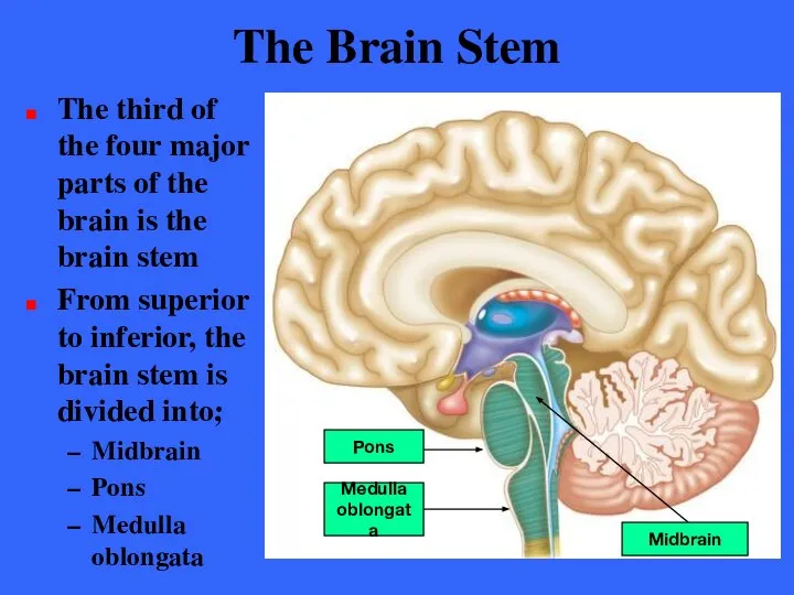
- 176. The Brain Stem The third of the four major parts of the brain is the brain
- 177. The Brain Stem Each region is roughly an inch long Together than constitute 2.5% of total
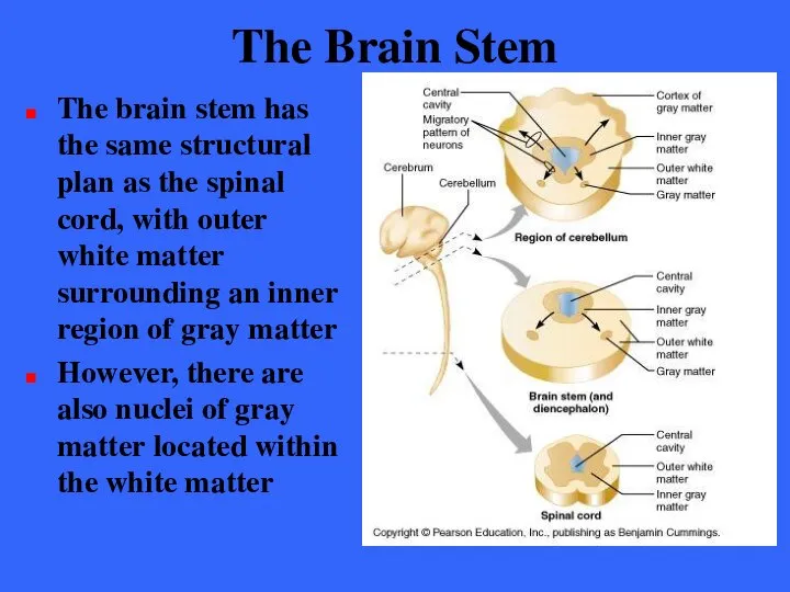
- 178. The Brain Stem The brain stem has the same structural plan as the spinal cord, with
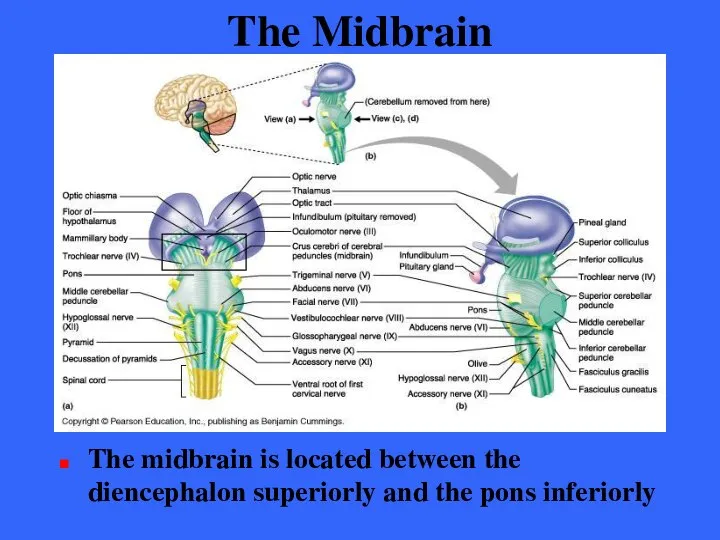
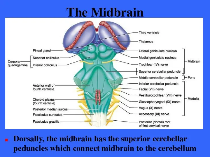
- 179. The Midbrain The midbrain is located between the diencephalon superiorly and the pons inferiorly
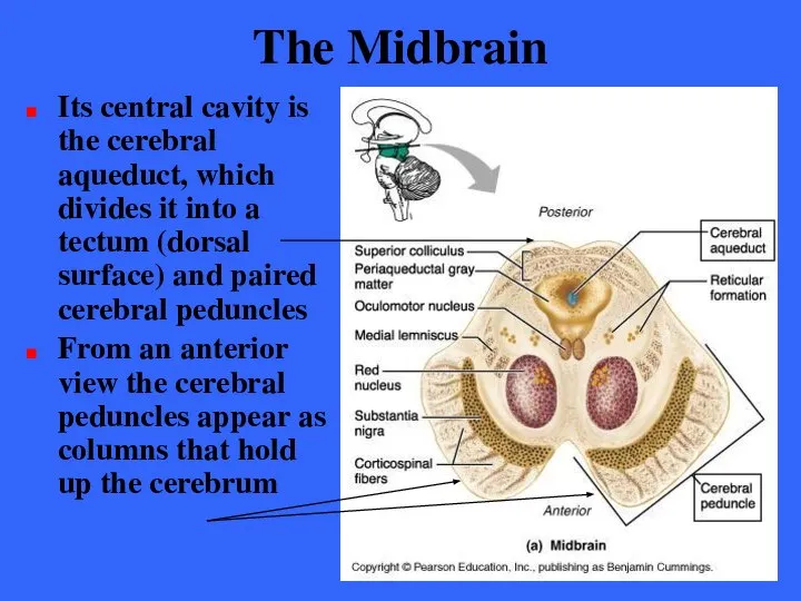
- 180. The Midbrain Its central cavity is the cerebral aqueduct, which divides it into a tectum (dorsal
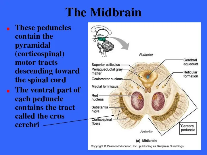
- 181. The Midbrain These peduncles contain the pyramidal (corticospinal) motor tracts descending toward the spinal cord The
- 182. The Midbrain Dorsally, the midbrain has the superior cerebellar peduncles which connect midbrain to the cerebellum
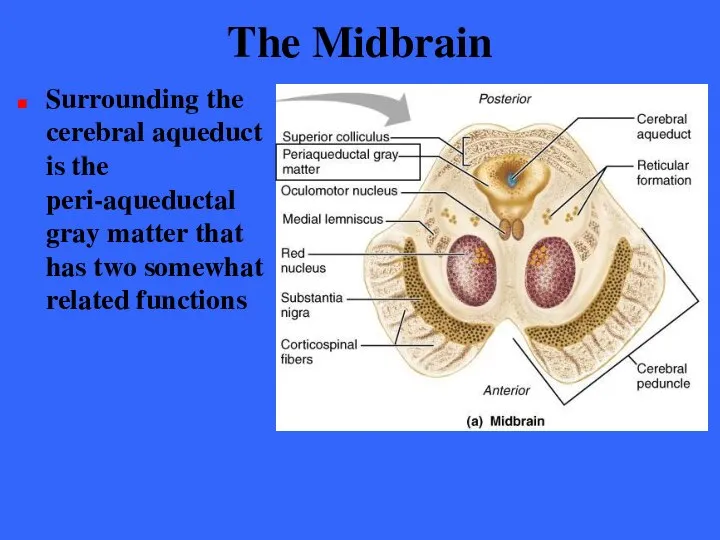
- 183. The Midbrain Surrounding the cerebral aqueduct is the peri-aqueductal gray matter that has two somewhat related
- 184. The Midbrain The periaqueductal gray matter is involved in the “fright-and-flight” sympathetic reaction The gray matter
- 185. The Midbrain The gray matter elicits A terror-induced increase in heart rate Skyrocketing blood pressure Wild
- 186. The Midbrain The periaqueductal gray matter also seems to mediate our response to visceral pain (as
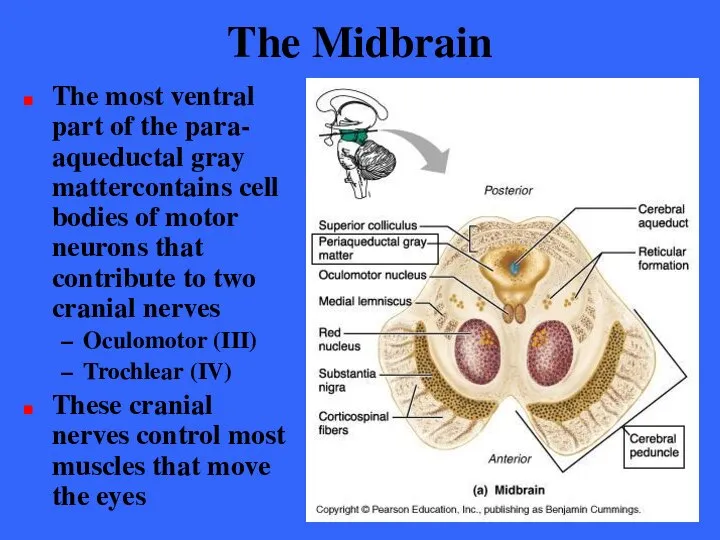
- 187. The Midbrain The most ventral part of the para- aqueductal gray mattercontains cell bodies of motor
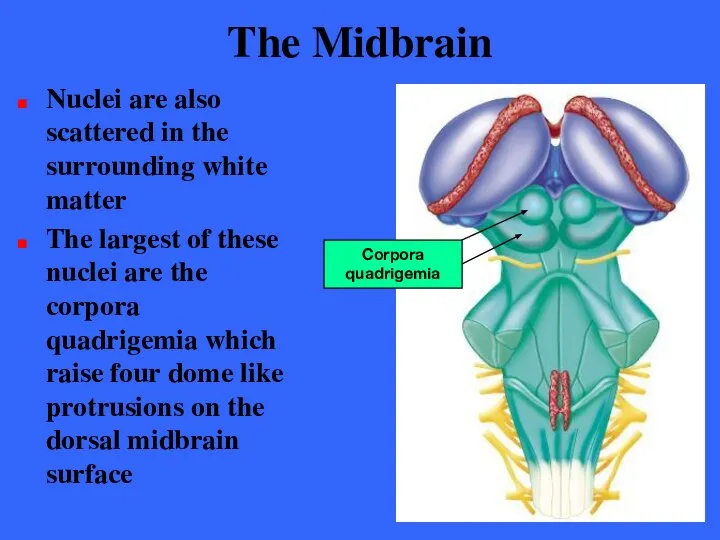
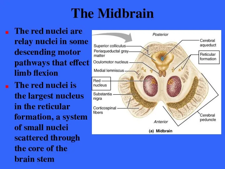
- 188. The Midbrain Nuclei are also scattered in the surrounding white matter The largest of these nuclei
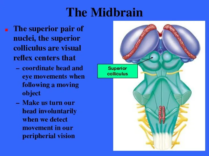
- 189. The Midbrain The superior pair of nuclei, the superior colliculus are visual reflex centers that coordinate
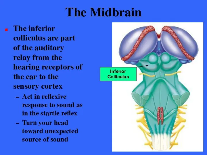
- 190. The Midbrain The inferior colliculus are part of the auditory relay from the hearing receptors of
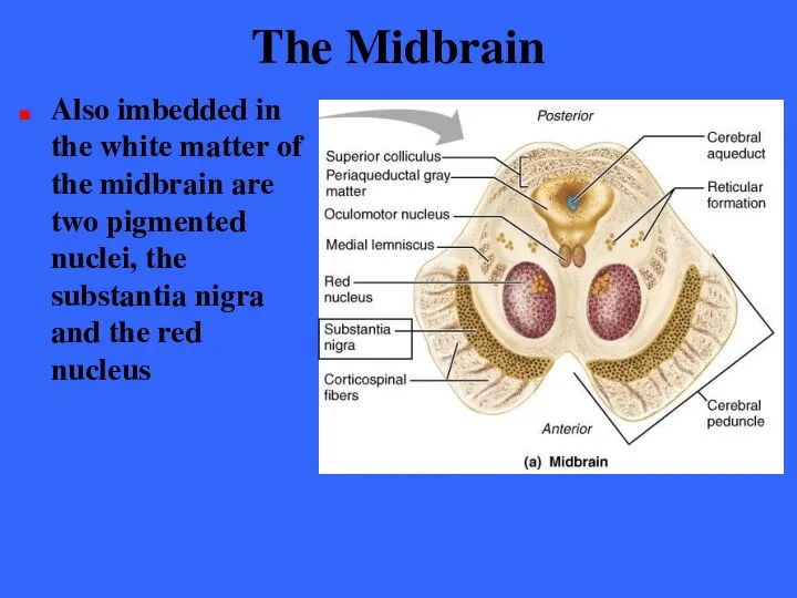
- 191. The Midbrain Also imbedded in the white matter of the midbrain are two pigmented nuclei, the
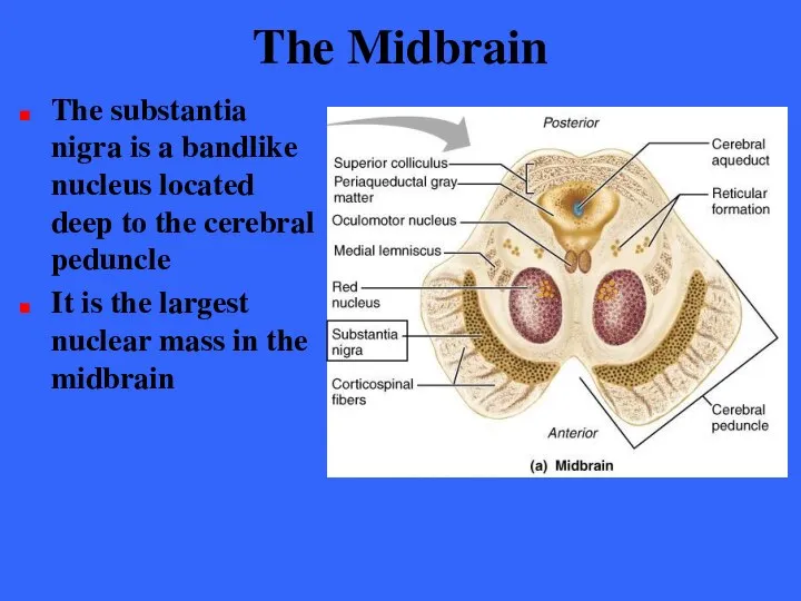
- 192. The Midbrain The substantia nigra is a bandlike nucleus located deep to the cerebral peduncle It
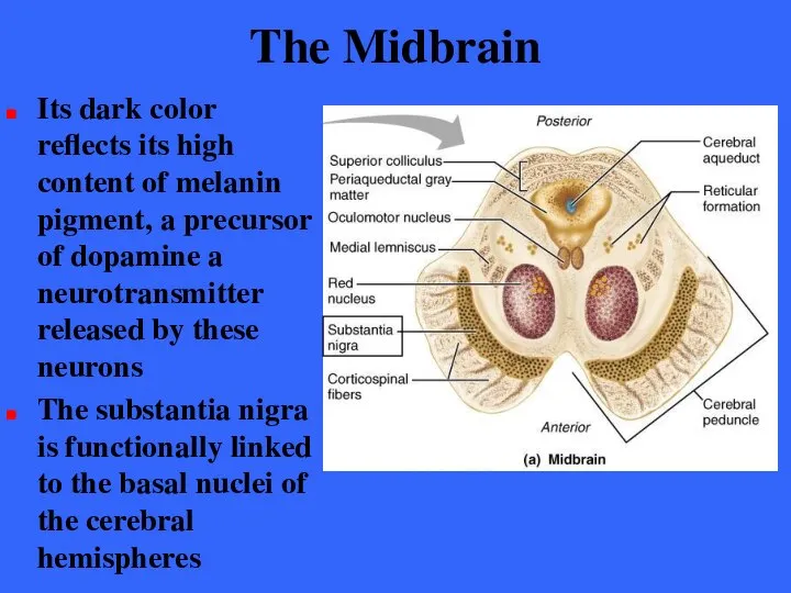
- 193. The Midbrain Its dark color reflects its high content of melanin pigment, a precursor of dopamine
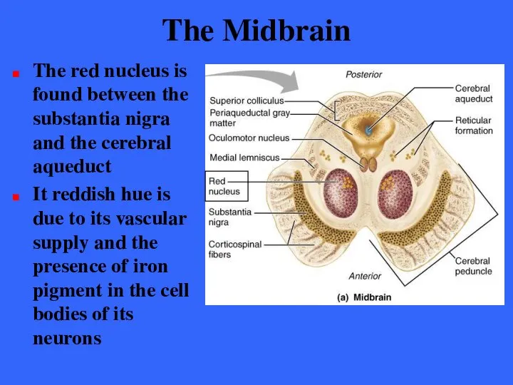
- 194. The Midbrain The red nucleus is found between the substantia nigra and the cerebral aqueduct It
- 195. The Midbrain The red nuclei are relay nuclei in some descending motor pathways that effect limb
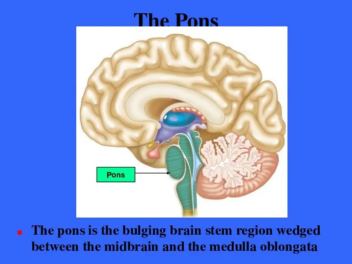
- 196. The Pons The pons is the bulging brain stem region wedged between the midbrain and the
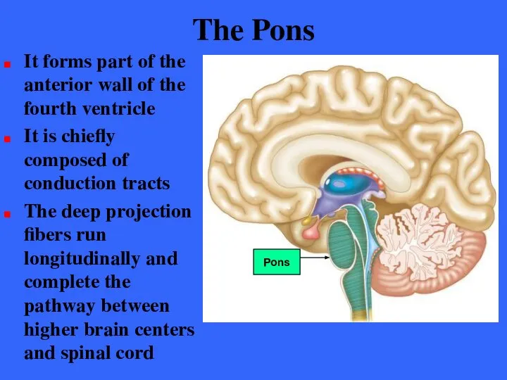
- 197. The Pons It forms part of the anterior wall of the fourth ventricle It is chiefly
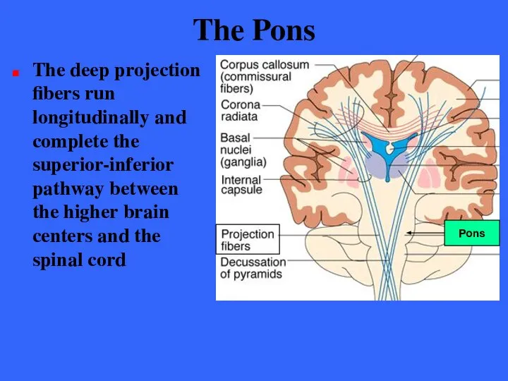
- 198. The Pons The deep projection fibers run longitudinally and complete the superior-inferior pathway between the higher
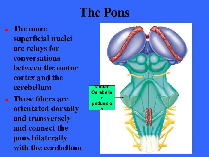
- 199. The Pons The more superficial nuclei are relays for conversations between the motor cortex and the
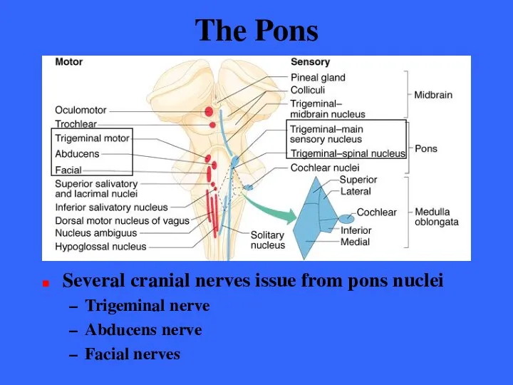
- 200. The Pons Several cranial nerves issue from pons nuclei Trigeminal nerve Abducens nerve Facial nerves
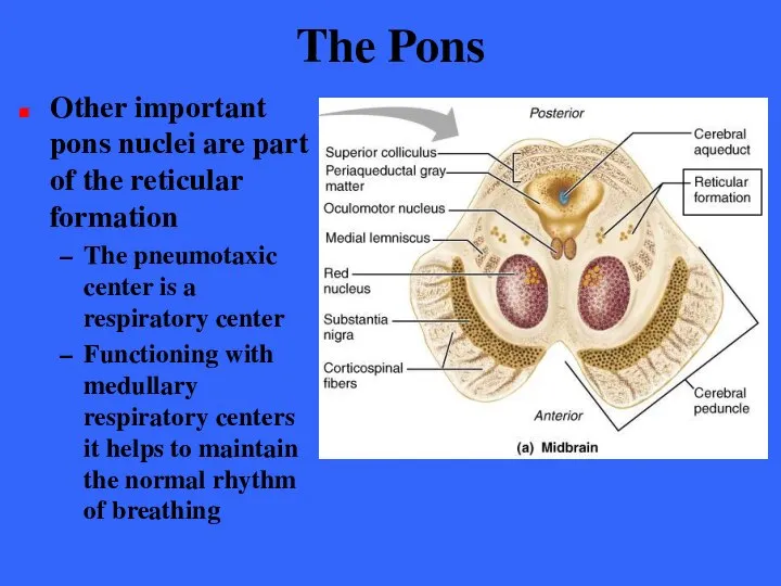
- 201. The Pons Other important pons nuclei are part of the reticular formation The pneumotaxic center is
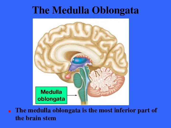
- 202. The Medulla Oblongata The medulla oblongata is the most inferior part of the brain stem Medulla
- 203. The Medulla Oblongata The medulla blends into the spinal cord at the level of the foramen
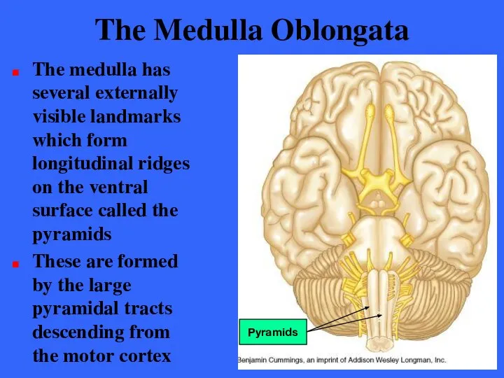
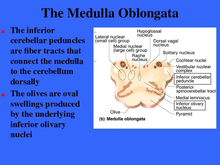
- 204. The Medulla Oblongata The medulla has several externally visible landmarks which form longitudinal ridges on the
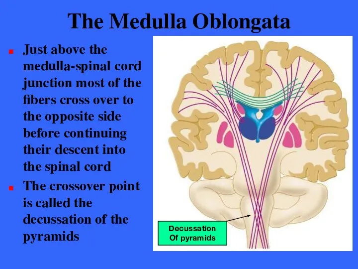
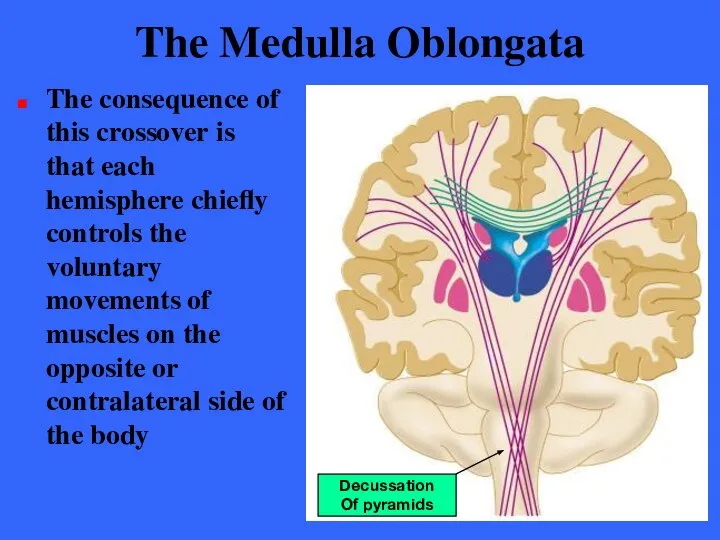
- 205. The Medulla Oblongata Just above the medulla-spinal cord junction most of the fibers cross over to
- 206. The Medulla Oblongata The consequence of this crossover is that each hemisphere chiefly controls the voluntary
- 207. The Medulla Oblongata The inferior cerebellar peduncles are fiber tracts that connect the medulla to the
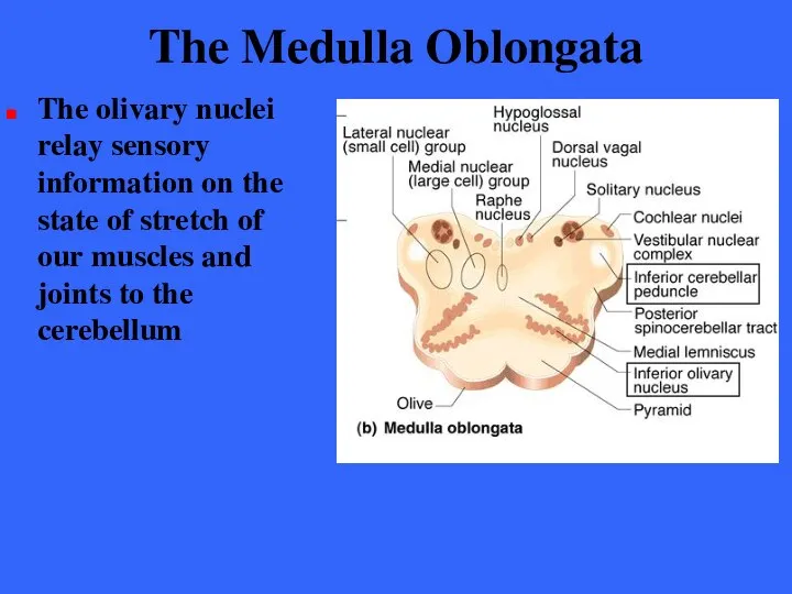
- 208. The Medulla Oblongata The olivary nuclei relay sensory information on the state of stretch of our
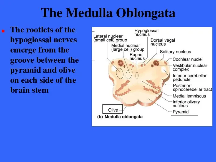
- 209. The Medulla Oblongata The rootlets of the hypoglossal nerves emerge from the groove between the pyramid
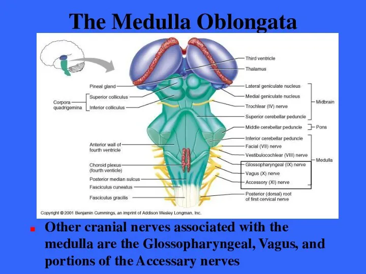
- 210. The Medulla Oblongata Other cranial nerves associated with the medulla are the Glossopharyngeal, Vagus, and portions
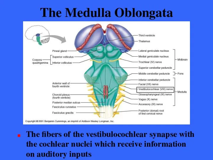
- 211. The Medulla Oblongata The fibers of the vestibulocochlear synapse with the cochlear nuclei which receive information
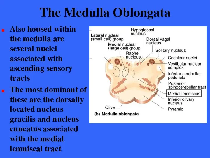
- 212. The Medulla Oblongata Also housed within the medulla are several nuclei associated with ascending sensory tracts
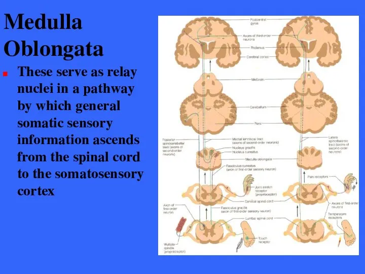
- 213. Medulla Oblongata These serve as relay nuclei in a pathway by which general somatic sensory information
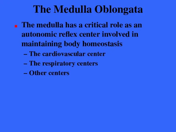
- 214. The Medulla Oblongata The medulla has a critical role as an autonomic reflex center involved in
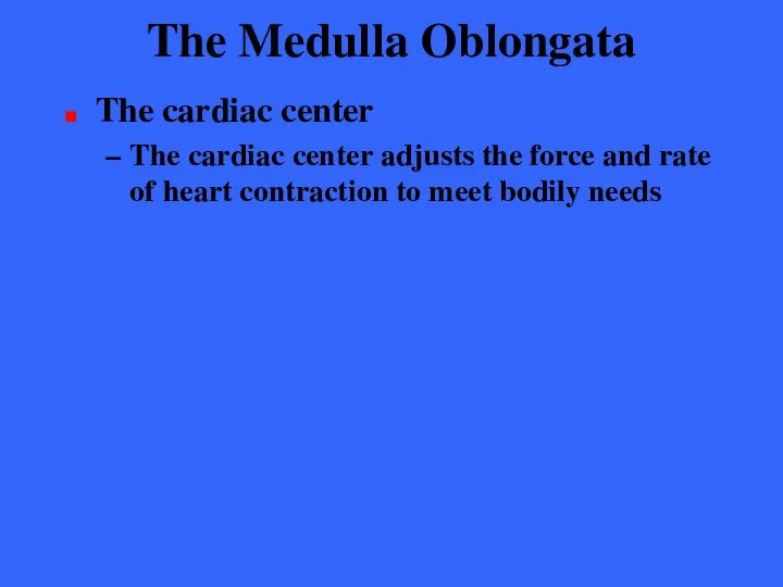
- 215. The Medulla Oblongata The cardiac center The cardiac center adjusts the force and rate of heart
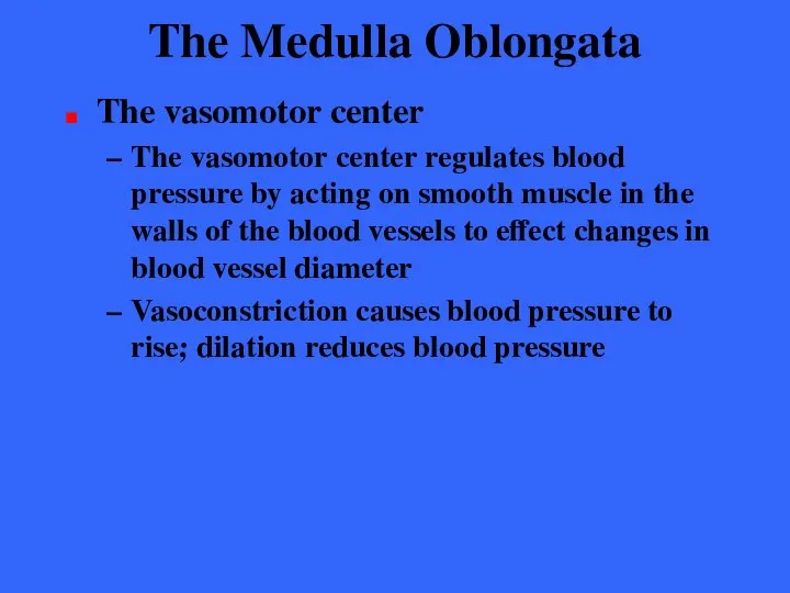
- 216. The Medulla Oblongata The vasomotor center The vasomotor center regulates blood pressure by acting on smooth
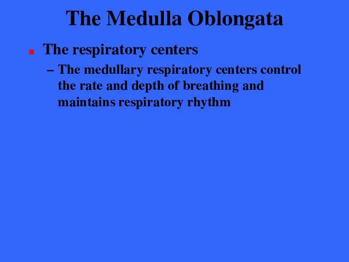
- 217. The Medulla Oblongata The respiratory centers The medullary respiratory centers control the rate and depth of
- 218. The Medulla Oblongata Other centers Additional centers regulate activities such as Vomiting Hiccuping Swallowing Coughing Sneezing
- 219. The Medulla Oblongata Many functions of the medulla overlap with those attributed to the hypothalamus The
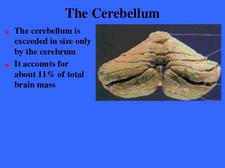
- 220. The Cerebellum The cerebellum is exceeded in size only by the cerebrum It accounts for about
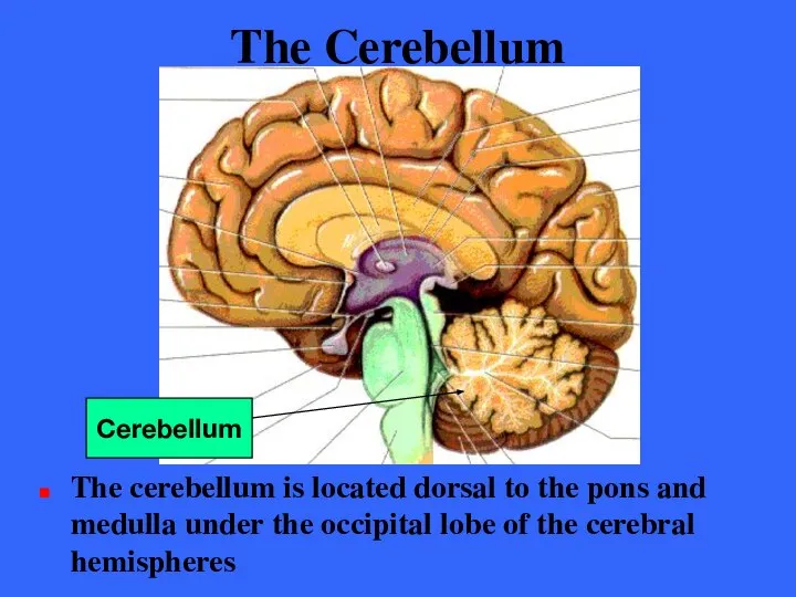
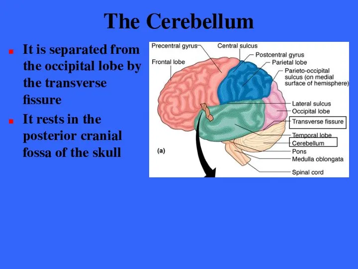
- 221. The Cerebellum The cerebellum is located dorsal to the pons and medulla under the occipital lobe
- 222. The Cerebellum It is separated from the occipital lobe by the transverse fissure It rests in
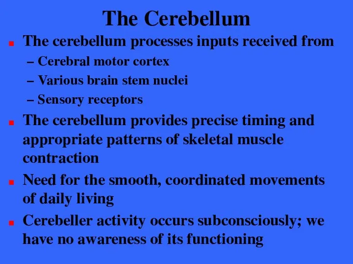
- 223. The Cerebellum The cerebellum processes inputs received from Cerebral motor cortex Various brain stem nuclei Sensory
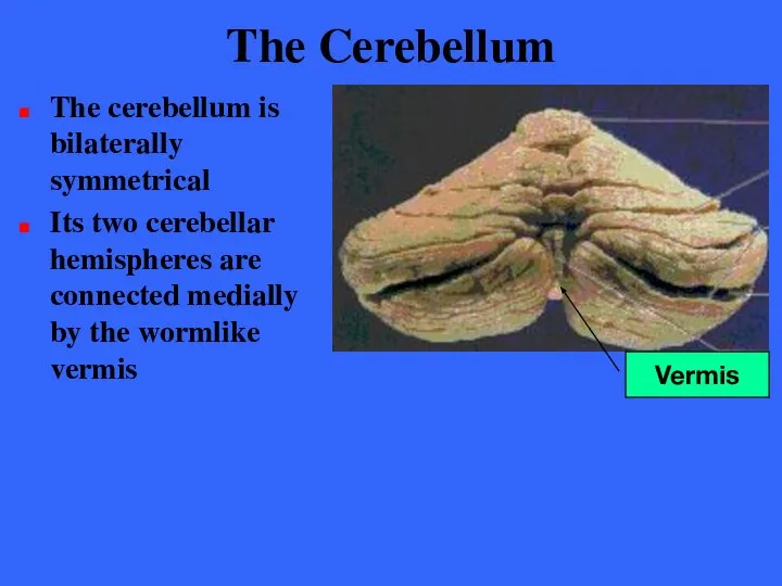
- 224. The Cerebellum The cerebellum is bilaterally symmetrical Its two cerebellar hemispheres are connected medially by the
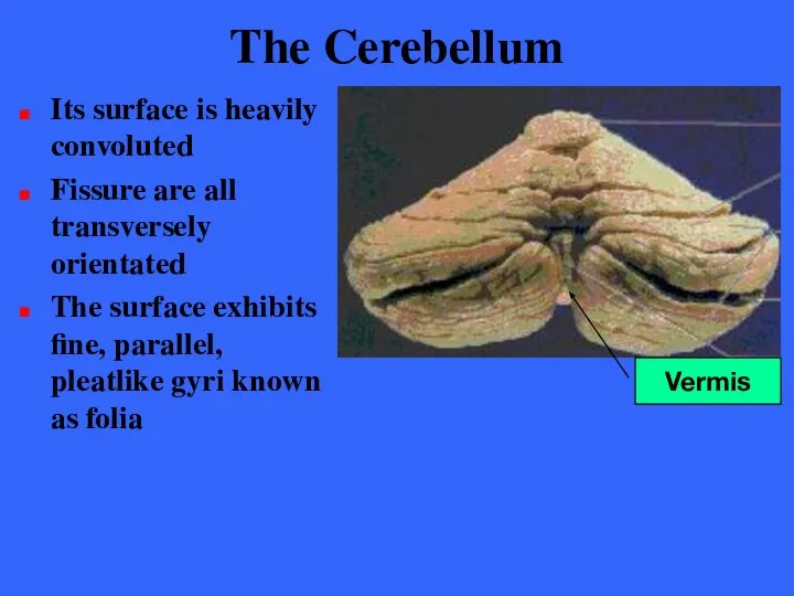
- 225. The Cerebellum Its surface is heavily convoluted Fissure are all transversely orientated The surface exhibits fine,
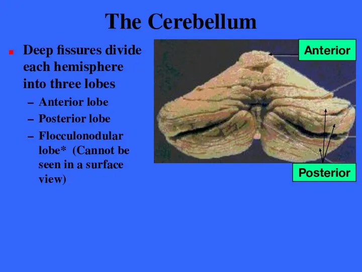
- 226. The Cerebellum Deep fissures divide each hemisphere into three lobes Anterior lobe Posterior lobe Flocculonodular lobe*
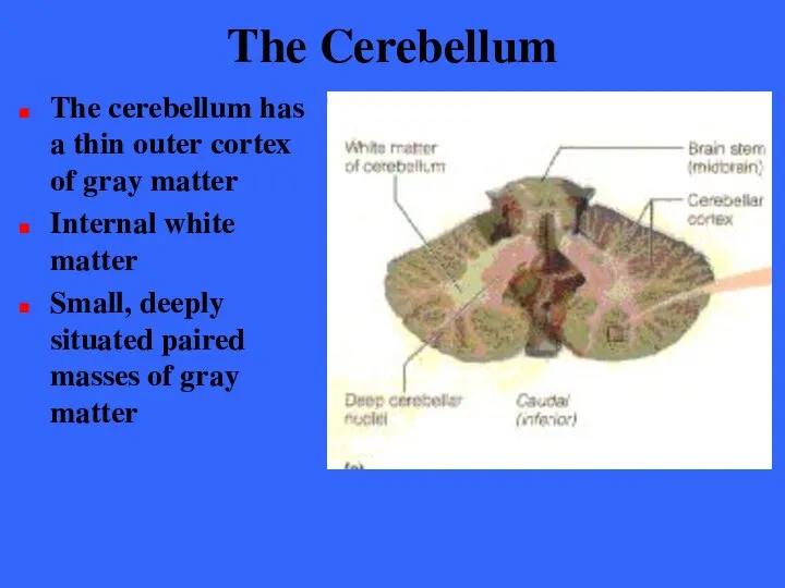
- 227. The Cerebellum The cerebellum has a thin outer cortex of gray matter Internal white matter Small,
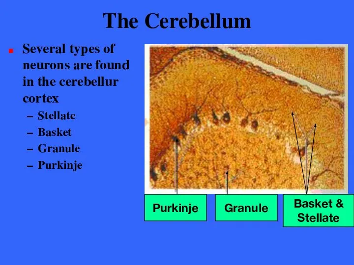
- 228. The Cerebellum Several types of neurons are found in the cerebellur cortex Stellate Basket Granule Purkinje
- 229. The Cerebellum The large Purkinje cells with their extensively branched dendrites are the only cortical neurons
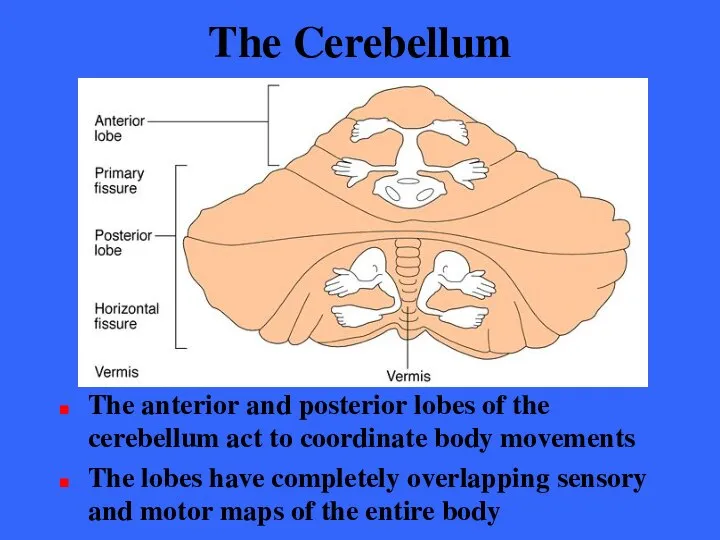
- 230. The Cerebellum The anterior and posterior lobes of the cerebellum act to coordinate body movements The
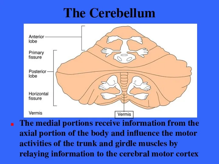
- 231. The Cerebellum The medial portions receive information from the axial portion of the body and influence
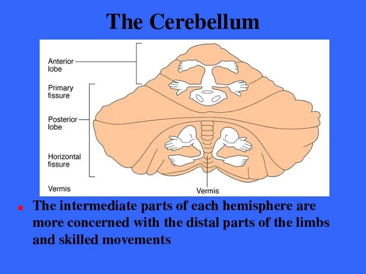
- 232. The Cerebellum The intermediate parts of each hemisphere are more concerned with the distal parts of
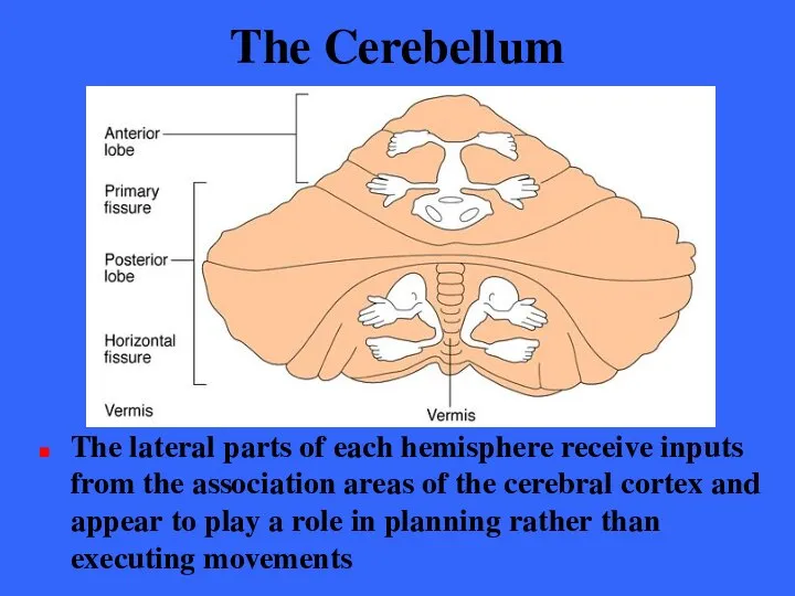
- 233. The Cerebellum The lateral parts of each hemisphere receive inputs from the association areas of the
- 234. The Cerebellum The small flocculonodular lobes receive inputs from the equilibrium apparatus of the inner ears,
- 235. The Cerebellum Three pairs of fiber tracts, cerebellur peduncles connect the the cerebellum to the brain
- 236. The Cerebellum Virtually all fibers entering and leaving the cerebellum are ipsilateral; from and to the
- 237. The Cerebellum The superior cerebellar peduncles connect the cerebellum and the midbrain. Fibers in these peduncles
- 238. The Cerebellum The middle cerebellar peduncles connect the pons the cerebellum.
- 239. The Cerebellum These fibers allow one-way communication from the pons to the cerebellar neurons, which enables
- 240. The Cerebellum The inferior cerebellar peduncles connect the cerebellum and the medulla
- 241. The Cerebellum These peduncles contain afferent tracts conveying sensory information to the cerebellum from (1) muscle
- 242. Cerebellar Processing - 1 The frontal motor association areas of the cerebral cortex indicates its intents
- 243. Cerebellar Processing - 2 At the same time, the cerebellum receives information from the proprioceptors throughout
- 244. Cerebellar Processing - 3 The cerebellar cortex assesses this information and calculates the best way to
- 245. Cerebellar Processing - 4 Via the superior peduncles, the cerebellum dispatches its “blueprint” for coordination to
- 246. The Cerebellum The cerebellum continually compares the higher brain’s intention with the body’s performance and sends
- 247. The Cerebellum Cerebellar injury results in the loss of muscle tone and clumsy, unsure movements, and
- 248. Functional Brain Systems Functional brain systems are networks of neurons that work together but span relatively
- 249. The Limbic System The limbic system is a group of structures located on the medial aspect
- 250. The Limbic System The limbic system encircles the upper part of the brain stem and includes
- 251. The Limbic System The observation that odors evoke emotional reactions and memories reflects the fact that
- 252. The Limbic System The limbic system is our emotional or affective brain Two parts seem especially
- 253. The Limbic System The amygdala contains the key nuclei for processing fear and then stimulating the
- 254. The Limbic System The amygdala also enables us to recognize menacing facial expression in others and
- 255. The Limbic System The cingulate gyrus allows us to shift between thoughts and to express our
- 256. The Limbic System The anterior part of the gyrus interprets pain as unpleasant and resolves mental
- 257. The Limbic System The limbic system also functions in consolidating and retrieving memories The structures involved,
- 258. The Limbic System The hippocampal formation encodes, consolidates, and later retrieves memories of facts and events
- 259. The Limbic System The amygdala forms memories of experiences that are based entirely on their emotional
- 260. The Limbic System The limbic system communicates with many other regions of the brain Most output
- 261. The Limbic system This fact explains why people under emotional stress experience visceral illnesses such as
- 262. The Limbic System We react emotionally to things we consciously understand to be happening We are
- 263. The Limbic System Communication between the cerebral cortex and the limbic system explains why emotions sometimes
- 264. The Reticular Formation The reticular formation extends through the central core of the medulla oblongata, pons,
- 265. The Reticular Formation It is an intricate system composed of loosely clustered neurons in what is
- 266. The Reticular Formation Reticular neurons can be localized into three broad columns along the length of
- 267. The Reticular Formation The outstanding feature of the reticular neurons is their far-flung axonal connections Individual
- 268. The Reticular Formation Certain reticular neurons send a continuous stream of impulses to the cerebrum (through
- 269. The Reticular Activating System The RAS synapses with all major ascending sensory tracts enhancing arousal of
- 270. Reticular Formation The RAS also acts as a filter to dampen repetitive, familiar, or weak signals
- 271. The Reticular Activating System The activity of the RAS is inhibited by sleep centers in the
- 272. The Reticular Formation The reticular formation also has a motor component Some if its motor nuclei
- 273. Protection of the Brain Nervous tissue is soft and vulnerable The brain is protected by Bony
- 274. Meninges The meninges are three connective tissue membranes that lie just external to the central nervous
- 275. Meninges The meningeal membranes Cover and protect the CNS organs Protect blood vessels and enclose venous
- 276. Meninges The meninges are three connective tissue membranes that lie just external to the central nervous
- 277. Meninges From external to internal, the meningeal layers are Dura mater Arachnoid Pia mater
- 278. The Dura Mater The leathery dura mater is by far the strongest of the meninges Where
- 279. The Dura Mater The periosteal layer is the superficial and lines the inner surface of the
- 280. The Dura Mater The brain’s dural layers are fused together except in certain areas where they
- 281. The Dura Mater In several places the meningeal dura mater extends inward to form flat septa
- 282. The Dura Mater The falx cerebri dips into the longitudinal fissue It attaches to the crista
- 283. The Dura Mater The falx cerebelli forms a midline partition that runs along the vermis of
- 284. The Dura Mater The tentorium cerebelli extends into the transverse fissue between the cerebral hemispheres and
- 285. The Arachnoid Mater The middle membrane forms a loose brain covering over the surface of the
- 286. The Arachnoid Mater The subarachnoid space is filled with cerebrospinal fluid and contains the largest blood
- 287. The Arachnoid Mater Arachnoid villi protrude through the overlying dura mater and into the dural sinuses
- 288. The Pia Mater The pia mater is a delicate connective tissue that is richly invested with
- 289. Cerebrospinal Fluid (CSF) CSF is found in and around the brain and spinal cord It forms
- 290. Cerebrospinal Fluid (CSF) CSF is a similar in composition to blood plasma, from which it arises
- 291. Choroid Plexus Choroid plexus hang from the roof of each ventricle These plexuses form CSF The
- 292. Choroid Plexus The capillaries of the choroid plexus are fairly permeable and tissue fluid filter continuously
- 293. Choroid Plexus The choroid plexus cells are joined by tight junctions and have ion pumps that
- 294. The Choroid Plexus In adults, the total CSF volume of about 150 ml is replaced every
- 295. CSF Circulation Most CSF enters the subarachnoid space via the apertures in the walls of the
- 296. CSF Circulation In the subarachnoid space the CSF bathes the outer surface of the brain and
- 297. Blood-Brain Barrier The barrier is a protective mechanism that helps maintain a stable environment for the
- 298. Blood-Brain Barrier Bloodborne substances within the brain’s capillaries are separated from the extra- cellular space and
- 299. Blood-Brain Barrier The capillary endothelial cells are joined almost seamlessly by tight junctions They are the
- 300. Blood-Brain Barrier The blood-brain barrier is a selective, rather than absolute barrier Nutrients, such as glucose,
- 301. Blood-Brain Barrier The barrier is ineffective against fats, fatty acids, oxygen, and carbon dioxide, and other
- 302. Homeostatic Imbalances Traumatic Brain Injuries Concussion / Contusion Cerebrovascular Accidents Strokes / Transient ischemic attacks Degenerative
- 304. Скачать презентацию
Introduction
Analogies; telephone switchboard; computer; miracle
A fantastically complex and flexible biological
Introduction
Analogies; telephone switchboard; computer; miracle
A fantastically complex and flexible biological
The Brain
The unimpressive appearance of the human brain give few hints
The Brain
The unimpressive appearance of the human brain give few hints
The Brain
Average adult male’s brain weighs about 1600 g (3.5 pounds)
Average
The Brain
Average adult male’s brain weighs about 1600 g (3.5 pounds)
Average
Embryonic Development
Starting in the third week of pregnancy, the ectoderm thickens
Embryonic Development
Starting in the third week of pregnancy, the ectoderm thickens
Embryonic Development
The neural plate then invaginates, forming a groove flanked by
Embryonic Development
The neural plate then invaginates, forming a groove flanked by
Development of Neural Tube
As the groove deepens the superior edges of
Development of Neural Tube
As the groove deepens the superior edges of
Development of Neural Tube
The neural tube is formed by the fourth
Development of Neural Tube
The neural tube is formed by the fourth
Development of Neural Tube
Small groups of neural fold cells migrate laterally
Development of Neural Tube
Small groups of neural fold cells migrate laterally
Development of Neural Tube
As soon as the neural tube is formed,
Development of Neural Tube
As soon as the neural tube is formed,
Primary Brain Vesicles
Constrictions in the neural tube appear to mark off
Primary Brain Vesicles
Constrictions in the neural tube appear to mark off
Secondary Brain Vesicles
By the fifth week, the five brain regions of
Secondary Brain Vesicles
By the fifth week, the five brain regions of
Secondary Brain Vesicles
Each of the five secondary brain vesicles develops rapidly
Secondary Brain Vesicles
Each of the five secondary brain vesicles develops rapidly
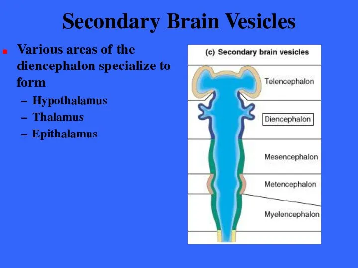
Secondary Brain Vesicles
Various areas of the diencephalon specialize to form
Hypothalamus
Thalamus
Epithalamus
Secondary Brain Vesicles
Various areas of the diencephalon specialize to form
Hypothalamus
Thalamus
Epithalamus
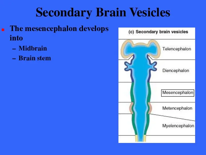
Secondary Brain Vesicles
The mesencephalon develops into
Midbrain
Brain stem
Secondary Brain Vesicles
The mesencephalon develops into
Midbrain
Brain stem
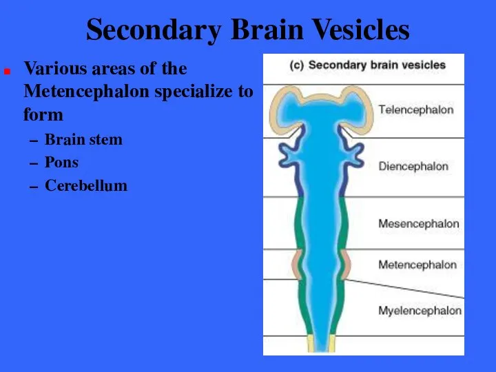
Secondary Brain Vesicles
Various areas of the Metencephalon specialize to form
Brain stem
Pons
Cerebellum
Secondary Brain Vesicles
Various areas of the Metencephalon specialize to form
Brain stem
Pons
Cerebellum
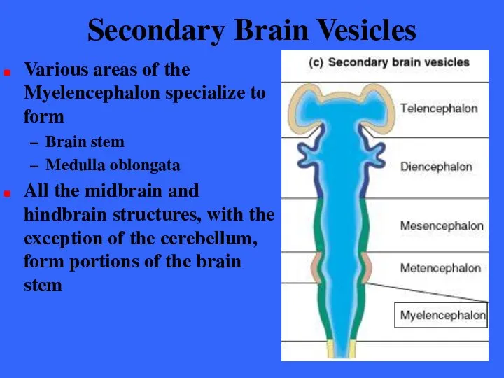
Secondary Brain Vesicles
Various areas of the Myelencephalon specialize to form
Brain stem
Medulla
Secondary Brain Vesicles
Various areas of the Myelencephalon specialize to form
Brain stem
Medulla
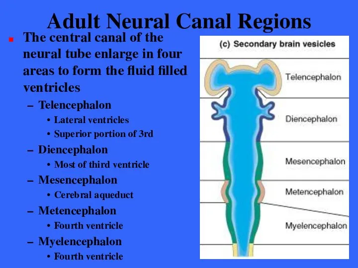
Adult Neural Canal Regions
The central canal of the neural tube enlarge
Adult Neural Canal Regions
The central canal of the neural tube enlarge
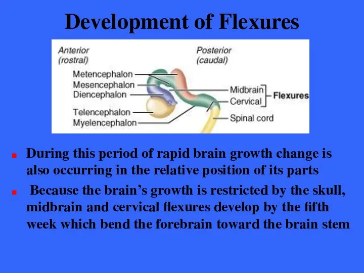
Development of Flexures
During this period of rapid brain growth change is
Development of Flexures
During this period of rapid brain growth change is
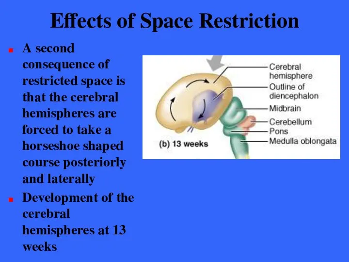
Effects of Space Restriction
A second consequence of restricted space is that
Effects of Space Restriction
A second consequence of restricted space is that
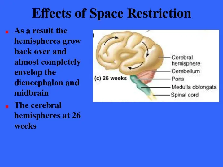
Effects of Space Restriction
As a result the hemispheres grow back over
Effects of Space Restriction
As a result the hemispheres grow back over
Effects of Space Restriction
Continued growth of the cerebral hemispheres causes their
Effects of Space Restriction
Continued growth of the cerebral hemispheres causes their

Effects of Space Restriction
The wrinkling of the hemispheres may result from
Effects of Space Restriction
The wrinkling of the hemispheres may result from
Regions of the Brain
The four main regions of the brain are:
Cerebral
Regions of the Brain
The four main regions of the brain are:
Cerebral
Gray and White Matter in CNS
The basic pattern of the CNS
Gray and White Matter in CNS
The basic pattern of the CNS
Gray and White Matter in CNS
The brain has the same basic
Gray and White Matter in CNS
The brain has the same basic
Gray and White Matter in CNS
The pattern of white and gray
Gray and White Matter in CNS
The pattern of white and gray
Ventricles of the Brain
The ventricles of the brain arise from the
Ventricles of the Brain
The ventricles of the brain arise from the
Ventricles of the Brain
The hollow ventricular chambers are filled with cerebrospinal
Ventricles of the Brain
The hollow ventricular chambers are filled with cerebrospinal
Ventricles of the Brain
The paired lateral ventricles are large C-shaped chambers
Ventricles of the Brain
The paired lateral ventricles are large C-shaped chambers
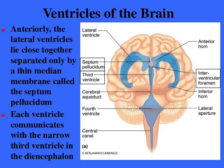
Ventricles of the Brain
Anteriorly, the lateral ventricles lie close together separated
Ventricles of the Brain
Anteriorly, the lateral ventricles lie close together separated
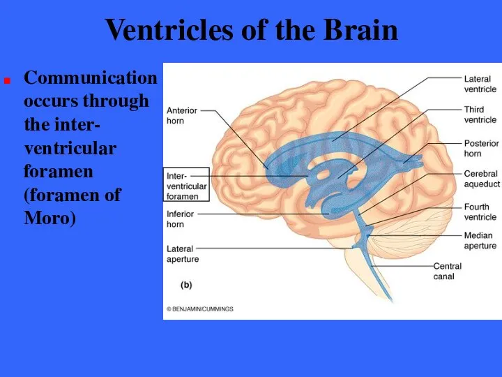
Ventricles of the Brain
Communication occurs through the inter- ventricular foramen (foramen
Ventricles of the Brain
Communication occurs through the inter- ventricular foramen (foramen
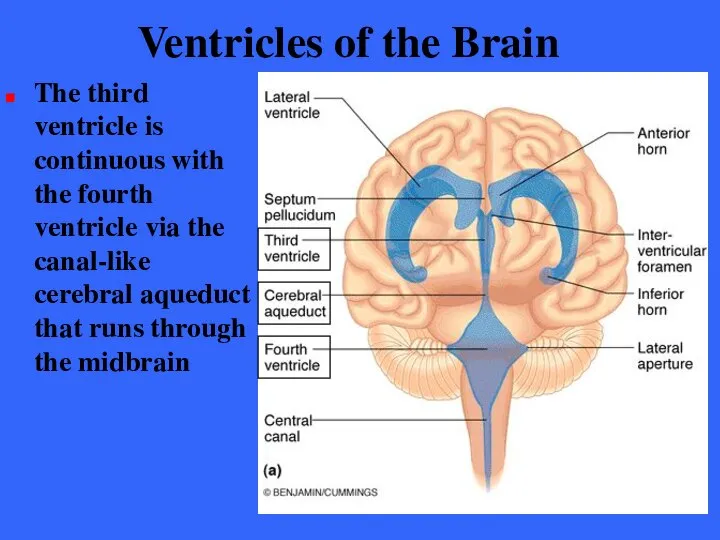
Ventricles of the Brain
The third ventricle is continuous with the fourth
Ventricles of the Brain
The third ventricle is continuous with the fourth
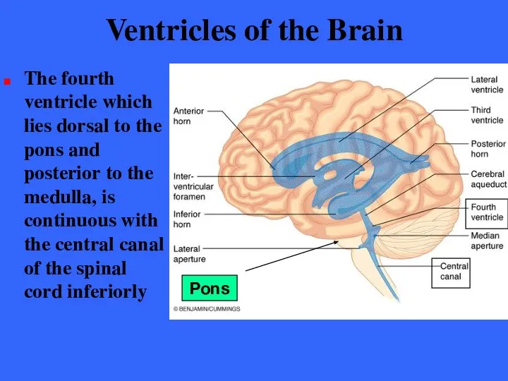
Ventricles of the Brain
The fourth ventricle which lies dorsal to the
Ventricles of the Brain
The fourth ventricle which lies dorsal to the
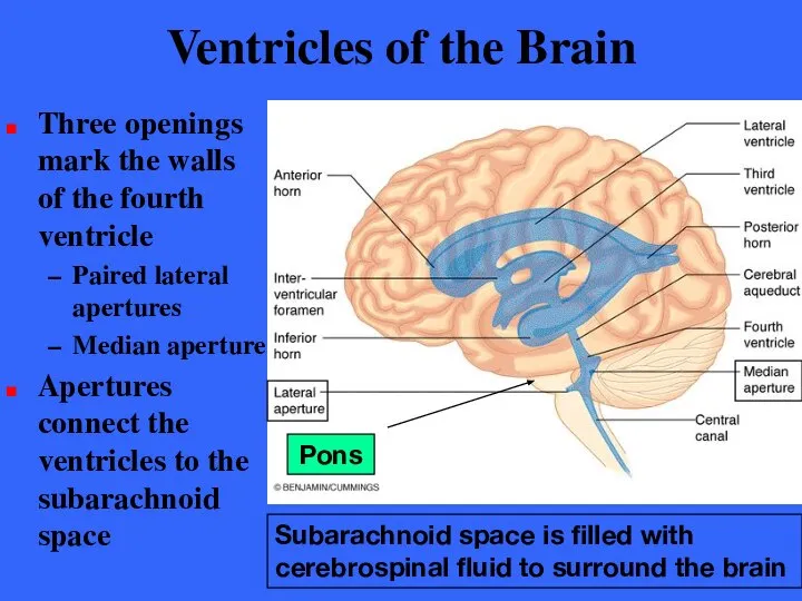
Ventricles of the Brain
Three openings mark the walls of the fourth
Ventricles of the Brain
Three openings mark the walls of the fourth
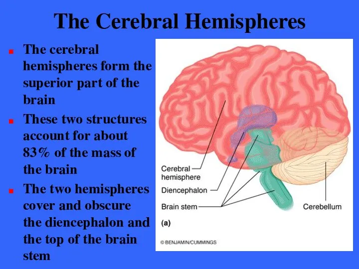
The Cerebral Hemispheres
The cerebral hemispheres form the superior part of the
The Cerebral Hemispheres
The cerebral hemispheres form the superior part of the
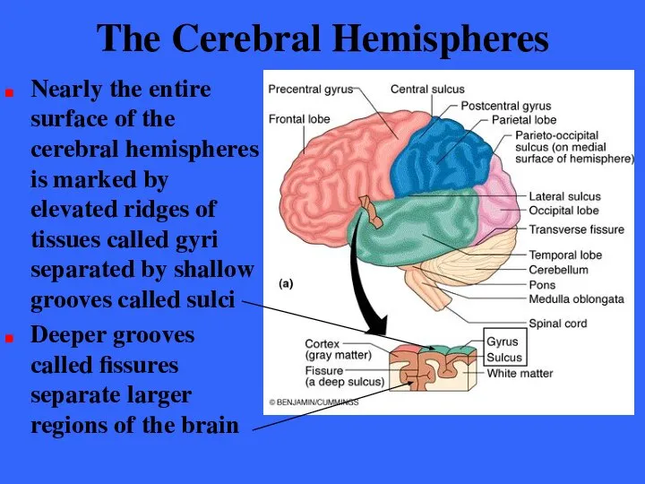
The Cerebral Hemispheres
Nearly the entire surface of the cerebral hemispheres is
The Cerebral Hemispheres
Nearly the entire surface of the cerebral hemispheres is
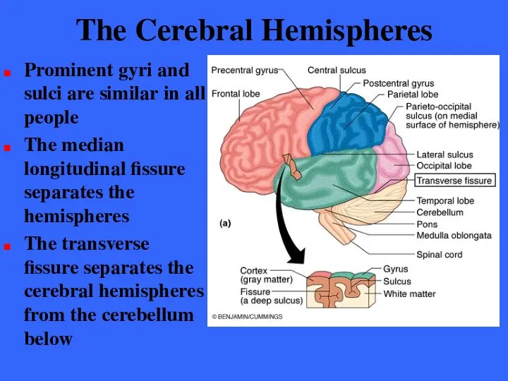
The Cerebral Hemispheres
Prominent gyri and sulci are similar in all people
The
The Cerebral Hemispheres
Prominent gyri and sulci are similar in all people
The
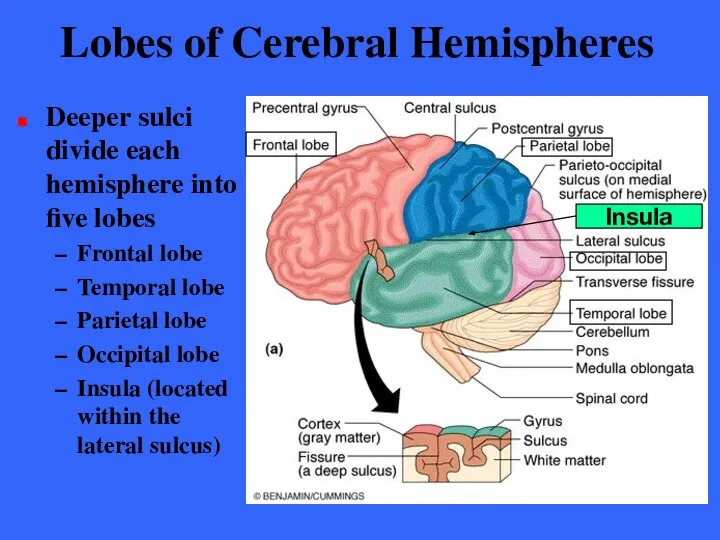
Lobes of Cerebral Hemispheres
Deeper sulci divide each hemisphere into five lobes
Frontal
Lobes of Cerebral Hemispheres
Deeper sulci divide each hemisphere into five lobes
Frontal
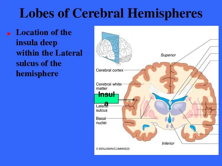
Lobes of Cerebral Hemispheres
Location of the insula deep within the Lateral
Lobes of Cerebral Hemispheres
Location of the insula deep within the Lateral
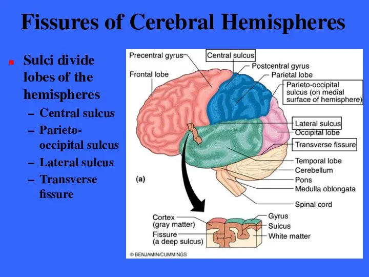
Fissures of Cerebral Hemispheres
Sulci divide lobes of the hemispheres
Central sulcus
Parieto-
Fissures of Cerebral Hemispheres
Sulci divide lobes of the hemispheres
Central sulcus
Parieto-
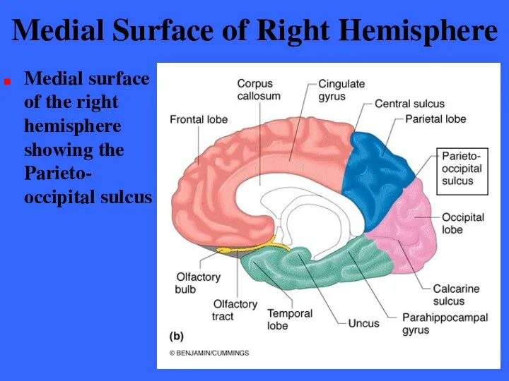
Medial Surface of Right Hemisphere
Medial surface of the right hemisphere showing
Medial Surface of Right Hemisphere
Medial surface of the right hemisphere showing
Position of Cerebral Hemispheres
The frontal lobes occupy the anterior cranial fossa
The
Position of Cerebral Hemispheres
The frontal lobes occupy the anterior cranial fossa
The
Cerebral Cortex
The cerebral cortex is the “executive suite” of the nervous
Cerebral Cortex
The cerebral cortex is the “executive suite” of the nervous
Cerebral Cortex
The cerebral cortex is gray matter composed of neuron cells
Cerebral Cortex
The cerebral cortex is gray matter composed of neuron cells
Cerebral Cortex
The cerebral cortex accounts for roughly 40% of total brain
Cerebral Cortex
The cerebral cortex accounts for roughly 40% of total brain
Cerebral Hemispheres
Coronal section of the brain which reveals the cerebral cortex,
Cerebral Hemispheres
Coronal section of the brain which reveals the cerebral cortex,
Cerebral Cortex
Research on the structure and function of the brain reveals
Cerebral Cortex
Research on the structure and function of the brain reveals
Cerebral Cortex - Generalizations
The cerebral cortex has three types of functional
Cerebral Cortex - Generalizations
The cerebral cortex has three types of functional
Cerebral Cortex - Generalizations
Although they are largely symmetrical in structure the
Cerebral Cortex - Generalizations
Although they are largely symmetrical in structure the
Motor Areas
Cortical areas controlling motor functions lie in the posterior part
Motor Areas
Cortical areas controlling motor functions lie in the posterior part
Primary Motor Cortex
The primary motor cortex is located in the precentral
Primary Motor Cortex
The primary motor cortex is located in the precentral
Pyramidal cells
These long axons, which project to the spinal cord, form
Pyramidal cells
These long axons, which project to the spinal cord, form
Pyramidal Tracts
The lateral corticospinal tract consists of the long axons of
Pyramidal Tracts
The lateral corticospinal tract consists of the long axons of
Motor Somatotopy
Body is represented spatially in the primary motor cortex
Motor Somatotopy
Body is represented spatially in the primary motor cortex
Motor Somatotopy
Motor innervation is contralateral; left primary motor controls right
Motor Somatotopy
Motor innervation is contralateral; left primary motor controls right
Motor Somatotopy
Damage to the localized areas of the primary motor
Motor Somatotopy
Damage to the localized areas of the primary motor
Premotor Cortex
The premotor cortex controls motor skills of repetitive or patterned
Premotor Cortex
The premotor cortex controls motor skills of repetitive or patterned
Premotor Cortex
The premotor cortex sends activating impulses to the primary motor
Premotor Cortex
The premotor cortex sends activating impulses to the primary motor
Premotor Cortex
This area appears to involved with motor planning
It controls voluntary
Premotor Cortex
This area appears to involved with motor planning
It controls voluntary
Premotor Cortex
Damage to the premotor area results in the loss of
Premotor Cortex
Damage to the premotor area results in the loss of
Broca’s area
The area has long been considered to be present in
Broca’s area
The area has long been considered to be present in
Broca’s area
Recent PET scans indicates that Broca’s area and a similar
Broca’s area
Recent PET scans indicates that Broca’s area and a similar
Frontal Eye Field
This cortical region controls the voluntary movements of the
Frontal Eye Field
This cortical region controls the voluntary movements of the
Sensory Areas
Areas concerned with the conscious awareness of sensation in the
Sensory Areas
Areas concerned with the conscious awareness of sensation in the
Primary Somato-sensory Cortex
Primary somato- sensory area resides in the postcentral
Primary Somato-sensory Cortex
Primary somato- sensory area resides in the postcentral
Synaptic Chain
Central axons of sensory (1st order) neurons enter dorsal root
Synaptic Chain
Central axons of sensory (1st order) neurons enter dorsal root
Primary Somato-sensory Cortex
In the cortex neurons process the sensory information
Primary Somato-sensory Cortex
In the cortex neurons process the sensory information
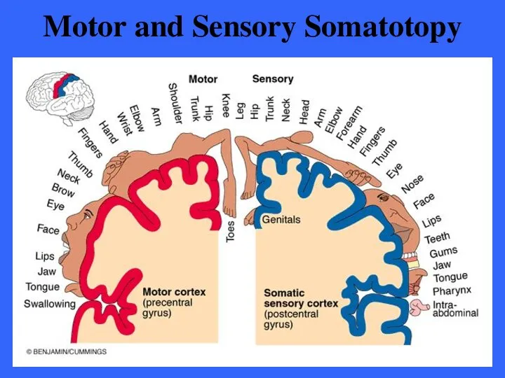
Motor and Sensory Somatotopy
Motor and Sensory Somatotopy
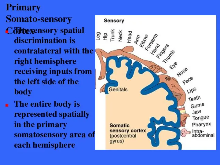
Primary Somato-sensory Cortex
The sensory spatial discrimination is contralateral with the right
Primary Somato-sensory Cortex
The sensory spatial discrimination is contralateral with the right
Primary Somato-sensory Cortex
The amount of sensory cortex devoted to a particular
Primary Somato-sensory Cortex
The amount of sensory cortex devoted to a particular
Primary Somatosensory Cortex
Damage to the primary somatisensory cortex destroys the conscious
Primary Somatosensory Cortex
Damage to the primary somatisensory cortex destroys the conscious
Somatosensory Association Area
The area lies just posterior to the primary somata-
Somatosensory Association Area
The area lies just posterior to the primary somata-
Somatosensory Association Area
The somatosensory association area forms a comprehensive evaluation of
Somatosensory Association Area
The somatosensory association area forms a comprehensive evaluation of
Somatosensory Association Area
Past associations allow you to recognize familiar objects (coins,
Somatosensory Association Area
Past associations allow you to recognize familiar objects (coins,
Primary Visual Cortex
The primary visual cortex (17) is located on the
Primary Visual Cortex
The primary visual cortex (17) is located on the
Primary Visual Cortex
Most of the primary visual cortex is located on
Primary Visual Cortex
Most of the primary visual cortex is located on
Primary Visual Cortex
The largest of all cortical sensory areas, the primary
Primary Visual Cortex
The largest of all cortical sensory areas, the primary
Primary Visual Cortex
Again, the right half of visual space is represented
Primary Visual Cortex
Again, the right half of visual space is represented
Primary Visual Cortex
The primary visual cortex is the first of a
Primary Visual Cortex
The primary visual cortex is the first of a
Visual Association Area
This area surrounds the primary visual area and encompasses
Visual Association Area
This area surrounds the primary visual area and encompasses
Visual Association Area
This area analyzes color, form and movement in light
Visual Association Area
This area analyzes color, form and movement in light
Visual Association Area
Recent neuroimaging has revealed that complex visual processing far
Visual Association Area
Recent neuroimaging has revealed that complex visual processing far
Visual Association Area
The ventral stream extends through the inferior part of
Visual Association Area
The ventral stream extends through the inferior part of
Visual Association Area
The dorsal stream extends through the posterior parietal cortex
Visual Association Area
The dorsal stream extends through the posterior parietal cortex
Visual Association Area
The dorsal stream in the parietal lobe is important
Visual Association Area
The dorsal stream in the parietal lobe is important
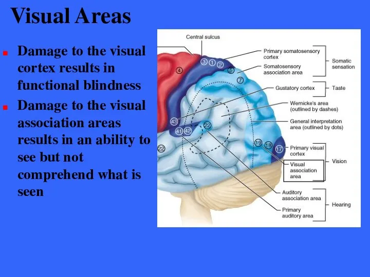
Visual Areas
Damage to the visual cortex results in functional blindness
Damage to
Visual Areas
Damage to the visual cortex results in functional blindness
Damage to
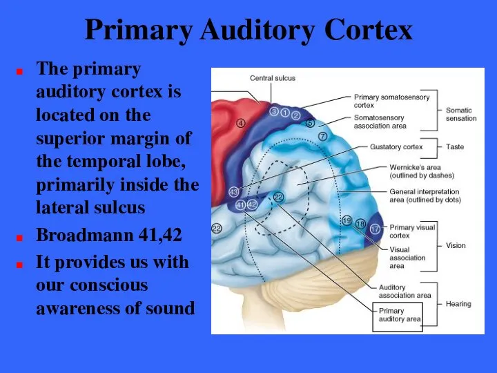
Primary Auditory Cortex
The primary auditory cortex is located on the superior
Primary Auditory Cortex
The primary auditory cortex is located on the superior
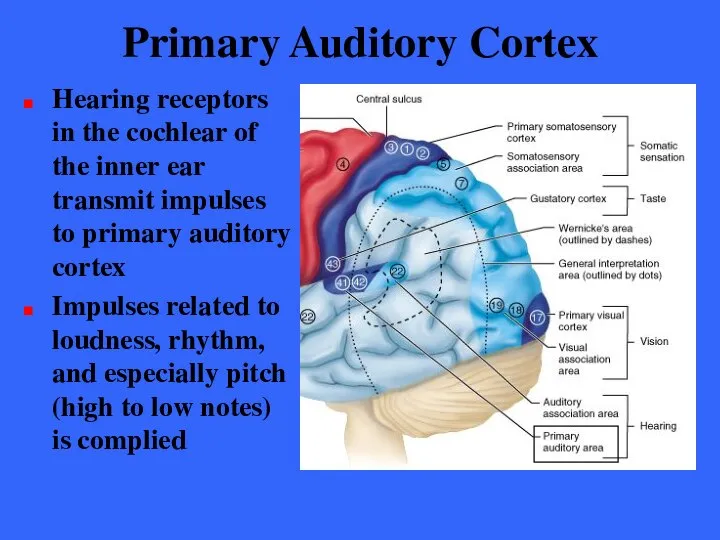
Primary Auditory Cortex
Hearing receptors in the cochlear of the inner ear
Primary Auditory Cortex
Hearing receptors in the cochlear of the inner ear
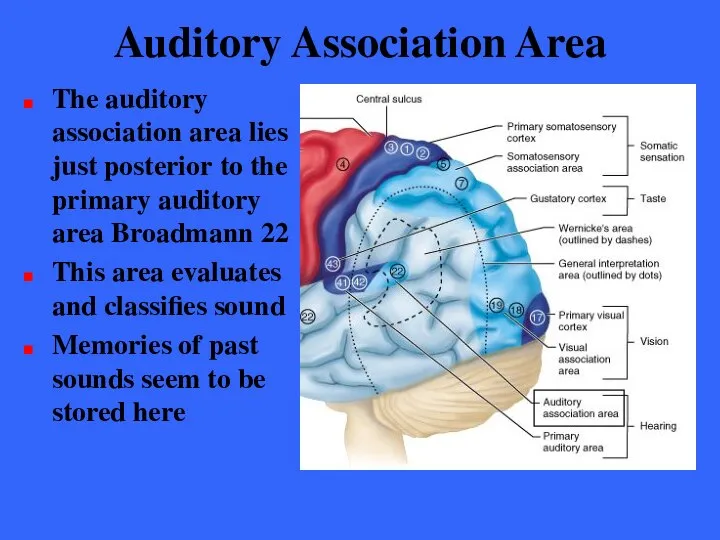
Auditory Association Area
The auditory association area lies just posterior to the
Auditory Association Area
The auditory association area lies just posterior to the
Auditory Association Area
In one hemisphere (usually the left), the auditory association
Auditory Association Area
In one hemisphere (usually the left), the auditory association
Auditory Association Area
Damage to Wernicke’s area interferes with the ability to
Auditory Association Area
Damage to Wernicke’s area interferes with the ability to
Gustatory (taste) Cortex
The gustatory cortex is involved in the conscious awareness
Gustatory (taste) Cortex
The gustatory cortex is involved in the conscious awareness
Vestibular (equilibrium) Cortex
The cortex is responsible for conscious aware-ness of the
Vestibular (equilibrium) Cortex
The cortex is responsible for conscious aware-ness of the
Olfactory Area
The primary olfactory cortex lie on the medial aspects of
Olfactory Area
The primary olfactory cortex lie on the medial aspects of
Olfactory Area
The olfactory nerves (Cranial nerve I) from the nasal cavity
Olfactory Area
The olfactory nerves (Cranial nerve I) from the nasal cavity
Olfactory Area
The olfactory cortex is part of a brain area called
Olfactory Area
The olfactory cortex is part of a brain area called
Olfactory Area
The piriform lobe, the olfactory tract, the olfactory bulb, and
Olfactory Area
The piriform lobe, the olfactory tract, the olfactory bulb, and
Olfactory Area
The rhinencephalon connects to the brain area that is involved
Olfactory Area
The rhinencephalon connects to the brain area that is involved
Olfactory Area
Part of the frontal lobe, the orbitofrontal cortex, is involved
Olfactory Area
Part of the frontal lobe, the orbitofrontal cortex, is involved
Association Areas
Association areas include all cortical areas other than primary sensory
Association Areas
Association areas include all cortical areas other than primary sensory
Association Areas
The term association area is fading from use and will
Association Areas
The term association area is fading from use and will

Prefrontal Cortex
The prefrontal cortex occupies the large region of the frontal
Prefrontal Cortex
The prefrontal cortex occupies the large region of the frontal
Prefrontal Cortex
Cognition is all aspects of thinking, perceiving and of intentionally
Prefrontal Cortex
Cognition is all aspects of thinking, perceiving and of intentionally
Prefrontal Cortex
The prefrontal cortex also is used for long- term planning,
Prefrontal Cortex
The prefrontal cortex also is used for long- term planning,
Prefrontal Cortex
The prefrontal cortex also seems to be related to mood
Prefrontal Cortex
The prefrontal cortex also seems to be related to mood

Prefrontal Cortex
Functional neuro-imaging techniques have begun to reveal the functions of
Prefrontal Cortex
Functional neuro-imaging techniques have begun to reveal the functions of

Prefrontal Cortex
The working memories of spatial relations are stored in the
Prefrontal Cortex
The working memories of spatial relations are stored in the
Prefrontal Cortex
Working memories of objects and faces are stored farther ventrally,
Prefrontal Cortex
Working memories of objects and faces are stored farther ventrally,
Prefrontal Cortex
More significant is the region that manages cognitive tasks by
Prefrontal Cortex
More significant is the region that manages cognitive tasks by
Prefrontal Cortex
The extreme anterior pole of the frontal cortex was found
Prefrontal Cortex
The extreme anterior pole of the frontal cortex was found
Prefrontal Cortex
The new findings suggest support for a general rule of
Prefrontal Cortex
The new findings suggest support for a general rule of
Prefrontal Cortex
The area just anterior to the corpus callosum may process
Prefrontal Cortex
The area just anterior to the corpus callosum may process
General Interpretation Area
The existence of this area within the brain is
General Interpretation Area
The existence of this area within the brain is
Language Area
The large area surrounding the lateral sulcus in the left
Language Area
The large area surrounding the lateral sulcus in the left
Language Area
Five areas have been identified with language; Broca’s area (speech
Language Area
Five areas have been identified with language; Broca’s area (speech
Language Area
The corresponding areas on the right hemisphere, although not involved
Language Area
The corresponding areas on the right hemisphere, although not involved
Insula
The insula is large and the functions of its cortex are
Insula
The insula is large and the functions of its cortex are
Lateralization of Cortical Function
We use both cerebral hemispheres for almost every
Lateralization of Cortical Function
We use both cerebral hemispheres for almost every
Lateralization of Cortical Function
In most people (Approx. 90%) the left hemisphere
Lateralization of Cortical Function
In most people (Approx. 90%) the left hemisphere
Lateralization of Cortical Function
Most individuals (90%) with left cerebral dominance are
Lateralization of Cortical Function
Most individuals (90%) with left cerebral dominance are
Lateralization of Cortical Function
The two cerebral hemispheres have perfect and almost
Lateralization of Cortical Function
The two cerebral hemispheres have perfect and almost
Cerebral White Matter
Communication within the brain is extensive
The cerebral white matter
Cerebral White Matter
Communication within the brain is extensive
The cerebral white matter
Cerebral White Matter
The white matter largely consists of myelinated fibers bundled
Cerebral White Matter
The white matter largely consists of myelinated fibers bundled
Cerebral White Matter
Commissures connect the hemispheres
Association fibers connect areas within hemispheres
Projection
Cerebral White Matter
Commissures connect the hemispheres
Association fibers connect areas within hemispheres
Projection
Cerebral White Matter
Commissures connect the corresponding areas of two hemispheres enabling
Cerebral White Matter
Commissures connect the corresponding areas of two hemispheres enabling
Cerebral White Matter
Association fibers transmit within a single hemisphere
Short fibers connect
Cerebral White Matter
Association fibers transmit within a single hemisphere
Short fibers connect
Cerebral White Matter
Projection fibers run vertically to connect levels of the
Cerebral White Matter
Projection fibers run vertically to connect levels of the
Cerebral White Matter
Ascending projection tracts pass between the thalamus and the
Cerebral White Matter
Ascending projection tracts pass between the thalamus and the
Cerebral White Matter
The fibers of the corona radiata fan out into
Cerebral White Matter
The fibers of the corona radiata fan out into
Basal Nuclei
In the cerebral white matter of each hemisphere are a
Basal Nuclei
In the cerebral white matter of each hemisphere are a
Basal Nuclei
The putamen and globus pallidus together form a mass called
Basal Nuclei
The putamen and globus pallidus together form a mass called
Basal Nuclei
The comma shaped caudate nucleus arches superiorly over the diencephalon
Basal Nuclei
The comma shaped caudate nucleus arches superiorly over the diencephalon
Basal Nuclei
The lentiform nucleus flanks the internal capsule laterally
Lentiform
nucleus
Basal Nuclei
The lentiform nucleus flanks the internal capsule laterally
Lentiform
nucleus
Basal Nuclei
Collectively the caudate nucleus and the lentiform nuclei are called
Basal Nuclei
Collectively the caudate nucleus and the lentiform nuclei are called
Basal Nuclei
The basal nuclei are functionally associated with the subthalamic nuclei
Basal Nuclei
The basal nuclei are functionally associated with the subthalamic nuclei
Basal Nuclei
The amygdaloid nucleus sits on the tail of the caudate
Basal Nuclei
The amygdaloid nucleus sits on the tail of the caudate
Basal Nuclei
Functionally, the basal nuclei can be viewed as complex neural
Basal Nuclei
Functionally, the basal nuclei can be viewed as complex neural
Basal Nuclei
The basal nuclei receive inputs from the entire cerebral cortex
Basal Nuclei
The basal nuclei receive inputs from the entire cerebral cortex
Basal Nuclei
Via relays the basal nuclei influence muscle movements directed by
Basal Nuclei
Via relays the basal nuclei influence muscle movements directed by
Basal Nuclei
The nuclei are involved in monitoring muscle movements that are
Basal Nuclei
The nuclei are involved in monitoring muscle movements that are
The Diencephanlon
Forms the central core of the forebrain and is surrounded
The Diencephanlon
Forms the central core of the forebrain and is surrounded
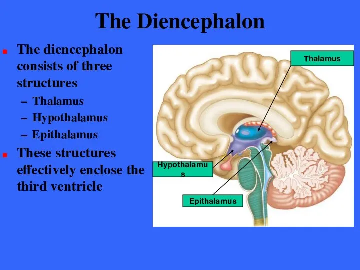
The Diencephalon
The diencephalon consists of three structures
Thalamus
Hypothalamus
Epithalamus
These structures effectively enclose the
The Diencephalon
The diencephalon consists of three structures
Thalamus
Hypothalamus
Epithalamus
These structures effectively enclose the
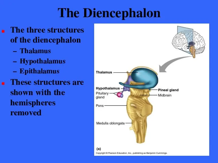
The Diencephalon
The three structures of the diencephalon
Thalamus
Hypothalamus
Epithalamus
These structures are shown
The Diencephalon
The three structures of the diencephalon
Thalamus
Hypothalamus
Epithalamus
These structures are shown
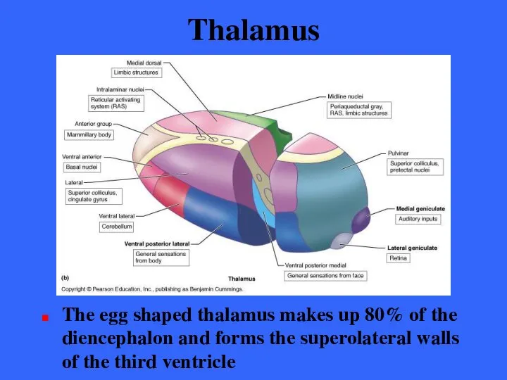
Thalamus
The egg shaped thalamus makes up 80% of the diencephalon and
Thalamus
The egg shaped thalamus makes up 80% of the diencephalon and
Thalamus
The thalamus is composed of bilateral masses of gray matter held
Thalamus
The thalamus is composed of bilateral masses of gray matter held
Thalamus
The thalamus has many different nuclei, most named for their location
Each
Thalamus
The thalamus has many different nuclei, most named for their location
Each
The Thalamus
Sensory inputs are not the only type of information relayed
The Thalamus
Sensory inputs are not the only type of information relayed
Thalamus
Afferent impulses from all senses and all parts of the body
Thalamus
Afferent impulses from all senses and all parts of the body
Thalamus
Impulses having to do with similar functions are grouped together and
Thalamus
Impulses having to do with similar functions are grouped together and
Thalamus
In addition to sensory inputs, virtually all inputs ascending to the
Thalamus
In addition to sensory inputs, virtually all inputs ascending to the
Thalamus
Lateral geniculate body
Visual relay from retina
Medial geniculate body
Auditory inputs
Anterior nuclear group
Regulation
Thalamus
Lateral geniculate body
Visual relay from retina
Medial geniculate body
Auditory inputs
Anterior nuclear group
Regulation
Thalamus
Pulvinar, medial dorsal and lateral nuclei are involved in the integration
Thalamus
Pulvinar, medial dorsal and lateral nuclei are involved in the integration
Thalamus
The thalamus plays a key role in mediating sensation, motor activities,
Thalamus
The thalamus plays a key role in mediating sensation, motor activities,
The Hypothalamus
The hypothalamus is located below the thalamus, capping the brain
The Hypothalamus
The hypothalamus is located below the thalamus, capping the brain
Hypothalamus
Merging into the midbrain inferiorly, it extends from the optic chiasma
Hypothalamus
Merging into the midbrain inferiorly, it extends from the optic chiasma
Mammillary Bodies
The mammillary bodies are paired nuclei that bulge anteriorly from
Mammillary Bodies
The mammillary bodies are paired nuclei that bulge anteriorly from
Hypothalamus
Between the optic chiasma and the mammillary bodies is the infundibulum
A
Hypothalamus
Between the optic chiasma and the mammillary bodies is the infundibulum
A
Hypothalamus
The hypothalamus contains about a dozen functionally important nuclei
Despite its small
Hypothalamus
The hypothalamus contains about a dozen functionally important nuclei
Despite its small
Autonomic Control Center
The hypothalamus regulates involuntary nervous activity by controlling the
Autonomic Control Center
The hypothalamus regulates involuntary nervous activity by controlling the
Center for Emotional Response
The hypothalamus has numerous connections with cortical association
Center for Emotional Response
The hypothalamus has numerous connections with cortical association
Center for Emotional Response
The hypothalamus acts through the autonomic nervous system
Center for Emotional Response
The hypothalamus acts through the autonomic nervous system
Body Temperature Regulation
The body’s thermostat is in the hypothalamus
The hypothalamus receives
Body Temperature Regulation
The body’s thermostat is in the hypothalamus
The hypothalamus receives
Body Temperature Regulation
Hypothalamic receptors in the preoptic region monitor the temperature
Body Temperature Regulation
Hypothalamic receptors in the preoptic region monitor the temperature
Body Temperature Regulation
According to signals received by the preoptic nuclei the
Body Temperature Regulation
According to signals received by the preoptic nuclei the
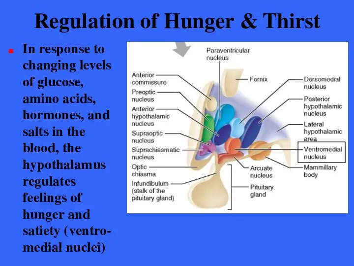
Regulation of Hunger & Thirst
In response to changing levels of glucose,
Regulation of Hunger & Thirst
In response to changing levels of glucose,
Regulation of Water Balance
When body fluids become too concentrated, hypothalamic neurons
Regulation of Water Balance
When body fluids become too concentrated, hypothalamic neurons
Regulation of Sleep-Wake Cycles
Acting with other brain regions, the hypothalamus helps
Regulation of Sleep-Wake Cycles
Acting with other brain regions, the hypothalamus helps
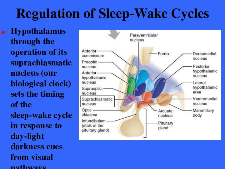
Regulation of Sleep-Wake Cycles
Hypothalamus through the operation of its suprachiasmatic nucleus
Regulation of Sleep-Wake Cycles
Hypothalamus through the operation of its suprachiasmatic nucleus
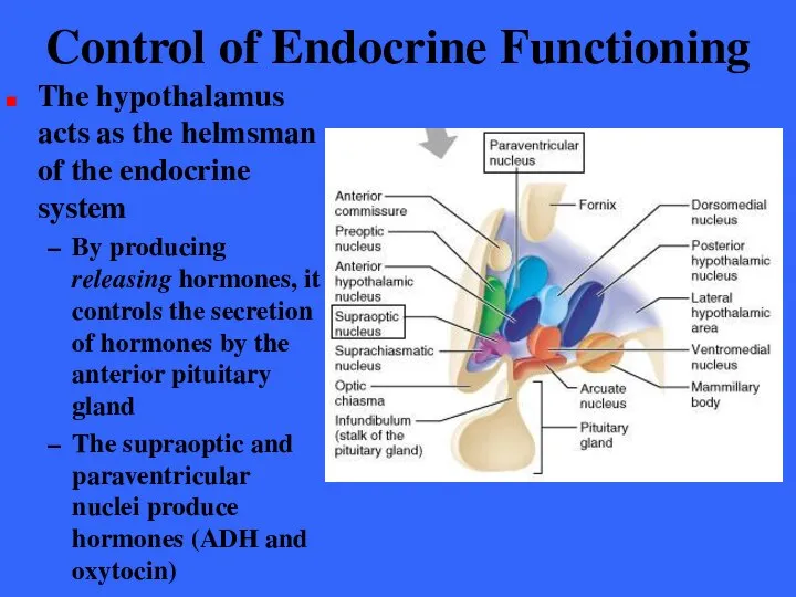
Control of Endocrine Functioning
The hypothalamus acts as the helmsman of
Control of Endocrine Functioning
The hypothalamus acts as the helmsman of
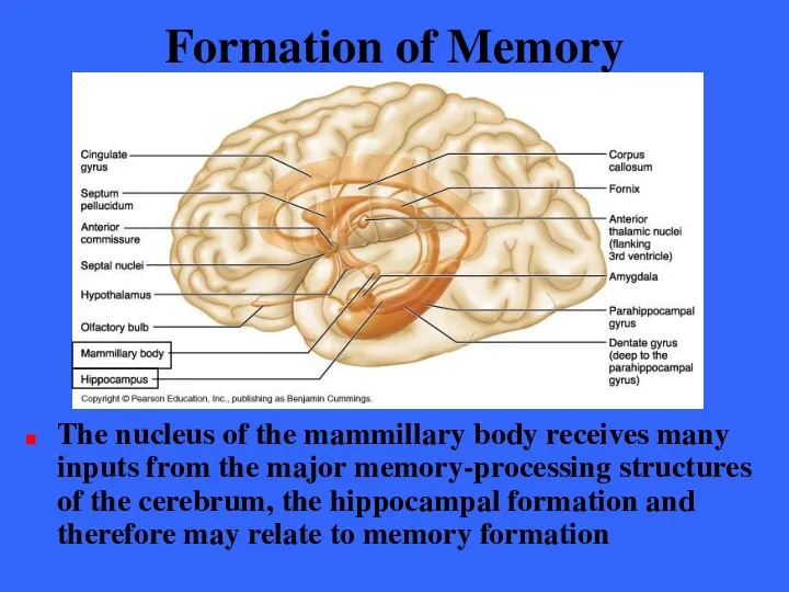
Formation of Memory
The nucleus of the mammillary body receives many inputs
Formation of Memory
The nucleus of the mammillary body receives many inputs
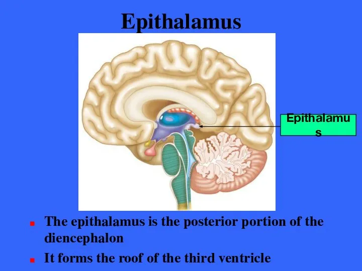
Epithalamus
The epithalamus is the posterior portion of the diencephalon
It forms the
Epithalamus
The epithalamus is the posterior portion of the diencephalon
It forms the
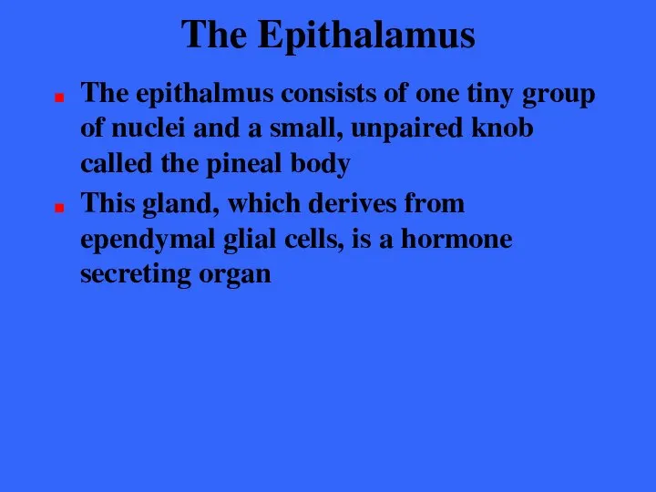
The Epithalamus
The epithalmus consists of one tiny group of nuclei and
The Epithalamus
The epithalmus consists of one tiny group of nuclei and
Epithalamus
The pineal gland extends from the posterior border of the epithalamus
Epithalamus
The pineal gland extends from the posterior border of the epithalamus
The Epithalamus
A cerebrospinal fluid-forming structure called a choroid plexus is also
The Epithalamus
A cerebrospinal fluid-forming structure called a choroid plexus is also
The Brain Stem
The third of the four major parts of the
The Brain Stem
The third of the four major parts of the
The Brain Stem
Each region is roughly an inch long
Together than constitute
The Brain Stem
Each region is roughly an inch long
Together than constitute
The Brain Stem
The brain stem has the same structural plan as
The Brain Stem
The brain stem has the same structural plan as
The Midbrain
The midbrain is located between the diencephalon superiorly and the
The Midbrain
The midbrain is located between the diencephalon superiorly and the
The Midbrain
Its central cavity is the cerebral aqueduct, which divides it
The Midbrain
Its central cavity is the cerebral aqueduct, which divides it
The Midbrain
These peduncles contain the pyramidal (corticospinal) motor tracts descending toward
The Midbrain
These peduncles contain the pyramidal (corticospinal) motor tracts descending toward
The Midbrain
Dorsally, the midbrain has the superior cerebellar peduncles which connect
The Midbrain
Dorsally, the midbrain has the superior cerebellar peduncles which connect
The Midbrain
Surrounding the cerebral aqueduct is the peri-aqueductal gray matter that
The Midbrain
Surrounding the cerebral aqueduct is the peri-aqueductal gray matter that
The Midbrain
The periaqueductal gray matter is involved in the “fright-and-flight” sympathetic
The Midbrain
The periaqueductal gray matter is involved in the “fright-and-flight” sympathetic
The Midbrain
The gray matter elicits
A terror-induced increase in heart rate
Skyrocketing
The Midbrain
The gray matter elicits
A terror-induced increase in heart rate
Skyrocketing
The Midbrain
The periaqueductal gray matter also seems to mediate our response
The Midbrain
The periaqueductal gray matter also seems to mediate our response
The Midbrain
The most ventral part of the para- aqueductal gray mattercontains
The Midbrain
The most ventral part of the para- aqueductal gray mattercontains
The Midbrain
Nuclei are also scattered in the surrounding white matter
The largest
The Midbrain
Nuclei are also scattered in the surrounding white matter
The largest
The Midbrain
The superior pair of nuclei, the superior colliculus are visual
The Midbrain
The superior pair of nuclei, the superior colliculus are visual
The Midbrain
The inferior colliculus are part of the auditory relay from
The Midbrain
The inferior colliculus are part of the auditory relay from
The Midbrain
Also imbedded in the white matter of the midbrain are
The Midbrain
Also imbedded in the white matter of the midbrain are
The Midbrain
The substantia nigra is a bandlike nucleus located deep to
The Midbrain
The substantia nigra is a bandlike nucleus located deep to
The Midbrain
Its dark color reflects its high content of melanin pigment,
The Midbrain
Its dark color reflects its high content of melanin pigment,
The Midbrain
The red nucleus is found between the substantia nigra and
The Midbrain
The red nucleus is found between the substantia nigra and
The Midbrain
The red nuclei are relay nuclei in some descending motor
The Midbrain
The red nuclei are relay nuclei in some descending motor
The Pons
The pons is the bulging brain stem region wedged between
The Pons
The pons is the bulging brain stem region wedged between
The Pons
It forms part of the anterior wall of the fourth
The Pons
It forms part of the anterior wall of the fourth
The Pons
The deep projection fibers run longitudinally and complete the superior-inferior
The Pons
The deep projection fibers run longitudinally and complete the superior-inferior
The Pons
The more superficial nuclei are relays for conversations between the
The Pons
The more superficial nuclei are relays for conversations between the
The Pons
Several cranial nerves issue from pons nuclei
Trigeminal nerve
Abducens nerve
Facial
The Pons
Several cranial nerves issue from pons nuclei
Trigeminal nerve
Abducens nerve
Facial
The Pons
Other important pons nuclei are part of the reticular formation
The
The Pons
Other important pons nuclei are part of the reticular formation
The
The Medulla Oblongata
The medulla oblongata is the most inferior part of
The Medulla Oblongata
The medulla oblongata is the most inferior part of
The Medulla Oblongata
The medulla blends into the spinal cord at the
The Medulla Oblongata
The medulla blends into the spinal cord at the
The Medulla Oblongata
The medulla has several externally visible landmarks which form
The Medulla Oblongata
The medulla has several externally visible landmarks which form
The Medulla Oblongata
Just above the medulla-spinal cord junction most of the
The Medulla Oblongata
Just above the medulla-spinal cord junction most of the
The Medulla Oblongata
The consequence of this crossover is that each hemisphere
The Medulla Oblongata
The consequence of this crossover is that each hemisphere
The Medulla Oblongata
The inferior cerebellar peduncles are fiber tracts that connect
The Medulla Oblongata
The inferior cerebellar peduncles are fiber tracts that connect
The Medulla Oblongata
The olivary nuclei relay sensory information on the state
The Medulla Oblongata
The olivary nuclei relay sensory information on the state
The Medulla Oblongata
The rootlets of the hypoglossal nerves emerge from the
The Medulla Oblongata
The rootlets of the hypoglossal nerves emerge from the
The Medulla Oblongata
Other cranial nerves associated with the medulla are the
The Medulla Oblongata
Other cranial nerves associated with the medulla are the
The Medulla Oblongata
The fibers of the vestibulocochlear synapse with the cochlear
The Medulla Oblongata
The fibers of the vestibulocochlear synapse with the cochlear
The Medulla Oblongata
Also housed within the medulla are several nuclei associated
The Medulla Oblongata
Also housed within the medulla are several nuclei associated
Medulla
Oblongata
These serve as relay nuclei in a pathway by which
Medulla
Oblongata
These serve as relay nuclei in a pathway by which
The Medulla Oblongata
The medulla has a critical role as an autonomic
The Medulla Oblongata
The medulla has a critical role as an autonomic
The Medulla Oblongata
The cardiac center
The cardiac center adjusts the force and
The Medulla Oblongata
The cardiac center
The cardiac center adjusts the force and
The Medulla Oblongata
The vasomotor center
The vasomotor center regulates blood pressure by
The Medulla Oblongata
The vasomotor center
The vasomotor center regulates blood pressure by
The Medulla Oblongata
The respiratory centers
The medullary respiratory centers control the rate
The Medulla Oblongata
The respiratory centers
The medullary respiratory centers control the rate
The Medulla Oblongata
Other centers
Additional centers regulate activities such as
Vomiting
Hiccuping
Swallowing
Coughing
Sneezing
The Medulla Oblongata
Other centers
Additional centers regulate activities such as
Vomiting
Hiccuping
Swallowing
Coughing
Sneezing
The Medulla Oblongata
Many functions of the medulla overlap with those attributed
The Medulla Oblongata
Many functions of the medulla overlap with those attributed
The Cerebellum
The cerebellum is exceeded in size only by the cerebrum
It
The Cerebellum
The cerebellum is exceeded in size only by the cerebrum
It
The Cerebellum
The cerebellum is located dorsal to the pons and medulla
The Cerebellum
The cerebellum is located dorsal to the pons and medulla
The Cerebellum
It is separated from the occipital lobe by the transverse
The Cerebellum
It is separated from the occipital lobe by the transverse
The Cerebellum
The cerebellum processes inputs received from
Cerebral motor cortex
Various brain
The Cerebellum
The cerebellum processes inputs received from
Cerebral motor cortex
Various brain
The Cerebellum
The cerebellum is bilaterally symmetrical
Its two cerebellar hemispheres are connected
The Cerebellum
The cerebellum is bilaterally symmetrical
Its two cerebellar hemispheres are connected
The Cerebellum
Its surface is heavily convoluted
Fissure are all transversely orientated
The surface
The Cerebellum
Its surface is heavily convoluted
Fissure are all transversely orientated
The surface
The Cerebellum
Deep fissures divide each hemisphere into three lobes
Anterior lobe
Posterior lobe
Flocculonodular
The Cerebellum
Deep fissures divide each hemisphere into three lobes
Anterior lobe
Posterior lobe
Flocculonodular
The Cerebellum
The cerebellum has a thin outer cortex of gray matter
Internal
The Cerebellum
The cerebellum has a thin outer cortex of gray matter
Internal
The Cerebellum
Several types of neurons are found in the cerebellur cortex
Stellate
Basket
Granule
Purkinje
Purkinje
Granule
Basket
The Cerebellum
Several types of neurons are found in the cerebellur cortex
Stellate
Basket
Granule
Purkinje
Purkinje
Granule
Basket
The Cerebellum
The large Purkinje cells with their extensively branched dendrites are
The Cerebellum
The large Purkinje cells with their extensively branched dendrites are
The Cerebellum
The anterior and posterior lobes of the cerebellum act to
The Cerebellum
The anterior and posterior lobes of the cerebellum act to
The Cerebellum
The medial portions receive information from the axial portion of
The Cerebellum
The medial portions receive information from the axial portion of
The Cerebellum
The intermediate parts of each hemisphere are more concerned with
The Cerebellum
The intermediate parts of each hemisphere are more concerned with
The Cerebellum
The lateral parts of each hemisphere receive inputs from the
The Cerebellum
The lateral parts of each hemisphere receive inputs from the
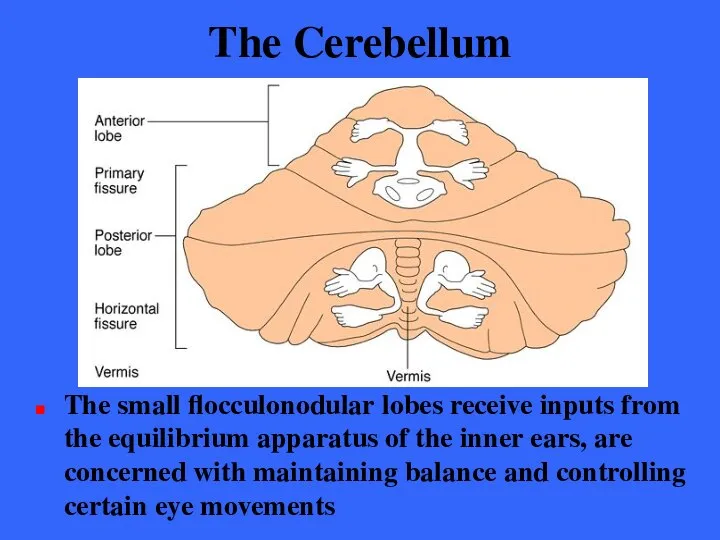
The Cerebellum
The small flocculonodular lobes receive inputs from the equilibrium apparatus
The Cerebellum
The small flocculonodular lobes receive inputs from the equilibrium apparatus
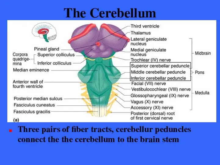
The Cerebellum
Three pairs of fiber tracts, cerebellur peduncles connect the the
The Cerebellum
Three pairs of fiber tracts, cerebellur peduncles connect the the

The Cerebellum
Virtually all fibers entering and leaving the cerebellum are ipsilateral;
The Cerebellum
Virtually all fibers entering and leaving the cerebellum are ipsilateral;
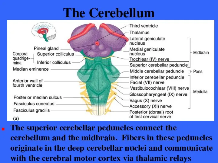
The Cerebellum
The superior cerebellar peduncles connect the cerebellum and the midbrain.
The Cerebellum
The superior cerebellar peduncles connect the cerebellum and the midbrain.

The Cerebellum
The middle cerebellar peduncles connect the pons the cerebellum.
The Cerebellum
The middle cerebellar peduncles connect the pons the cerebellum.
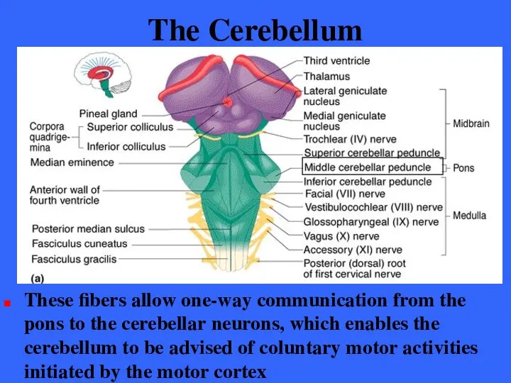
The Cerebellum
These fibers allow one-way communication from the pons to the
The Cerebellum
These fibers allow one-way communication from the pons to the
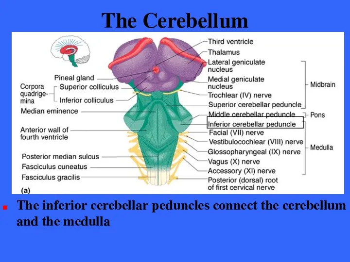
The Cerebellum
The inferior cerebellar peduncles connect the cerebellum and the medulla
The Cerebellum
The inferior cerebellar peduncles connect the cerebellum and the medulla
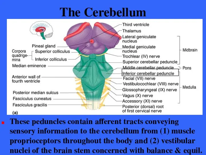
The Cerebellum
These peduncles contain afferent tracts conveying sensory information to the
The Cerebellum
These peduncles contain afferent tracts conveying sensory information to the
Cerebellar Processing - 1
The frontal motor association areas of the cerebral
Cerebellar Processing - 1
The frontal motor association areas of the cerebral
Cerebellar Processing - 2
At the same time, the cerebellum receives information
Cerebellar Processing - 2
At the same time, the cerebellum receives information
Cerebellar Processing - 3
The cerebellar cortex assesses this information and calculates
Cerebellar Processing - 3
The cerebellar cortex assesses this information and calculates
Cerebellar Processing - 4
Via the superior peduncles, the cerebellum dispatches its
Cerebellar Processing - 4
Via the superior peduncles, the cerebellum dispatches its
The Cerebellum
The cerebellum continually compares the higher brain’s intention with the
The Cerebellum
The cerebellum continually compares the higher brain’s intention with the
The Cerebellum
Cerebellar injury results in the loss of muscle tone and
The Cerebellum
Cerebellar injury results in the loss of muscle tone and

Functional Brain Systems
Functional brain systems are networks of neurons that work
Functional Brain Systems
Functional brain systems are networks of neurons that work
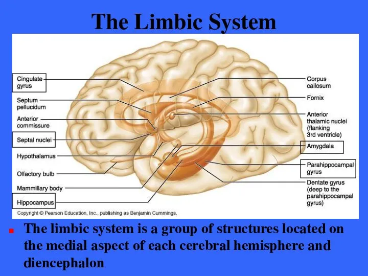
The Limbic System
The limbic system is a group of structures located
The Limbic System
The limbic system is a group of structures located
The Limbic System
The limbic system encircles the upper part of the
The Limbic System
The limbic system encircles the upper part of the
The Limbic System
The observation that odors evoke emotional reactions and memories
The Limbic System
The observation that odors evoke emotional reactions and memories
The Limbic System
The limbic system is our emotional or affective brain
Two
The Limbic System
The limbic system is our emotional or affective brain
Two
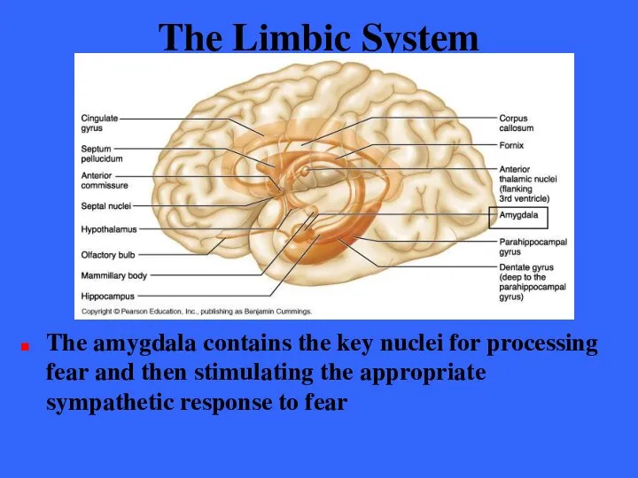
The Limbic System
The amygdala contains the key nuclei for processing fear
The Limbic System
The amygdala contains the key nuclei for processing fear
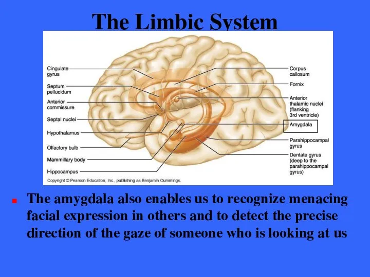
The Limbic System
The amygdala also enables us to recognize menacing facial
The Limbic System
The amygdala also enables us to recognize menacing facial
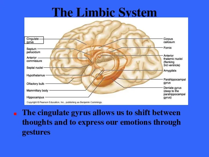
The Limbic System
The cingulate gyrus allows us to shift between thoughts
The Limbic System
The cingulate gyrus allows us to shift between thoughts
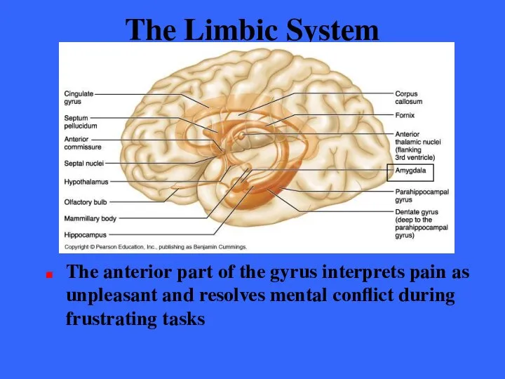
The Limbic System
The anterior part of the gyrus interprets pain as
The Limbic System
The anterior part of the gyrus interprets pain as
The Limbic System
The limbic system also functions in consolidating and retrieving
The Limbic System
The limbic system also functions in consolidating and retrieving
The Limbic System
The hippocampal formation encodes, consolidates, and later retrieves memories
The Limbic System
The hippocampal formation encodes, consolidates, and later retrieves memories
The Limbic System
The amygdala forms memories of experiences that are based
The Limbic System
The amygdala forms memories of experiences that are based
The Limbic System
The limbic system communicates with many other regions of
The Limbic System
The limbic system communicates with many other regions of
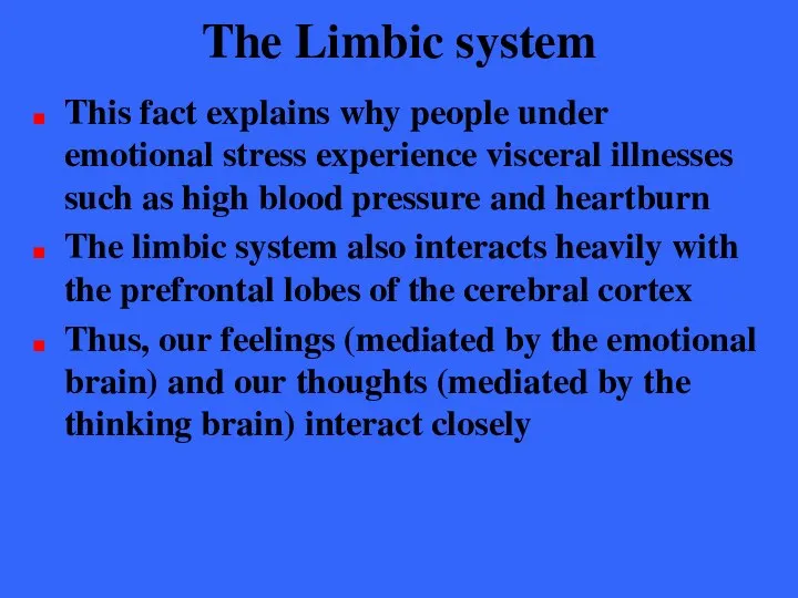
The Limbic system
This fact explains why people under emotional stress experience
The Limbic system
This fact explains why people under emotional stress experience
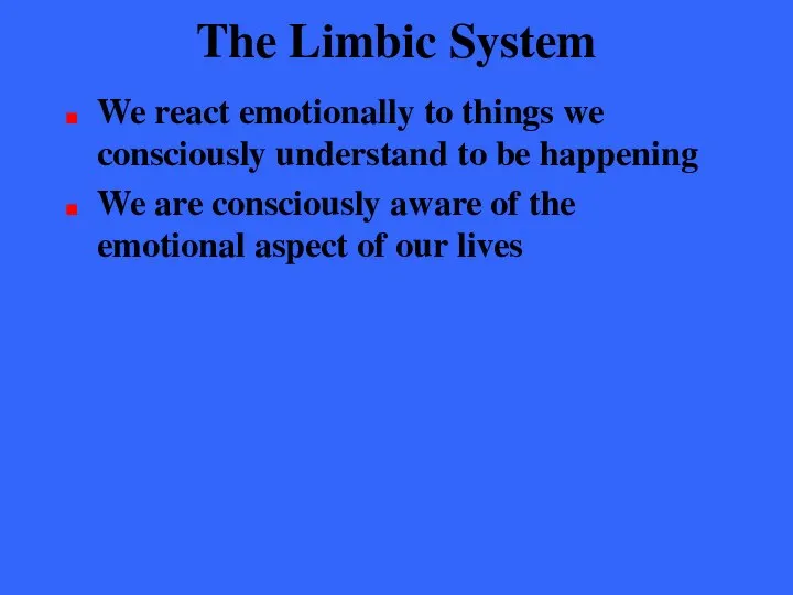
The Limbic System
We react emotionally to things we consciously understand to
The Limbic System
We react emotionally to things we consciously understand to
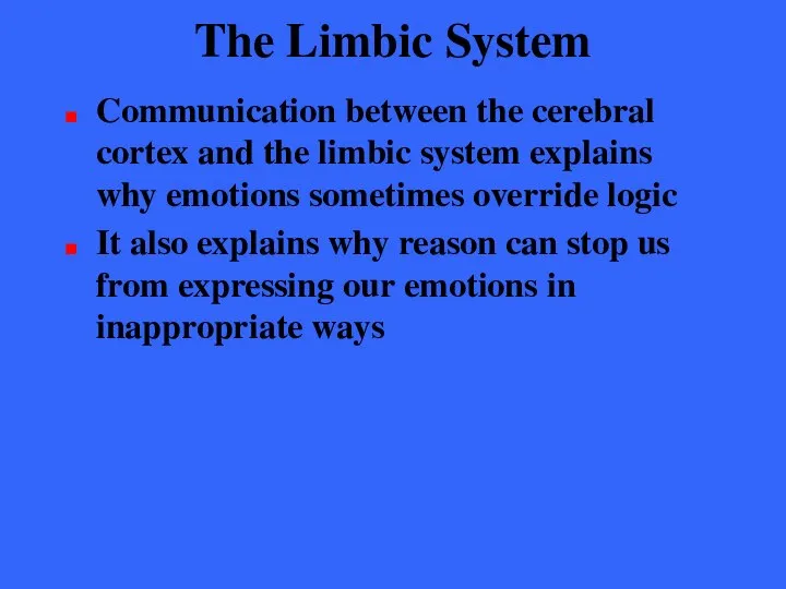
The Limbic System
Communication between the cerebral cortex and the limbic system
The Limbic System
Communication between the cerebral cortex and the limbic system
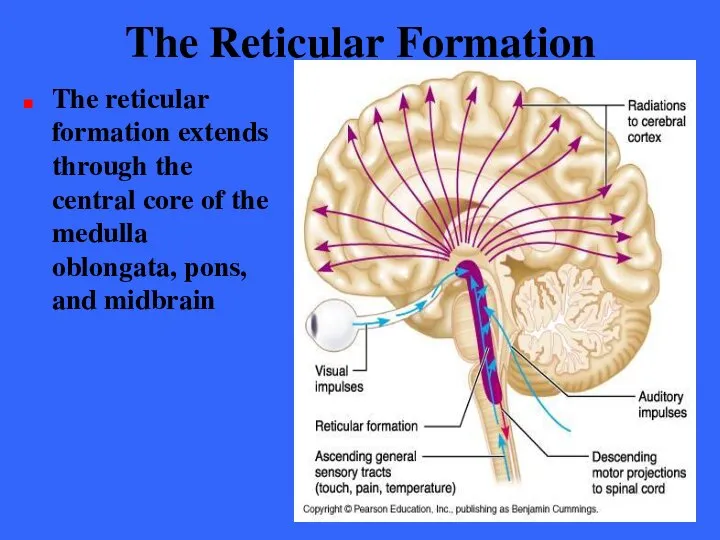
The Reticular Formation
The reticular formation extends through the central core of
The Reticular Formation
The reticular formation extends through the central core of
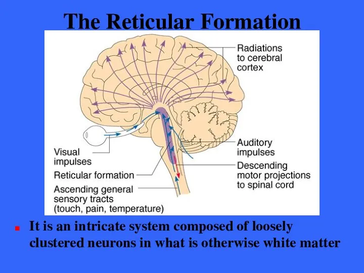
The Reticular Formation
It is an intricate system composed of loosely clustered
The Reticular Formation
It is an intricate system composed of loosely clustered

The Reticular Formation
Reticular neurons can be localized into three broad columns
The Reticular Formation
Reticular neurons can be localized into three broad columns

The Reticular Formation
The outstanding feature of the reticular neurons is their
The Reticular Formation
The outstanding feature of the reticular neurons is their
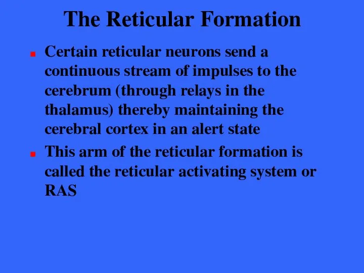
The Reticular Formation
Certain reticular neurons send a continuous stream of impulses
The Reticular Formation
Certain reticular neurons send a continuous stream of impulses
The Reticular Activating System
The RAS synapses with all major ascending sensory
The Reticular Activating System
The RAS synapses with all major ascending sensory
Reticular Formation
The RAS also acts as a filter to dampen repetitive,
Reticular Formation
The RAS also acts as a filter to dampen repetitive,
The Reticular Activating System
The activity of the RAS is inhibited
The Reticular Activating System
The activity of the RAS is inhibited
The Reticular Formation
The reticular formation also has a motor component
Some if
The Reticular Formation
The reticular formation also has a motor component
Some if
Protection of the Brain
Nervous tissue is soft and vulnerable
The brain is
Protection of the Brain
Nervous tissue is soft and vulnerable
The brain is
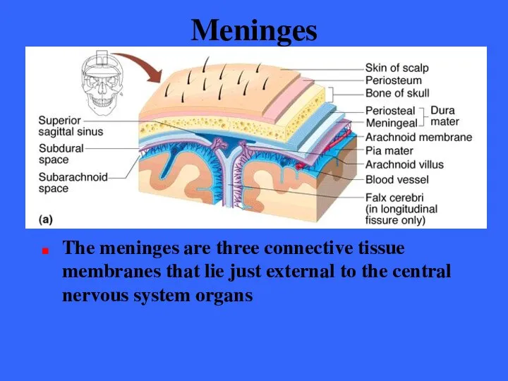
Meninges
The meninges are three connective tissue membranes that lie just external
Meninges
The meninges are three connective tissue membranes that lie just external
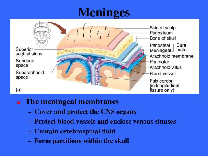
Meninges
The meningeal membranes
Cover and protect the CNS organs
Protect blood vessels
Meninges
The meningeal membranes
Cover and protect the CNS organs
Protect blood vessels

Meninges
The meninges are three connective tissue membranes that lie just external
Meninges
The meninges are three connective tissue membranes that lie just external
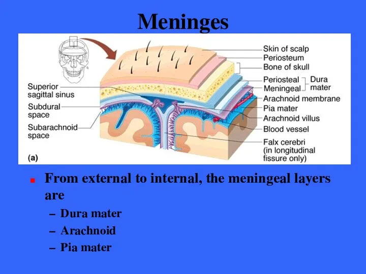
Meninges
From external to internal, the meningeal layers are
Dura mater
Arachnoid
Pia mater
Meninges
From external to internal, the meningeal layers are
Dura mater
Arachnoid
Pia mater
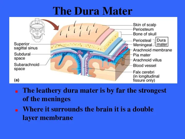
The Dura Mater
The leathery dura mater is by far the strongest
The Dura Mater
The leathery dura mater is by far the strongest
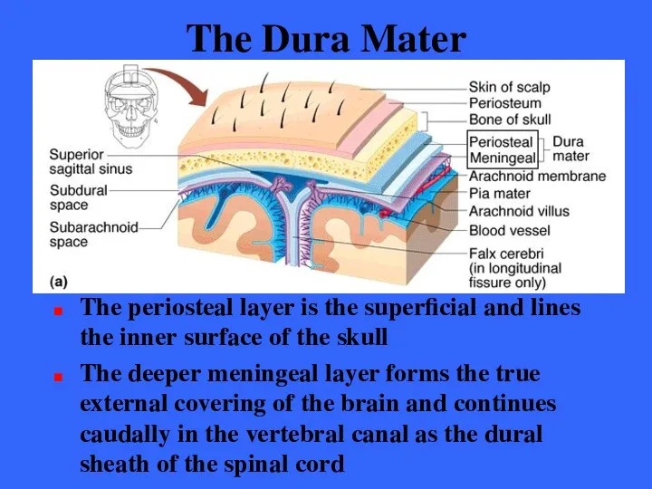
The Dura Mater
The periosteal layer is the superficial and lines the
The Dura Mater
The periosteal layer is the superficial and lines the
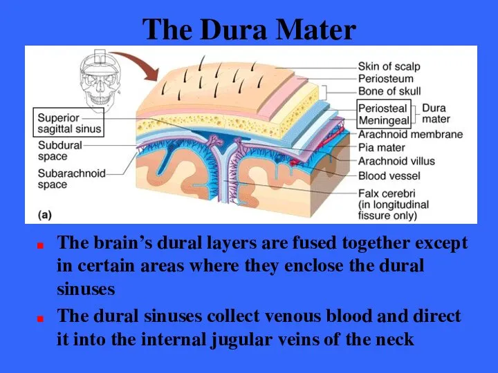
The Dura Mater
The brain’s dural layers are fused together except in
The Dura Mater
The brain’s dural layers are fused together except in
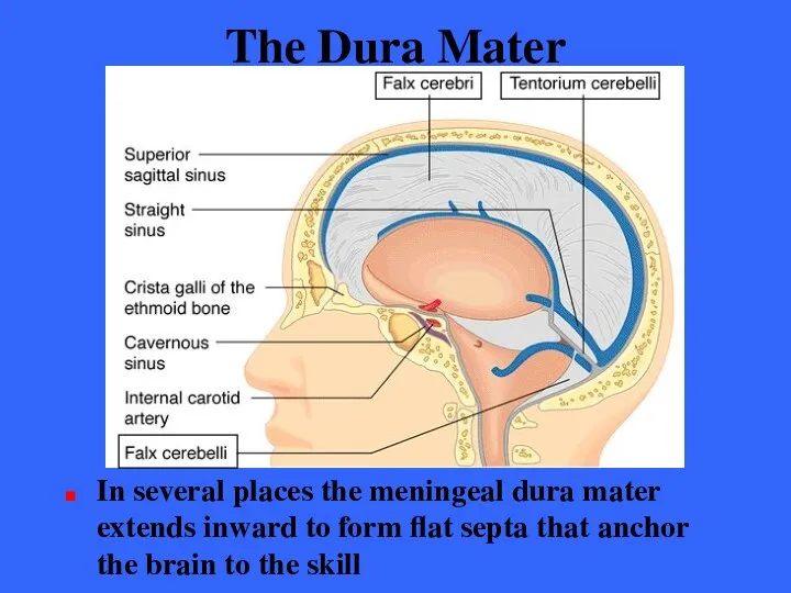
The Dura Mater
In several places the meningeal dura mater extends inward
The Dura Mater
In several places the meningeal dura mater extends inward
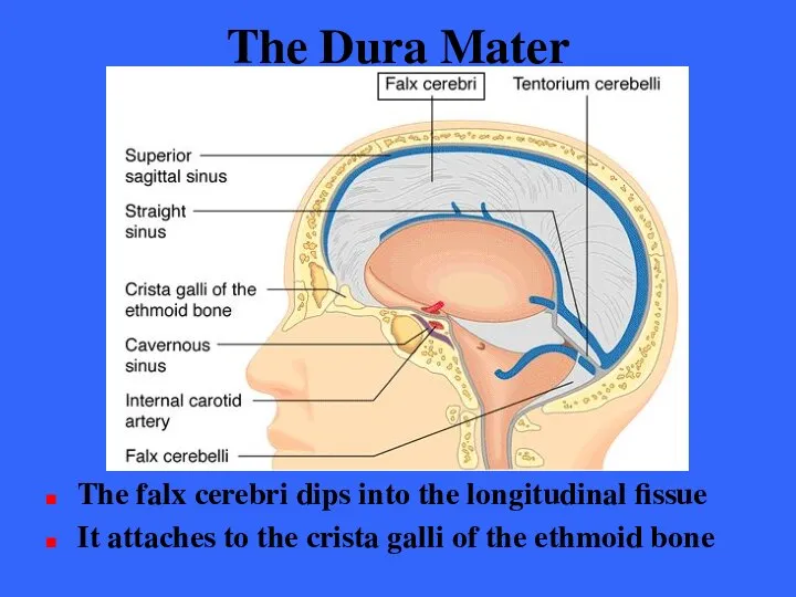
The Dura Mater
The falx cerebri dips into the longitudinal fissue
It attaches
The Dura Mater
The falx cerebri dips into the longitudinal fissue
It attaches
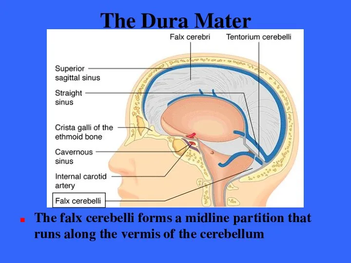
The Dura Mater
The falx cerebelli forms a midline partition that runs
The Dura Mater
The falx cerebelli forms a midline partition that runs
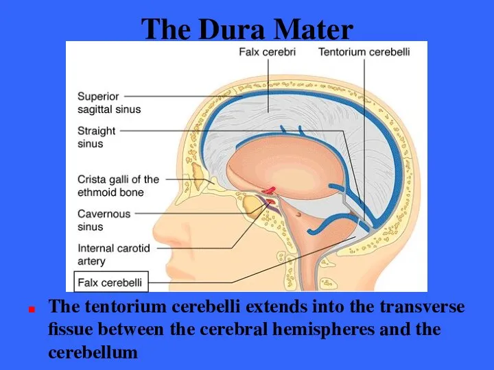
The Dura Mater
The tentorium cerebelli extends into the transverse fissue between
The Dura Mater
The tentorium cerebelli extends into the transverse fissue between
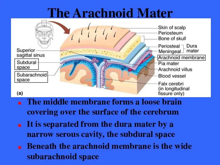
The Arachnoid Mater
The middle membrane forms a loose brain covering over
The Arachnoid Mater
The middle membrane forms a loose brain covering over
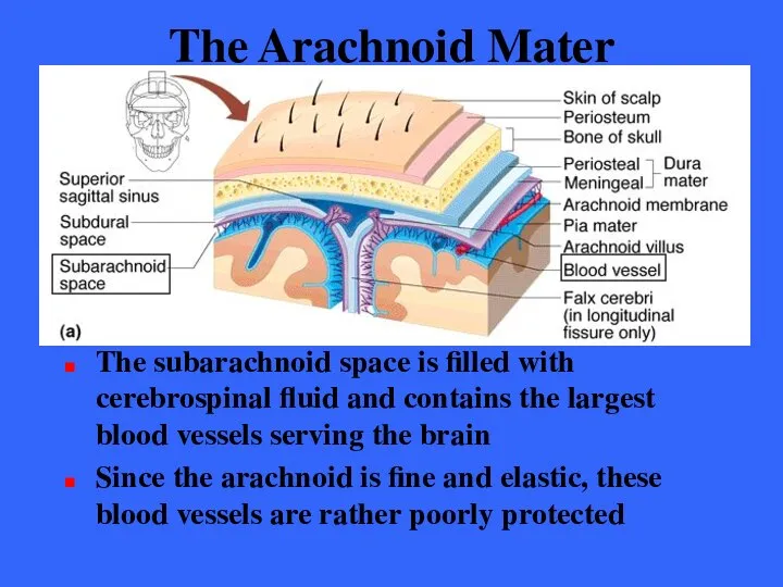
The Arachnoid Mater
The subarachnoid space is filled with cerebrospinal fluid and
The Arachnoid Mater
The subarachnoid space is filled with cerebrospinal fluid and
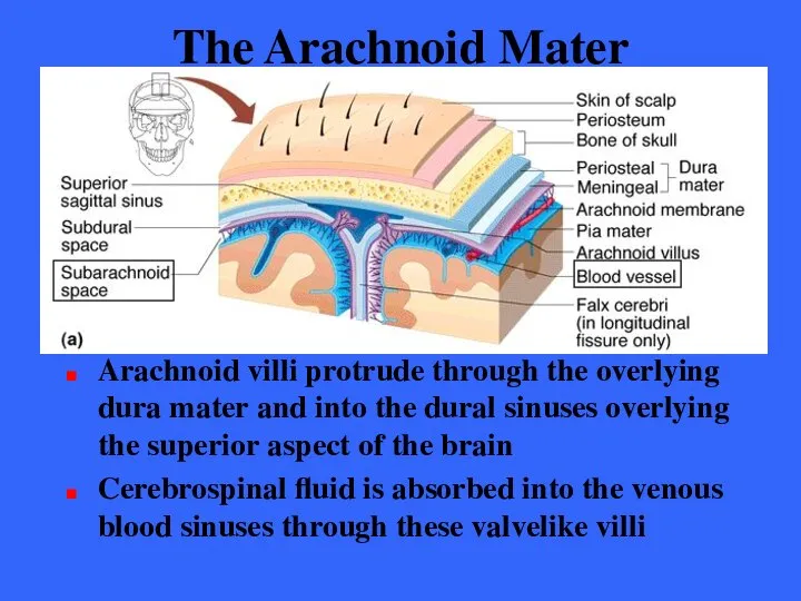
The Arachnoid Mater
Arachnoid villi protrude through the overlying dura mater and
The Arachnoid Mater
Arachnoid villi protrude through the overlying dura mater and
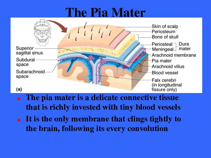
The Pia Mater
The pia mater is a delicate connective tissue that
The Pia Mater
The pia mater is a delicate connective tissue that
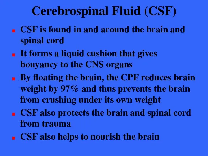
Cerebrospinal Fluid (CSF)
CSF is found in and around the brain and
Cerebrospinal Fluid (CSF)
CSF is found in and around the brain and
Cerebrospinal Fluid (CSF)
CSF is a similar in composition to blood plasma,
Cerebrospinal Fluid (CSF)
CSF is a similar in composition to blood plasma,
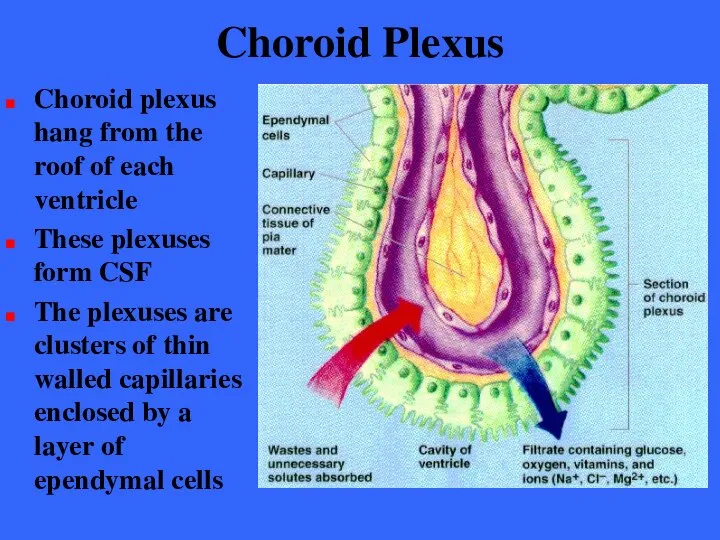
Choroid Plexus
Choroid plexus hang from the roof of each ventricle
These plexuses
Choroid Plexus
Choroid plexus hang from the roof of each ventricle
These plexuses
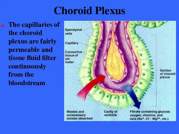
Choroid Plexus
The capillaries of the choroid plexus are fairly permeable and
Choroid Plexus
The capillaries of the choroid plexus are fairly permeable and
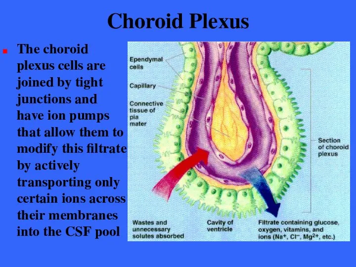
Choroid Plexus
The choroid plexus cells are joined by tight junctions and
Choroid Plexus
The choroid plexus cells are joined by tight junctions and
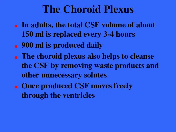
The Choroid Plexus
In adults, the total CSF volume of about 150
The Choroid Plexus
In adults, the total CSF volume of about 150
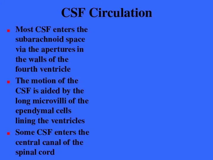
CSF Circulation
Most CSF enters the subarachnoid space via the apertures in
CSF Circulation
Most CSF enters the subarachnoid space via the apertures in
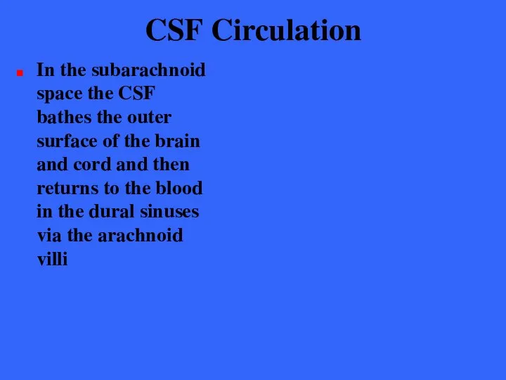
CSF Circulation
In the subarachnoid space the CSF bathes the outer surface
CSF Circulation
In the subarachnoid space the CSF bathes the outer surface
Blood-Brain Barrier
The barrier is a protective mechanism that helps maintain a
Blood-Brain Barrier
The barrier is a protective mechanism that helps maintain a
Blood-Brain Barrier
Bloodborne substances within the brain’s capillaries are separated from the
Blood-Brain Barrier
Bloodborne substances within the brain’s capillaries are separated from the
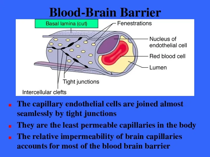
Blood-Brain Barrier
The capillary endothelial cells are joined almost seamlessly by tight
Blood-Brain Barrier
The capillary endothelial cells are joined almost seamlessly by tight
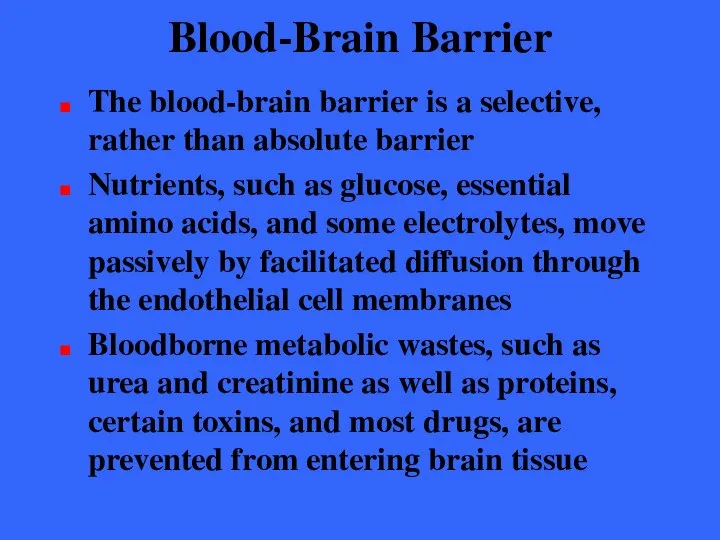
Blood-Brain Barrier
The blood-brain barrier is a selective, rather than absolute barrier
Nutrients,
Blood-Brain Barrier
The blood-brain barrier is a selective, rather than absolute barrier
Nutrients,
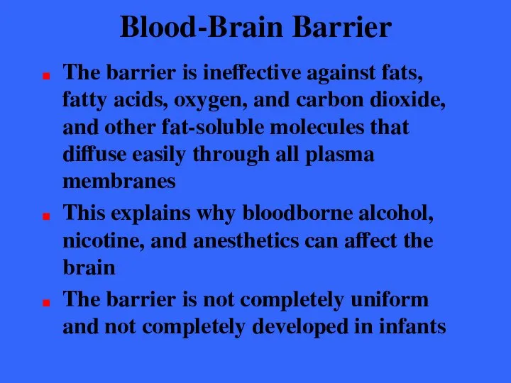
Blood-Brain Barrier
The barrier is ineffective against fats, fatty acids, oxygen, and
Blood-Brain Barrier
The barrier is ineffective against fats, fatty acids, oxygen, and
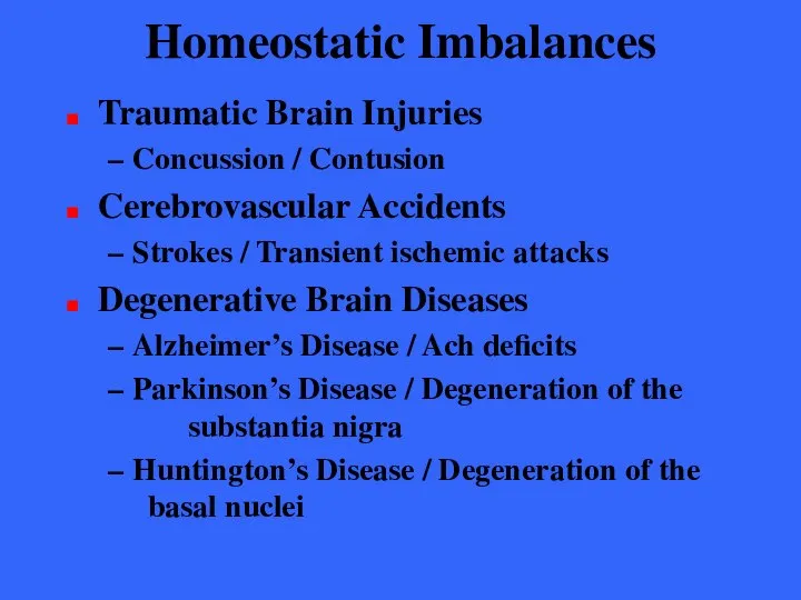
Homeostatic Imbalances
Traumatic Brain Injuries
Concussion / Contusion
Cerebrovascular Accidents
Strokes / Transient ischemic
Homeostatic Imbalances
Traumatic Brain Injuries
Concussion / Contusion
Cerebrovascular Accidents
Strokes / Transient ischemic
 Анализ и управление требованиями. Документирование. Контроль. (Часть 4)
 Формирование навыков чтения у младших школьников Абзалутдинова Маргарита Инсафовна учитель начальных классов МОУ «СОШ №60» г
 Шаблоны информационных технологий как механизм реализации стратегии компании Николай Михайловский NTR Lab, Генеральный директ
 Сканирующий тахеометр Trimble SX10
 ІНТЕЛЕКТУАЛЬНА ВЛАl
  Презентация
 Конфигурациялық тұрақты сақтау құрылғысы
 «Здоровьесберегающая среда классной комнаты» Выполнили ученики и родители 7 – А класса МОУ СОШ №1 Г. Покров (помогали ученики
  Системный анализ: его роль в становлении теории политической системы. Модель политической системы Истона Подготовила Ермакова
  учитель: Громова С.А. МСОШ № 2 имени Е.В.Камышева город Гагарин
 Ингибирование ферментативной активности
 Презентация на тему "Центр Интеллектуального развития "ЮВЕНТА"" - скачать презентации по Педагогике
 Анализ радиальной и латеральной структуры ландшафтов
 Презентация по МХК Авангардное искусство
  Вернакулярные ландшафты Джексона
 Людина і спорт
 «Иуда Искариот» Л. Андреева и Евангельский сюжет
 Производство домиков для котов, когтеточек, комплексов из дерева
 Диоды Ганна
 Точность деталей сборочных единиц изделий
 Презентация на тему "Выпуски школьной газеты за 2006-2007г." - скачать презентации по Педагогике
 JavaScript. Основы программирования
 Презентация "Кацусика Хокусай "36 видов фудзи"" - скачать презентации по МХК
 Основное назначение библиoтeки jQuery
 Новогодние обычаи и традиции
 Культура города Казани
 Виды наследования
 Презентация Microsoft Office PowerPoint