- Dermatology. Basal Cell Carcinoma (BCC)

Содержание
- 2. 3.001
- 3. Basal Cell Carcinoma (BCC) Age: usually >35 years More frequent in males Mostly on sun-exposed areas:
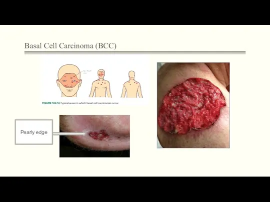
- 4. Basal Cell Carcinoma (BCC) Pearly edge
- 5. Implantation dermoid cysts as the result of implantation of epidermal fragments into the dermis by a
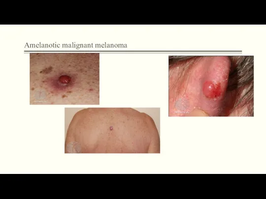
- 6. Amelanotic malignant melanoma Amelanotic melanoma is a form of melanoma The malignant cells have little to
- 7. Amelanotic malignant melanoma
- 8. External angular dermoids Looks like subcutaneous lumps at the lateral angle of the eye
- 9. Keratoacanthoma Tumour of keratinocytes Occur singly on light-exposed areas Raised crater with central keratin plug Grows
- 10. 3.002
- 11. Malignant melanoma
- 12. Melanoma Typical age range 30–50 years (average 40 years) Can occur anywhere on the body—more common:
- 13. Melanoma Prognosis thickness (Breslow classification) level or depth (worse in level IV or V) site (worse
- 14. Management points for naevi and melanomas
- 15. Neuropathic ulcer A neuropathic ulcer is one that occurs as a result of peripheral neuropathy Neuropathic
- 16. Diabetic foot ulcer A feature is a deep, punched-out lesion over pressure points. The ulcers may
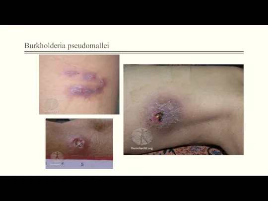
- 17. Burkholderia pseudomallei Melioidosis is an uncommon tropical disease caused by the bacterium, Burkholderia pseudomallei a soil
- 18. Burkholderia pseudomallei
- 19. Necrotising fasciitis Necrotising fasciitis is a very serious bacterial infection of the soft tissue and fascia
- 21. Скачать презентацию
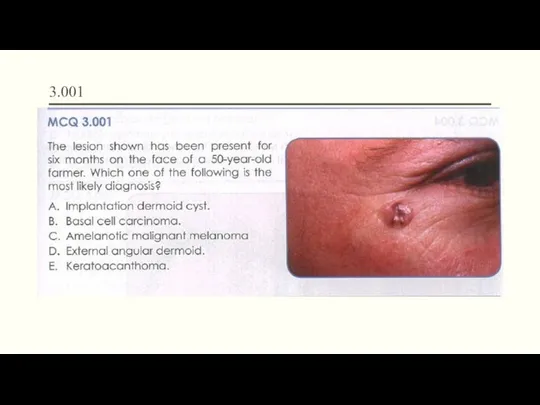
3.001
3.001
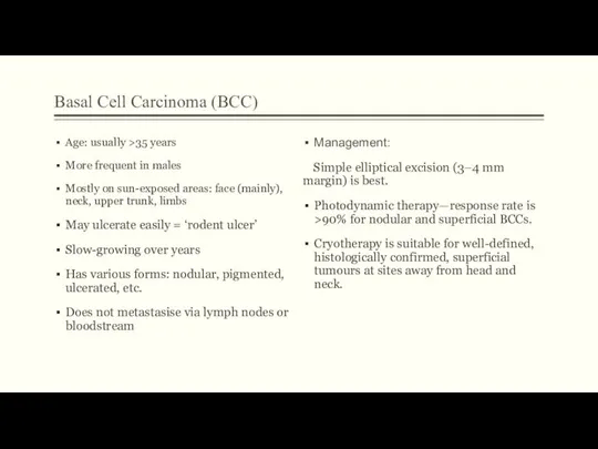
Basal Cell Carcinoma (BCC)
Age: usually >35 years
More frequent in males
Mostly on
Basal Cell Carcinoma (BCC)
Age: usually >35 years
More frequent in males
Mostly on
Basal Cell Carcinoma (BCC)
Pearly edge
Basal Cell Carcinoma (BCC)
Pearly edge
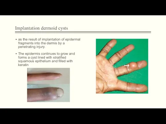
Implantation dermoid cysts
as the result of implantation of epidermal fragments into
Implantation dermoid cysts
as the result of implantation of epidermal fragments into
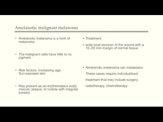
Amelanotic malignant melanoma
Amelanotic melanoma is a form of melanoma
The malignant cells
Amelanotic malignant melanoma
Amelanotic melanoma is a form of melanoma
The malignant cells
Amelanotic malignant melanoma
Amelanotic malignant melanoma
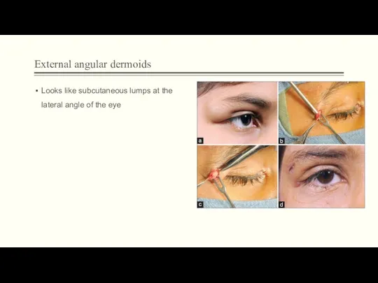
External angular dermoids
Looks like subcutaneous lumps at the lateral angle of
External angular dermoids
Looks like subcutaneous lumps at the lateral angle of
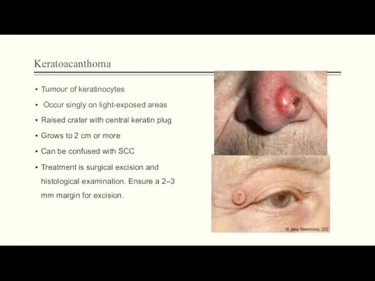
Keratoacanthoma
Tumour of keratinocytes
Occur singly on light-exposed areas
Raised crater with central
Keratoacanthoma
Tumour of keratinocytes
Occur singly on light-exposed areas
Raised crater with central
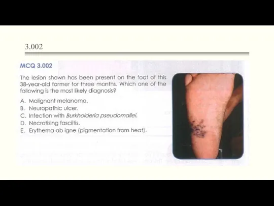
3.002
3.002
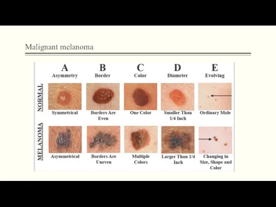
Malignant melanoma
Malignant melanoma
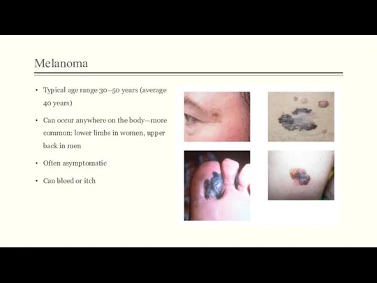
Melanoma
Typical age range 30–50 years (average 40 years)
Can occur anywhere on
Melanoma
Typical age range 30–50 years (average 40 years)
Can occur anywhere on
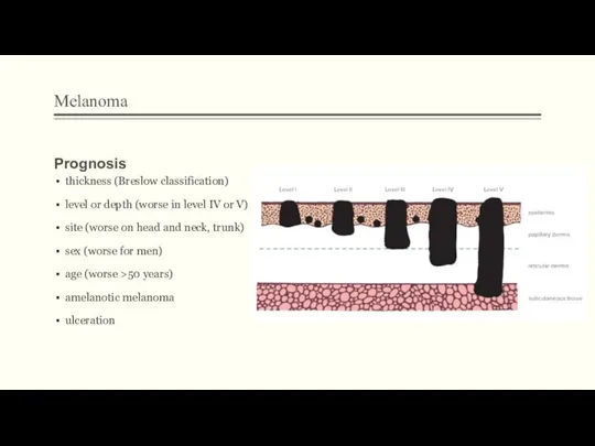
Melanoma
Prognosis
thickness (Breslow classification)
level or depth (worse in level IV or V)
Melanoma
Prognosis
thickness (Breslow classification)
level or depth (worse in level IV or V)
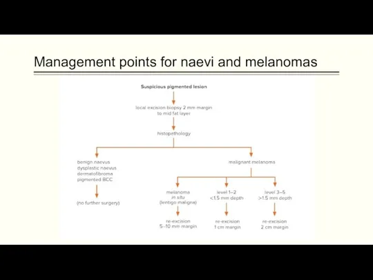
Management points for naevi and melanomas
Management points for naevi and melanomas
Neuropathic ulcer
A neuropathic ulcer is one that occurs as a result
Neuropathic ulcer
A neuropathic ulcer is one that occurs as a result
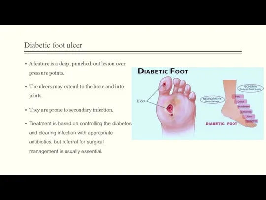
Diabetic foot ulcer
A feature is a deep, punched-out lesion over pressure
Diabetic foot ulcer
A feature is a deep, punched-out lesion over pressure
Burkholderia pseudomallei
Melioidosis is an uncommon tropical disease caused by the bacterium,
Burkholderia pseudomallei
Melioidosis is an uncommon tropical disease caused by the bacterium,
Burkholderia pseudomallei
Burkholderia pseudomallei
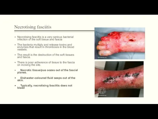
Necrotising fasciitis
Necrotising fasciitis is a very serious bacterial infection of the
Necrotising fasciitis
Necrotising fasciitis is a very serious bacterial infection of the
 Рефлюкстік гастро-эзофагальдІ синдром
 Порядок установления инвалидности и степени утраты трудоспособности
 Лекарственные растения и травы в ветеринарии
 Память. Процессы памяти. Виды памяти. Индивидуальные особенности памяти
 Фрейдизм
 Нарушения углеводного обмена
 Китайская медицина
 Ведение беременности и родов при узком тазе
 Медицинский туризм
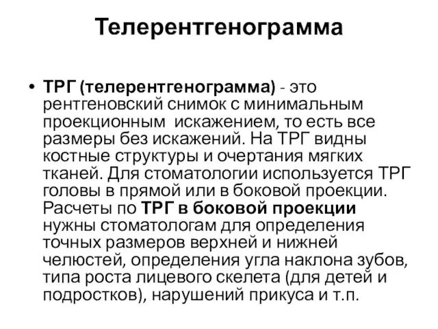 Телерентгенограмма. Виды ТРГ
 Косыночные повязки
 Клинико-психологическое сопровождение пожилых людей страдающих инволлюционным паранойдом
 Т-клеточные лимфомы кожи
 Введение. Общая рецептура
 Сепсис у новорожденных
 Личность. Структура личности
 Болезни женских половых органов и молочных желез
 Наследственные заболевания легких у детей
 Медикаментозная терапия высокорезистентной трофобластической опухоли
 Медицинская реабилитация при плоскостопии
 Оказания НП при травмах. Шины Крамера ( перелом костей голени)
 Балалар мен жасөспірімдердің әлеуметтік жағдайының дене дамуына әсері
 Заболевания органов дыхания. Доврачебная помощь при нарушениях дыхания
 Дифференциальная диагностика желтух у детей раннего возраста
 Внутрішньоутробні інфекції
 Остановка кровотечения жгутом
 Психиатрическое интервью
 Қазақстан республикасының денсаулық сақтау жүйесіне интеграцияланған қолданыстағы ақпараттық жүйелерді шолу. Фармацевтика