- Tuberculosis

Содержание
- 2. Tuberculosis is a chronic communicable disease with specific granulomatous inflammation caused by a variety of tubercle
- 3. Mycobacterium tuberculosis (95 %) M. bоvis (5 %)
- 4. Mode of transmission By inhalation into the respiratory tract. Ingestion. Through ingestion into GI tract leads
- 5. 5
- 6. Features of Primary Tuberculosis Development of disease at the first getting of the activator into the
- 7. PRIMARY COMPLEX OF TUBERCULOSIS "Ghon complex“ consists of Pulmonary component so called Primary affect or primary
- 8. There is small tan-yellow subpleural granuloma in the mid-lung field on the right (1). In the
- 9. It is 1-2 cm solitary area of caseous pneumonia surrounding by perifocal serous inflammation. A central
- 10. Primary tuberculosis of alimentary tract Tuberculous mesenterial lymphadenitis
- 11. Calcified pulmonary lymph nodes in tuberculosis Calcification
- 12. Hematogenous generalization of Primary Tuberculosis
- 13. Classifications of hematogenous tuberculosis Generalized hematogenous tuberculosis: а) The most acute tubercular sepsis. b) Acute general
- 14. On closer inspection, the granulomas have areas of caseous necrosis with formation of the small cavernes.
- 15. Miliary pulmonary tuberculosis The focal nature of granulomatous inflammation is demonstrated in this microscopic section of
- 16. Miliary tuberculosis of the spleen 41
- 17. RENAL TUBERCULOSIS 39
- 18. MILIARY TUBERCULOSIS IN LIVER 40
- 19. Forms or stages of the secondary tuberculosis: 1.Acute local tuberculosis. 2.Fibrous-local tuberculosis. 3.Infiltrative tuberculosis. 4.Tuberculoma. 5.Caseous
- 20. Acute local tuberculosis There are several 1 cm diameter, partially calcified foci (dry, crumbly, and white)
- 21. Fibrous-local tuberculosis
- 22. Infiltrative tuberculosis
- 23. Tuberculoma
- 24. Caseous pneumonia
- 25. Greyish-white wall of the cavity 2 to 3 mm thick Acute cavernous tuberculosis Wall of acute
- 26. Fibrous – cavernous tuberculosis
- 27. Fibrotic scar in the wall of tuberculous cavity consists of fibroblast, collagen, and scattered Langerhans giant
- 28. Cirrhotic tuberculosis
- 29. Complications and causes of death Scarring and calcification. Pneumothorax. Empyema. Pleural fibrosis and adhesions, with associated
- 30. Pleural fibrosis and adhesions, with associated pleurisy, sharp pleuritic pain, and shortness of breath. Pneumothorax
- 31. Acute hemorrhage due to erosion of vessels.
- 33. Скачать презентацию
Tuberculosis is a chronic communicable disease with specific granulomatous inflammation caused
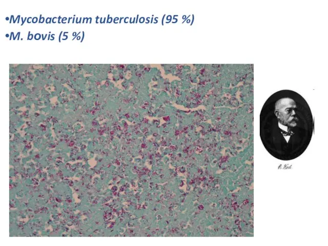
Mycobacterium tuberculosis (95 %)
M. bоvis (5 %)
Mycobacterium tuberculosis (95 %)
M. bоvis (5 %)
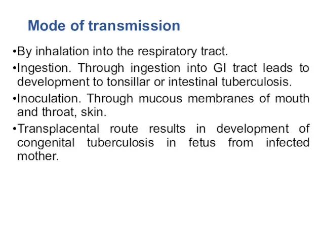
Mode of transmission
By inhalation into the respiratory tract.
Ingestion. Through ingestion into
Mode of transmission
By inhalation into the respiratory tract.
Ingestion. Through ingestion into
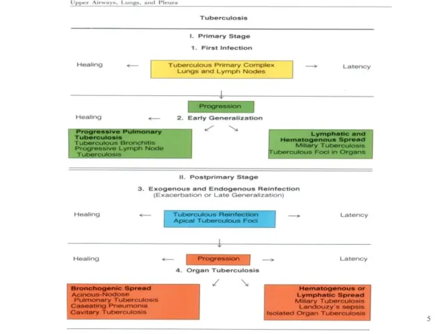
5
5
Features of Primary Tuberculosis
Development of disease at the first getting of
Features of Primary Tuberculosis
Development of disease at the first getting of
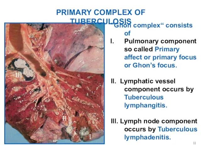
PRIMARY COMPLEX OF TUBERCULOSIS
"Ghon complex“ consists of
Pulmonary component so called Primary
PRIMARY COMPLEX OF TUBERCULOSIS
"Ghon complex“ consists of
Pulmonary component so called Primary
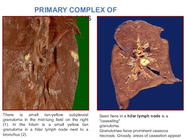
There is small tan-yellow subpleural granuloma in the mid-lung field on
There is small tan-yellow subpleural granuloma in the mid-lung field on
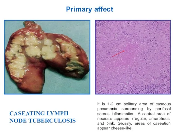
It is 1-2 cm solitary area of caseous pneumonia surrounding by
It is 1-2 cm solitary area of caseous pneumonia surrounding by
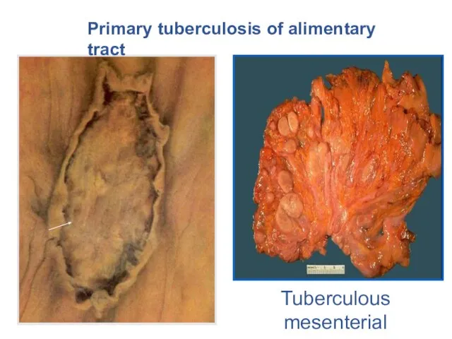
Primary tuberculosis of alimentary tract
Tuberculous mesenterial lymphadenitis
Primary tuberculosis of alimentary tract
Tuberculous mesenterial lymphadenitis
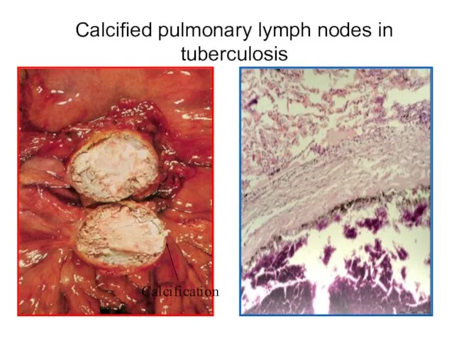
Calcified pulmonary lymph nodes in tuberculosis
Calcification
Calcified pulmonary lymph nodes in tuberculosis
Calcification

Hematogenous generalization of Primary Tuberculosis
Hematogenous generalization of Primary Tuberculosis
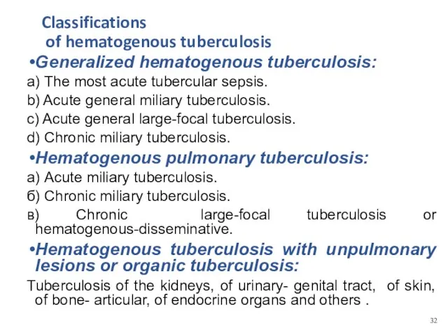
Classifications
of hematogenous tuberculosis
Generalized hematogenous tuberculosis:
а) The most acute tubercular
Classifications
of hematogenous tuberculosis
Generalized hematogenous tuberculosis:
а) The most acute tubercular
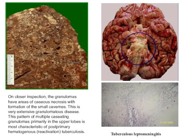
On closer inspection, the granulomas have areas of caseous necrosis with
On closer inspection, the granulomas have areas of caseous necrosis with
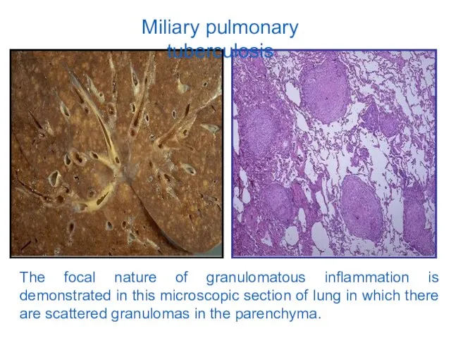
Miliary pulmonary tuberculosis
The focal nature of granulomatous inflammation is demonstrated in
Miliary pulmonary tuberculosis
The focal nature of granulomatous inflammation is demonstrated in
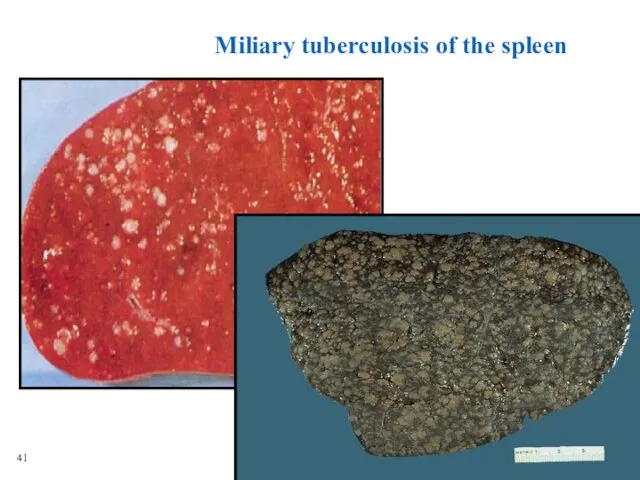
Miliary tuberculosis of the spleen
41
Miliary tuberculosis of the spleen
41
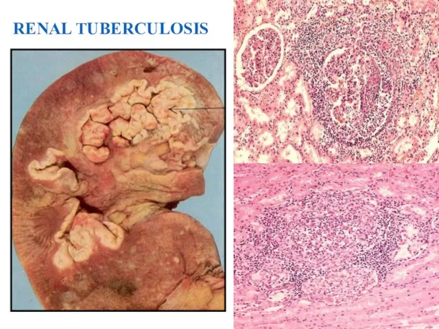
RENAL TUBERCULOSIS
39
RENAL TUBERCULOSIS
39
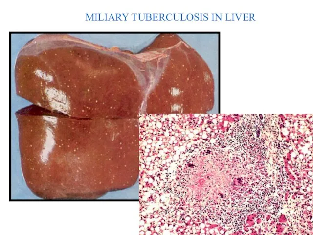
MILIARY TUBERCULOSIS IN LIVER
40
MILIARY TUBERCULOSIS IN LIVER
40
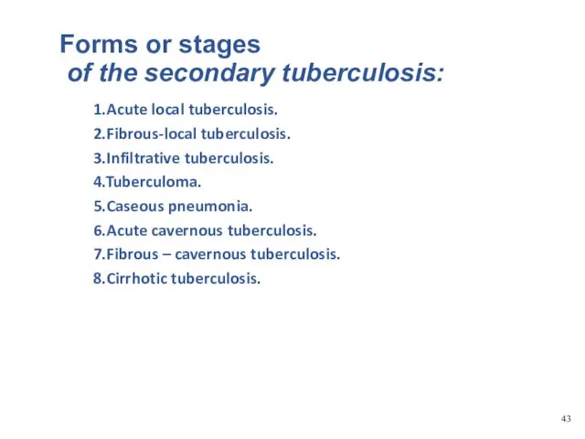
Forms or stages
of the secondary tuberculosis:
1.Acute local tuberculosis.
2.Fibrous-local tuberculosis.
3.Infiltrative tuberculosis.
4.Tuberculoma.
5.Caseous
Forms or stages
of the secondary tuberculosis:
1.Acute local tuberculosis.
2.Fibrous-local tuberculosis.
3.Infiltrative tuberculosis.
4.Tuberculoma.
5.Caseous
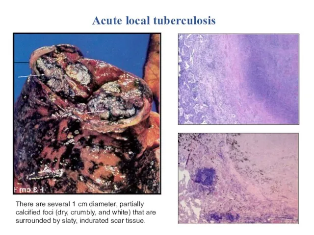
Acute local tuberculosis
There are several 1 cm diameter, partially calcified foci
Acute local tuberculosis
There are several 1 cm diameter, partially calcified foci
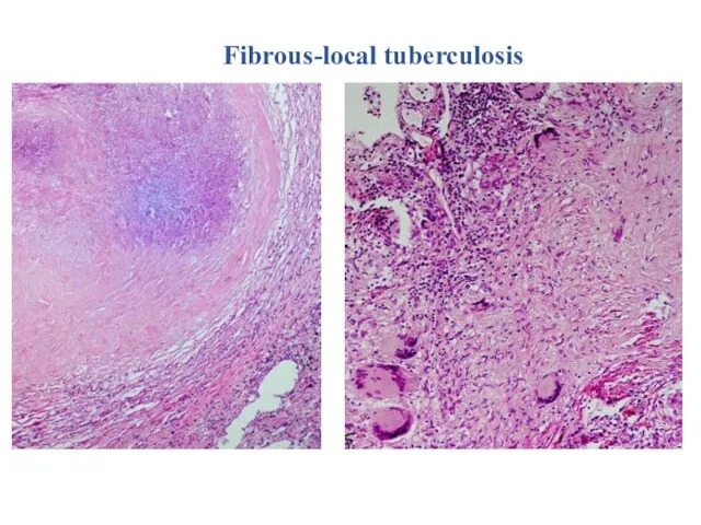
Fibrous-local tuberculosis
Fibrous-local tuberculosis
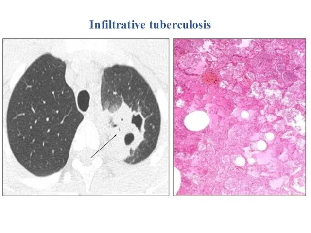
Infiltrative tuberculosis
Infiltrative tuberculosis
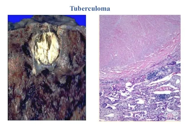
Tuberculoma
Tuberculoma
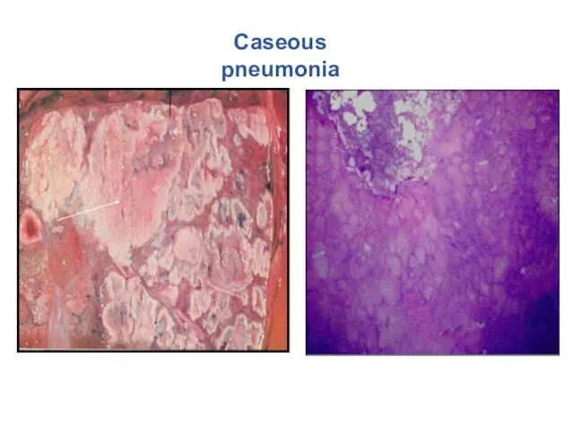
Caseous pneumonia
Caseous pneumonia
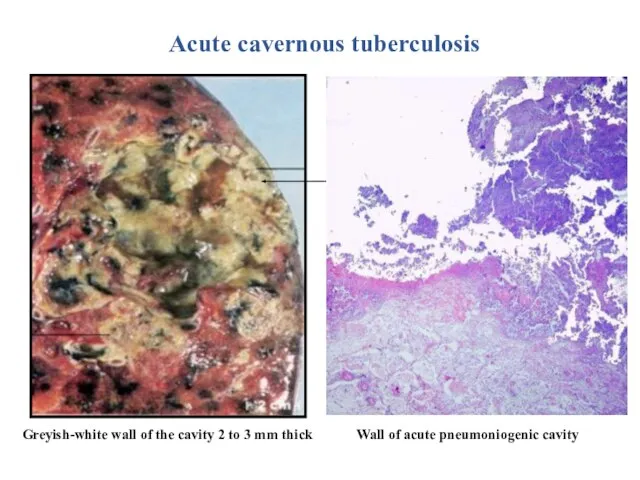
Greyish-white wall of the cavity 2 to 3 mm thick
Acute
Greyish-white wall of the cavity 2 to 3 mm thick
Acute
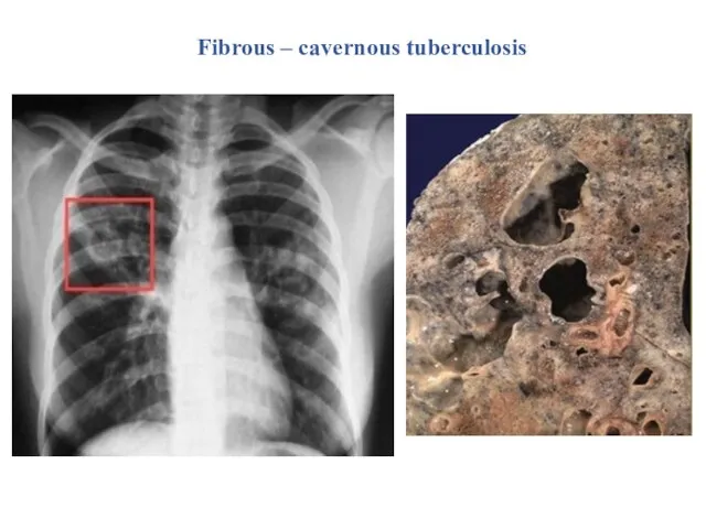
Fibrous – cavernous tuberculosis
Fibrous – cavernous tuberculosis
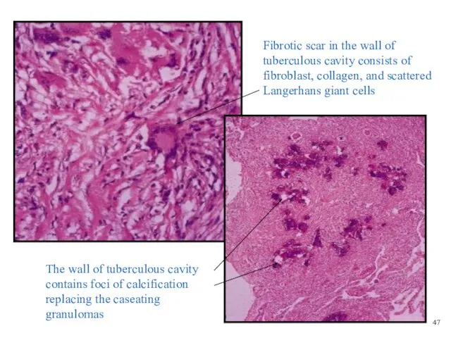
Fibrotic scar in the wall of tuberculous cavity consists of fibroblast,
Fibrotic scar in the wall of tuberculous cavity consists of fibroblast,
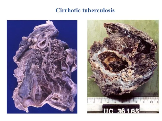
Cirrhotic tuberculosis
Cirrhotic tuberculosis
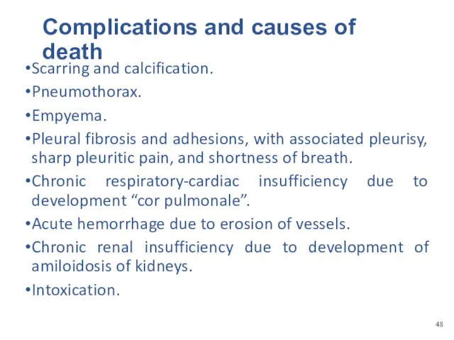
Complications and causes of death
Scarring and calcification.
Pneumothorax.
Empyema.
Pleural fibrosis and adhesions, with
Complications and causes of death
Scarring and calcification.
Pneumothorax.
Empyema.
Pleural fibrosis and adhesions, with
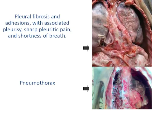
Pleural fibrosis and adhesions, with associated pleurisy, sharp pleuritic pain, and
Pleural fibrosis and adhesions, with associated pleurisy, sharp pleuritic pain, and
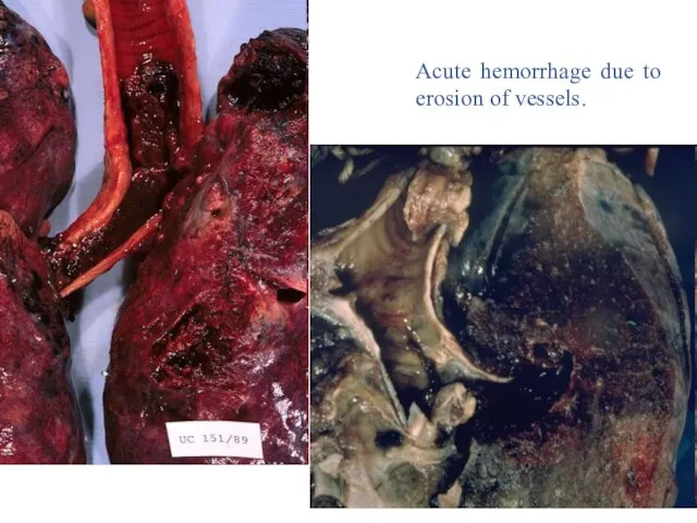
Acute hemorrhage due to erosion of vessels.
Acute hemorrhage due to erosion of vessels.
 Внутренняя среда организм. Кровь и её состав
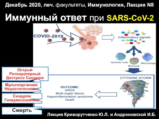 Иммунный ответ при SARS-CoV-2
 Национальные особенности деловой культуры европейских стран
 Неэпителиальные опухоли яичников
 Лучевые методы исследования желудочно-кишечного тракта
 Увеиты
 Клинические задачи
 ЛФК при заболеваниях ОДА
 Физиология и диагностика беременности. Влияние вредных факторов на плод
 Пародонт ауруларының жалпы емі. Фармакотерапия
 Организация и структура системы первичной медико-санитарной помощи
 Рубцы
Рубцы Многообразие противоинфекционных препаратов
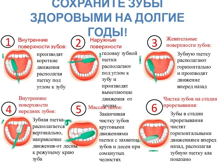 Сохраните зубы здоровыми на долгие годы!
 Лихорадка
 Bayer HealthCare
 Методологические принципы современной психологии. Тема № 2
 Новые стандарты онкологической помощи по ОМС
 Медицина в Канаде
 Гипоксия (кислородное голодание)
 Общие правила транспортировки пострадавшего
 Постреанимационная болезнь
 Головная боль после пункции твердой мозговой оболочки
 Первичная профилактика патологического старения
 Кабинет педагога-психолога
 Амбулаторлы-поликлиникалық қызмет немесе бірншілік медициналық санитарлық көмек жұмыстарын ұйымдастыру қағидалары
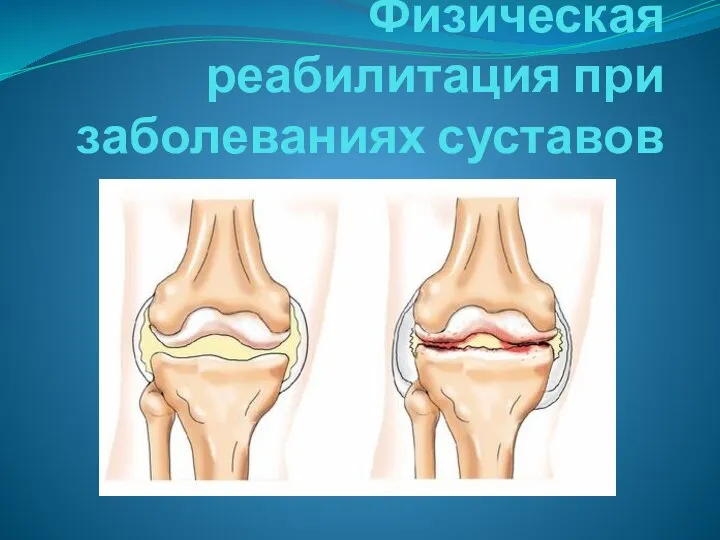 Физическая реабилитация при заболеваниях суставов
 Созылмалы бүйрек ауруларының үдеу механизмі