- Chapter 23 - Part 1 Lecture Outline

Содержание
- 2. The Respiratory System Respiration is gas exchange: O2 for CO2 Occurs between atmosphere and body cells
- 3. Aerobic Cellular Respiration Oxygen Carbon Dioxide ATP
- 4. 23.1 Introduction to the Respiratory System State the functions of the respiratory system. Distinguish between the
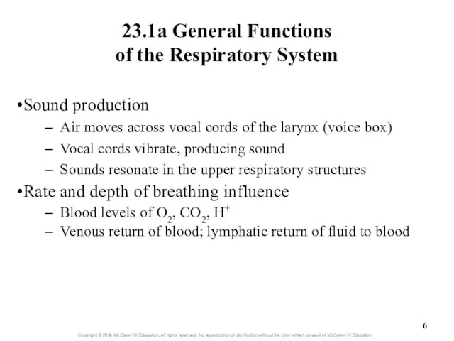
- 5. 23.1a General Functions of the Respiratory System Air passageway Air moves from atmosphere to alveoli as
- 6. 23.1a General Functions of the Respiratory System Sound production Air moves across vocal cords of the
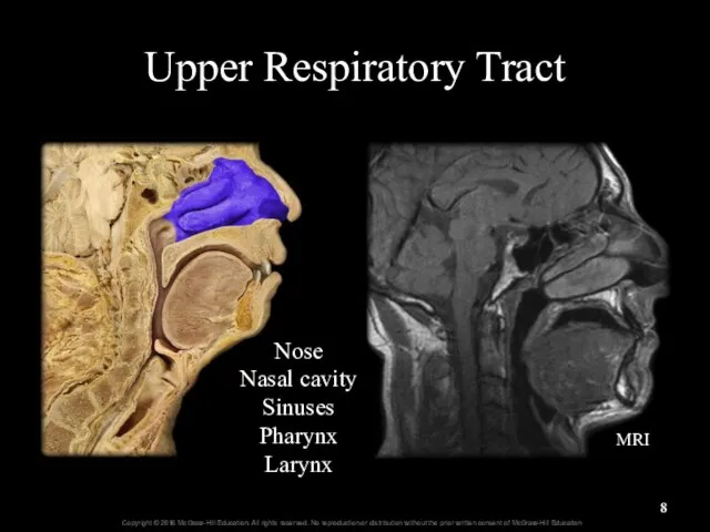
- 7. 23.1b General Organization of the Respiratory System Structural organization Upper respiratory tract Larynx and above Lower
- 8. Upper Respiratory Tract Nose Nasal cavity Sinuses Pharynx Larynx MRI
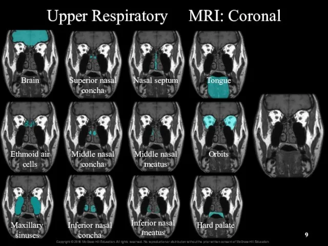
- 9. Upper Respiratory MRI: Coronal Brain Ethmoid air cells Maxillary sinuses Superior nasal concha Middle nasal concha
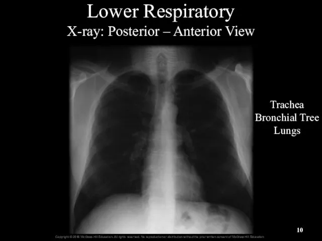
- 10. Lower Respiratory X-ray: Posterior – Anterior View Trachea Bronchial Tree Lungs
- 11. General Anatomy of the Respiratory System Figure 23.1
- 12. 23.1c Respiratory Mucosa Mucosa = mucous membrane: respiratory lining Epithelium resting on a basement membrane Underlying
- 13. Respiratory Mucosa Figure 23.2a
- 14. Respiratory Mucosa Figure 23.2b
- 15. Respiratory Epithelium High Magnification Respiratory epithelium Cilia Columnar epithelial cells Goblet cells Basal cells Basement membrane
- 16. 23.1c Respiratory Mucosa Mucous secretions Produced from secretions of Goblet cells of epithelial lining Mucous and
- 17. What did you learn? What is the difference between the conducting and respiratory zones? How does
- 18. 23.2 Upper Respiratory Tract Describe the structure and function of the nose. Provide a general description
- 19. 23.2a Nose and Nasal Cavity Nose: first conducting structure for inhaled air Formed by bone, hyaline
- 20. Upper Respiratory Tract Figure 23.3a,b
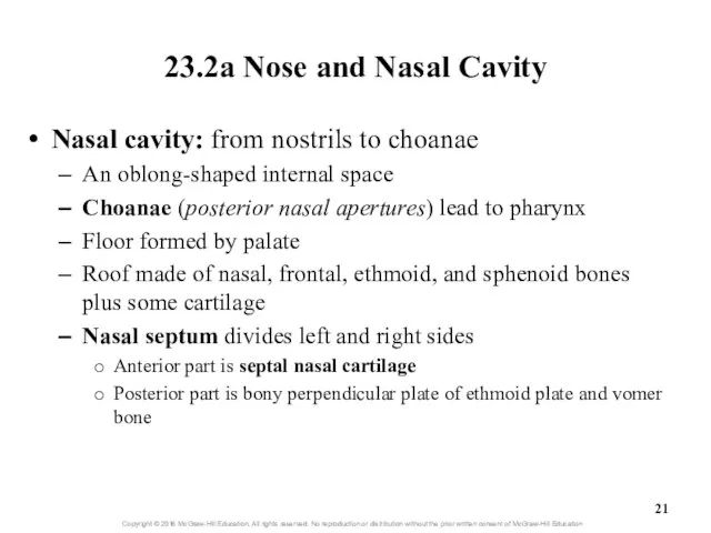
- 21. 23.2a Nose and Nasal Cavity Nasal cavity: from nostrils to choanae An oblong-shaped internal space Choanae
- 22. Nasal Cavity and Choanae Nares Nasal Cavity Nasal Septum Nasopharynx Soft Palate Uvula Choanae Hard Palate
- 23. Nasal Septum Perpendicular plate of ethmoid Vomer
- 24. Nasal Septum Vomer Perpendicular plate Septal nasal cartilage
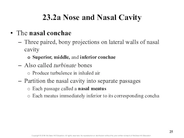
- 25. 23.2a Nose and Nasal Cavity The nasal conchae Three paired, bony projections on lateral walls of
- 26. Nasal vestibule Choana Nasal septum Hard palate Nares Nose Superior nasal concha Middle nasal concha Inferior
- 27. Nasal Conchae-MRI Superior Middle Inferior Septum
- 28. 23.2a Nose and Nasal Cavity Nasal cavity parts Nasal vestibule: just inside nostrils Lined by skin
- 29. Olfactory Region 16- Olfactory tract Olfactory bulb Olfactory nerves Olfactory mucosa
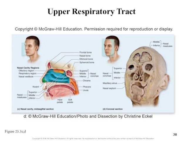
- 30. Upper Respiratory Tract Figure 23.3c,d
- 31. 23.2a Nose and Nasal Cavity Nasolacrimal ducts Drain lacrimal secretions from eye surfaces to nasal cavity
- 32. Clinical View: Runny Nose Rhinorrhea (runny nose) occurs as a result of Increased production of mucus
- 33. 23.2b Paranasal Sinuses Paranasal sinuses: spaces within skull bones Named for specific bone in which they
- 34. Maxillary Frontal Ethmoid Sphenoid Paranasal Sinuses
- 35. 23.2b Paranasal Sinuses Lined by pseudostratified ciliated columnar epithelium Mucus swept into pharynx and swallowed Figure
- 36. Paranasal sinuses
- 37. Clinical View: Sinus Infections and Sinus Headaches Respiratory infection or allergy can cause inflammation of the
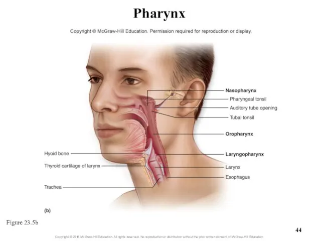
- 38. 23.2c Pharynx Pharynx (throat) Funnel-shaped passageway posterior to nasal cavity, oral cavity, and larynx Lateral walls
- 39. Nasopharynx Oropharynx Laryngopharynx Pharynx (throat)
- 40. Pharynx Oropharynx Nasopharynx Laryngopharynx
- 41. 23.2c Pharynx Nasopharynx: most superior part of pharynx Posterior to nasal cavity, superior to soft palate
- 42. 23.2c Pharynx Oropharynx: middle pharyngeal region Posterior to oral cavity Extends from soft palate to hyoid
- 43. 23.2c Pharynx Laryngopharynx: inferior, narrow region of pharynx Posterior to the larynx From level of hyoid
- 44. Pharynx Figure 23.5b
- 45. What did you learn? What are vibrissae? Between which conchae is the middle nasal meatus located?
- 46. 23.3 Lower Respiratory Tract Describe the general functions and structure of the larynx. Explain how the
- 47. 23.3 Lower Respiratory Tract (continued) Explain the processes of bronchoconstriction and bronchodilation. Describe the structure and
- 48. 23.3 Lower Respiratory Tract Includes conducting pathways from larynx to terminal bronchioles Includes structures involved in
- 49. 23.3a Larynx Larynx (voice box) Cylindrical airway between laryngopharynx and trachea Several functions Air passageway (usually
- 50. 22.3a Larynx Several functions (continued) Participates in sneeze and cough reflexes Help remove irritants from nasal
- 51. Larynx Epiglottis Vocal cords
- 52. Larynx
- 53. 23.3a Larynx Larynx anatomy Laryngeal inlet (laryngeal aperture) connects pharynx and larynx Larynx formed and supported
- 54. 23.3a Larynx Larynx anatomy (continued) Thyroid cartilage: large, shield-shaped Forms lateral and anterior walls of larynx
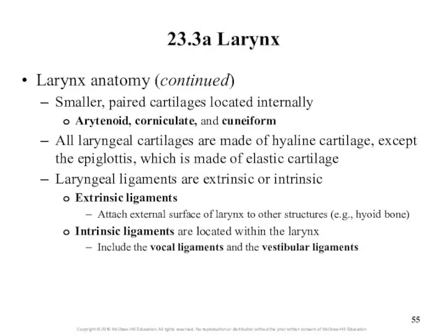
- 55. 23.3a Larynx Larynx anatomy (continued) Smaller, paired cartilages located internally Arytenoid, corniculate, and cuneiform All laryngeal
- 56. Larynx - Anterior Thyroid cartilage Cricoid cartilage Epiglottis Circothyroid ligament Thyrohyoid membrane
- 57. Larynx - Posterior Laryngeal cartilages Epiglottis Thyroid Cricoid Arytenoid Corniculate
- 58. Larynx Figure 23.6
- 59. Larynx - Lateral Epiglottis Thyroid cartilage Vestibular fold Vocal fold
- 60. 23.3a Larynx Larynx anatomy: ligaments (continued) Vocal ligaments extend between thyroid and arytenoid cartilages Composed primarily
- 61. Vocal Folds Figure 23.7a
- 62. Vocal Folds Figure 23.7b
- 63. 23.3a Larynx Larynx anatomy (continued) Extrinsic skeletal muscles Stabilize larynx and help it move during swallowing
- 64. 23.3a Larynx Sound production: vocal cord vibration Intrinsic laryngeal muscles narrow opening of rima glottidis Air
- 65. 23.3a Larynx Sound production (continued) Other structures are also necessary for speech Pharynx, nasal and oral
- 66. Clinical View: Laryngitis Inflammation of the larynx Symptoms of hoarse voice, sore throat, sometimes fever Caused
- 67. 23.3b Trachea Gross anatomy of trachea (windpipe) Flexible, slightly rigid, tubular organ Goes from larynx to
- 68. 23.3b Trachea Gross anatomy of the trachea (continued) Carina: internal ridge at inferior end of trachea
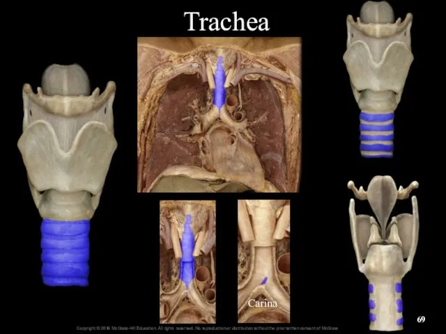
- 69. Trachea Carina
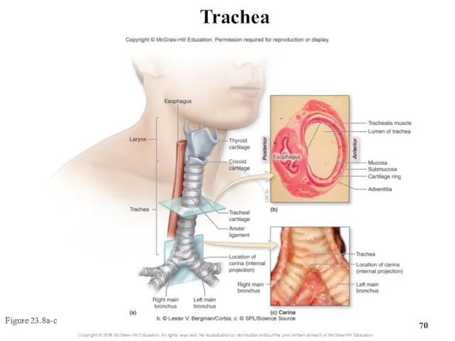
- 70. Trachea Figure 23.8a-c
- 71. 23.3b Trachea Histology of the tracheal wall Layers, inner to outer Mucosa: pseudostratified ciliated columnar epithelium
- 72. Trachea Low Magnification Epithelium Lamina propria Submucosa Perichondrium Cartilage
- 73. Trachea High Magnification Tracheal epithelium Cilia Ciliated cells Goblet cells Nuclei of basal cells Basement membrane
- 74. 23.3c Bronchial Tree Bronchial tree: system of highly branched air passages Originates at main bronchi, branches
- 75. Chest X-ray and Bronchogram
- 76. Trachea and Main (Primary) Bronchi
- 77. 23.3c Bronchial Tree Gross anatomy of the bronchial tree (continued) Each main bronchus branches into lobar
- 78. Bronchial Tree Figure 23.9
- 79. Clinical View: Bronchitis Inflammation of the bronchi caused by bacterial or viral infection or inhaled irritants
- 80. 23.3c Bronchial Tree Histology of the bronchial tree Main bronchi are supported by incomplete rings of
- 81. Structure of the Bronchial Wall Figure 23.10
- 82. Clinical View: Asthma Episodes of bronchoconstriction, wheezing, coughing, shortness of breath, and excess mucus Asthmatic with
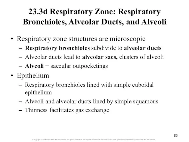
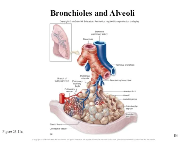
- 83. 23.3d Respiratory Zone: Respiratory Bronchioles, Alveolar Ducts, and Alveoli Respiratory zone structures are microscopic Respiratory bronchioles
- 84. Bronchioles and Alveoli Figure 23.11a
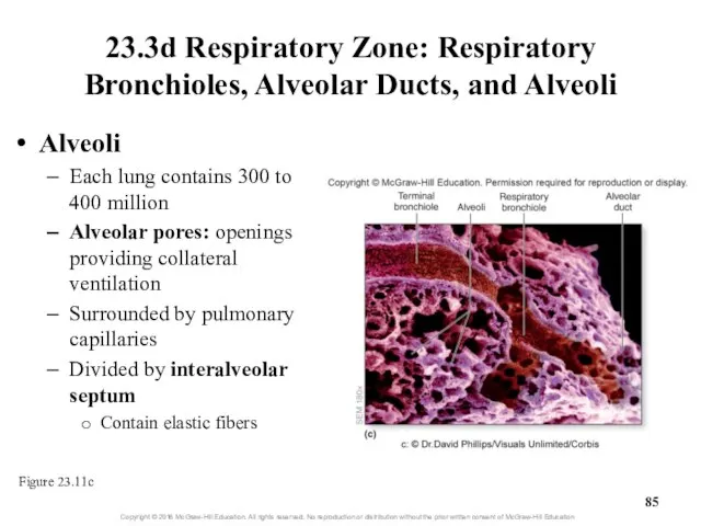
- 85. 23.3d Respiratory Zone: Respiratory Bronchioles, Alveolar Ducts, and Alveoli Alveoli Each lung contains 300 to 400
- 86. 23.3d Respiratory Zone: Respiratory Bronchioles, Alveolar Ducts, and Alveoli Cell types of alveolar wall Simple squamous
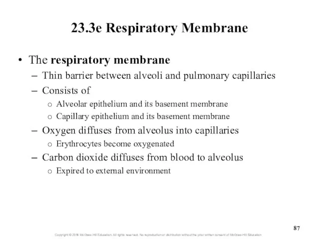
- 87. 23.3e Respiratory Membrane The respiratory membrane Thin barrier between alveoli and pulmonary capillaries Consists of Alveolar
- 88. Alveoli and Respiratory Membrane Figure 23.12
- 89. What did you learn? What makes one speech sound have a higher pitch than another? How
- 90. 23.4 Lungs Describe the location and general structure of the lungs. Compare and contrast the right
- 91. 23.4 Lungs (continued) Describe the pleural membranes and pleural cavity. Explain the function of serous fluid
- 92. 23.4a Gross Anatomy of the Lung Lungs are in thorax on either side of mediastinum House
- 93. Rt. and Lt. Lungs
- 94. Lungs
- 95. Chest X-ray and Bronchogram
- 96. 23.4a Gross Anatomy of the Lung Lung surfaces Costal surface adjacent to ribs Mediastinal surface adjacent
- 97. Position of the Lungs Figure 23.13
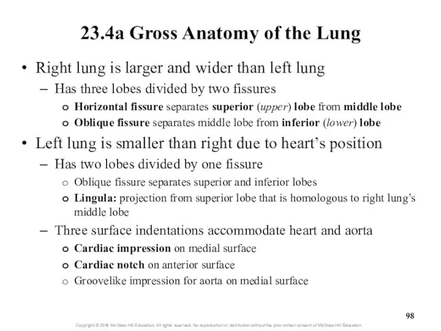
- 98. 23.4a Gross Anatomy of the Lung Right lung is larger and wider than left lung Has
- 99. Right Lung Superior lobe Middle lobe Inferior lobe Horizontal fissure Oblique fissure
- 100. Left Lung Oblique fissure Superior lobe Inferior lobe
- 101. Cardiac Impression
- 102. Figure 23.14 Lungs
- 103. 23.4a Gross Anatomy of the Lung Each lung has multiple bronchopulmonary segments 10 segments in right
- 104. Bronchopulmonary Segments and Lobules of the Lungs Figure 23.15
- 105. Clinical View: Smoking Causes respiratory changes with increased chance of Respiratory infections Cellular or genetic changes
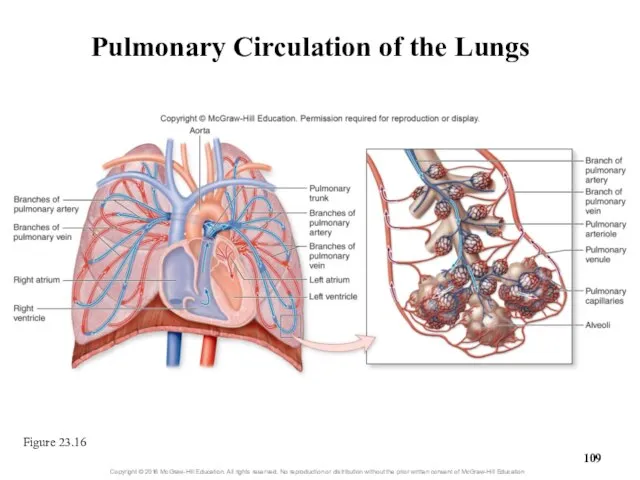
- 106. 23.4b Circulation to and Innervation of the Lungs Blood supply Two types of circulation in the
- 107. Pulmonary Circulation Pulmonary trunk Lt. Pulmonary artery Lt. Lung Lt. Pulmonary veins Rt. Pulmonary veins Lt.
- 108. 23.4b Circulation to and Innervation of the Lungs Blood supply (continued) Bronchial circulation transports oxygenated blood
- 109. Pulmonary Circulation of the Lungs Figure 23.16
- 110. Clinical Views: Lung Cancer Highly aggressive malignancy with early metastasis Originates in respiratory epithelium Caused by
- 111. 23.4b Circulation to and Innervation of the Lungs Lymph drainage Lymph vessels and nodes located: Within
- 112. 23.4b Circulation to and Innervation of the Lungs Innervation of the respiratory system Autonomic nervous system
- 113. 23.4c Pleura Membranes and Pleural Cavity Pleura: serous membrane Outer lining of lung surfaces and adjacent
- 114. Pleura Membranes Visceral pleura Parietal pleura Pleural cavity
- 115. Parietal and Visceral Pleurae
- 116. 23.4c Pleura Membranes and Pleural Cavity Pleural cavity Located between visceral and parietal serous membranes When
- 117. Clinical View: Pleurisy and Pleural Effusion Pleurisy = inflammation of the pleural membranes Severe chest pain
- 118. 23.4d How Lungs Remain Inflated Intrapleural pressure (between membranes) is low Chest wall configured to expand
- 119. Pleural Membranes and Pressures Associated with Lungs Figure 23.17
- 120. Clinical Views: Pneumothorax and Atelectasis Pneumothorax = free air in the pleural cavity Air introduced externally—penetrating
- 122. Скачать презентацию
The Respiratory System
Respiration is gas exchange: O2 for CO2
Occurs between
The Respiratory System
Respiration is gas exchange: O2 for CO2
Occurs between
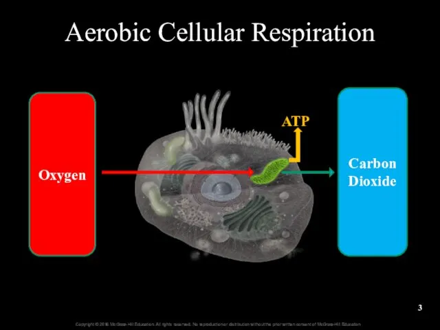
Aerobic Cellular Respiration
Oxygen
Carbon Dioxide
ATP
Aerobic Cellular Respiration
Oxygen
Carbon Dioxide
ATP
23.1
Introduction to the Respiratory System
State the functions of the respiratory
23.1
Introduction to the Respiratory System
State the functions of the respiratory
23.1a General Functions
of the Respiratory System
Air passageway
Air moves from atmosphere
23.1a General Functions
of the Respiratory System
Air passageway
Air moves from atmosphere
23.1a General Functions of the Respiratory System
Sound production
Air moves across vocal
23.1a General Functions of the Respiratory System
Sound production
Air moves across vocal

23.1b General Organization
of the Respiratory System
Structural organization
Upper respiratory tract
Larynx and
23.1b General Organization
of the Respiratory System
Structural organization
Upper respiratory tract
Larynx and
Upper Respiratory Tract
Nose
Nasal cavity
Sinuses
Pharynx
Larynx
MRI
Upper Respiratory Tract
Nose
Nasal cavity
Sinuses
Pharynx
Larynx
MRI
Upper Respiratory MRI: Coronal
Brain
Ethmoid air cells
Maxillary sinuses
Superior nasal concha
Middle nasal concha
Inferior
Upper Respiratory MRI: Coronal
Brain
Ethmoid air cells
Maxillary sinuses
Superior nasal concha
Middle nasal concha
Inferior
Lower Respiratory
X-ray: Posterior – Anterior View
Trachea
Bronchial Tree
Lungs
Lower Respiratory
X-ray: Posterior – Anterior View
Trachea
Bronchial Tree
Lungs
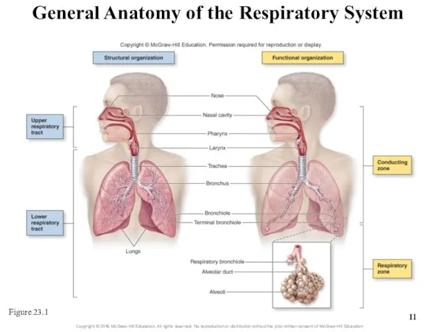
General Anatomy of the Respiratory System
Figure 23.1
General Anatomy of the Respiratory System
Figure 23.1
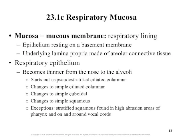
23.1c Respiratory Mucosa
Mucosa = mucous membrane: respiratory lining
Epithelium resting on a
23.1c Respiratory Mucosa
Mucosa = mucous membrane: respiratory lining
Epithelium resting on a
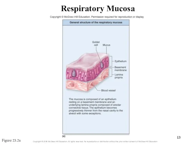
Respiratory Mucosa
Figure 23.2a
Respiratory Mucosa
Figure 23.2a

Respiratory Mucosa
Figure 23.2b
Respiratory Mucosa
Figure 23.2b

Respiratory Epithelium
High Magnification
Respiratory epithelium
Cilia
Columnar epithelial cells
Goblet cells
Basal cells
Basement membrane
Lamina propria
Respiratory Epithelium
High Magnification
Respiratory epithelium
Cilia
Columnar epithelial cells
Goblet cells
Basal cells
Basement membrane
Lamina propria
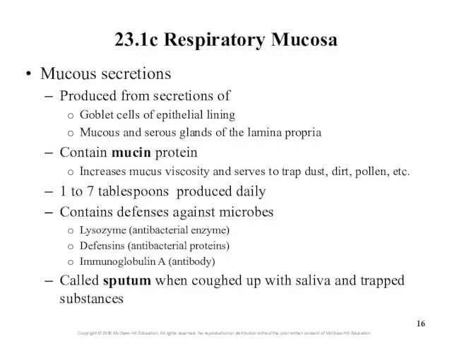
23.1c Respiratory Mucosa
Mucous secretions
Produced from secretions of
Goblet cells of epithelial lining
Mucous
23.1c Respiratory Mucosa
Mucous secretions
Produced from secretions of
Goblet cells of epithelial lining
Mucous
What did you learn?
What is the difference between the conducting and
What did you learn?
What is the difference between the conducting and
23.2
Upper Respiratory Tract
Describe the structure and function of the nose.
Provide
23.2
Upper Respiratory Tract
Describe the structure and function of the nose.
Provide
23.2a Nose and Nasal Cavity
Nose: first conducting structure for inhaled air
Formed
23.2a Nose and Nasal Cavity
Nose: first conducting structure for inhaled air
Formed
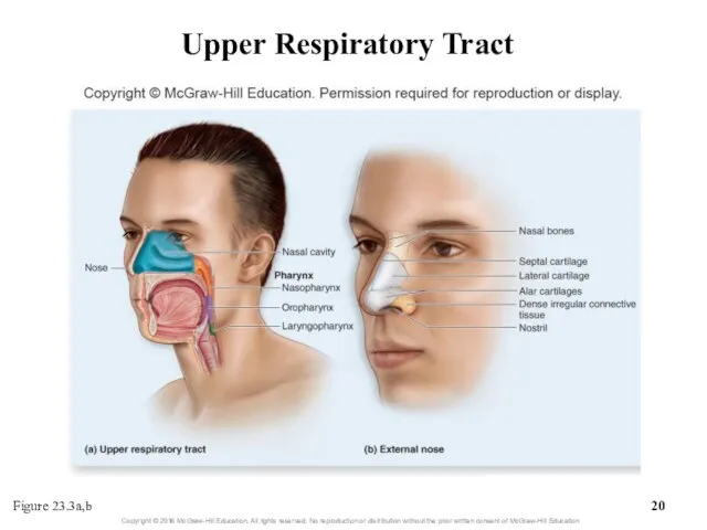
Upper Respiratory Tract
Figure 23.3a,b
Upper Respiratory Tract
Figure 23.3a,b
23.2a Nose and Nasal Cavity
Nasal cavity: from nostrils to choanae
An
23.2a Nose and Nasal Cavity
Nasal cavity: from nostrils to choanae
An

Nasal Cavity and Choanae
Nares
Nasal Cavity
Nasal Septum
Nasopharynx
Soft Palate
Uvula
Choanae
Hard Palate
Nasal Cavity and Choanae
Nares
Nasal Cavity
Nasal Septum
Nasopharynx
Soft Palate
Uvula
Choanae
Hard Palate
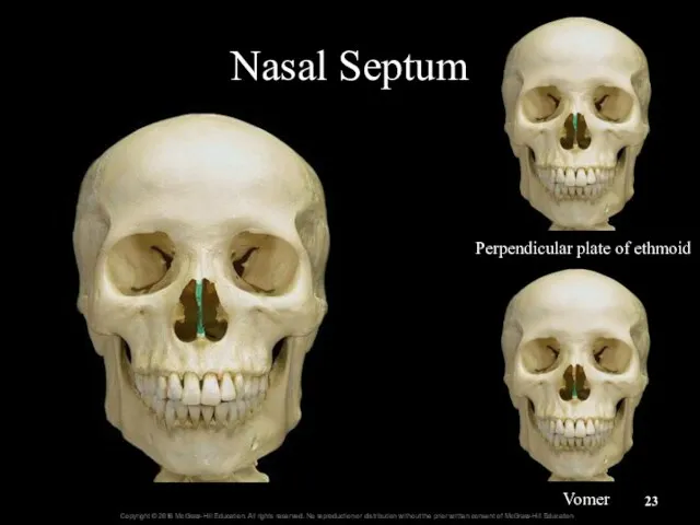
Nasal Septum
Perpendicular plate of ethmoid
Vomer
Nasal Septum
Perpendicular plate of ethmoid
Vomer
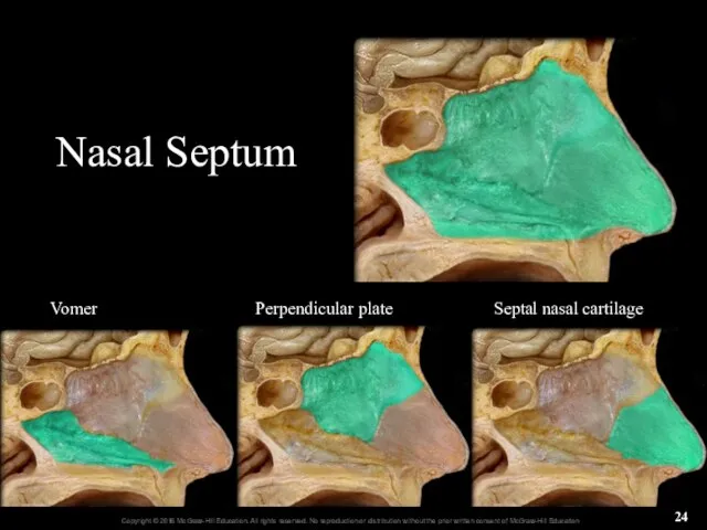
Nasal Septum
Vomer
Perpendicular plate
Septal nasal cartilage
Nasal Septum
Vomer
Perpendicular plate
Septal nasal cartilage
23.2a Nose and Nasal Cavity
The nasal conchae
Three paired, bony projections on
23.2a Nose and Nasal Cavity
The nasal conchae
Three paired, bony projections on
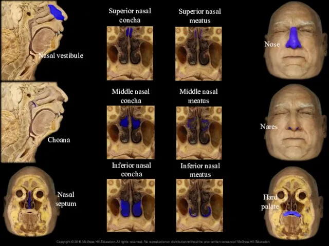
Nasal vestibule
Choana
Nasal septum
Hard palate
Nares
Nose
Superior nasal concha
Middle nasal concha
Inferior nasal concha
Superior nasal
Nasal vestibule
Choana
Nasal septum
Hard palate
Nares
Nose
Superior nasal concha
Middle nasal concha
Inferior nasal concha
Superior nasal
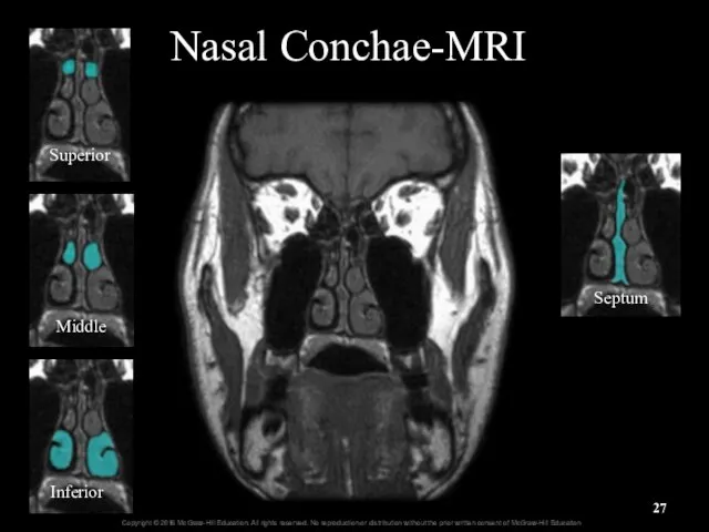
Nasal Conchae-MRI
Superior
Middle
Inferior
Septum
Nasal Conchae-MRI
Superior
Middle
Inferior
Septum
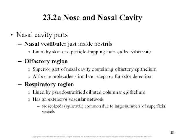
23.2a Nose and Nasal Cavity
Nasal cavity parts
Nasal vestibule: just inside nostrils
Lined
23.2a Nose and Nasal Cavity
Nasal cavity parts
Nasal vestibule: just inside nostrils
Lined
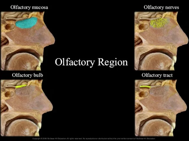
Olfactory Region
16-
Olfactory tract
Olfactory bulb
Olfactory nerves
Olfactory mucosa
Olfactory Region
16-
Olfactory tract
Olfactory bulb
Olfactory nerves
Olfactory mucosa
Upper Respiratory Tract
Figure 23.3c,d
Upper Respiratory Tract
Figure 23.3c,d
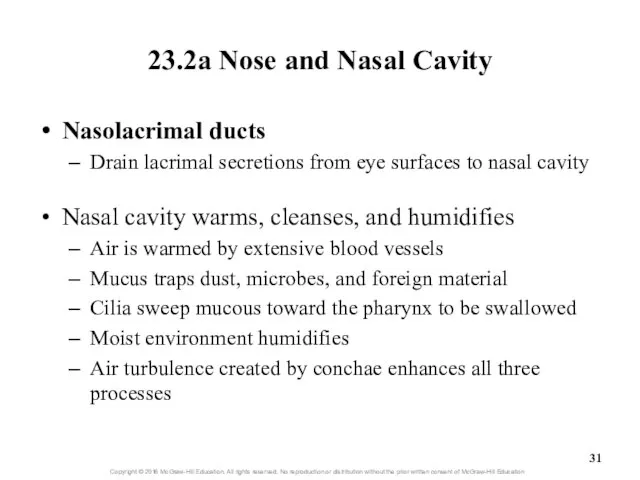
23.2a Nose and Nasal Cavity
Nasolacrimal ducts
Drain lacrimal secretions from eye surfaces
23.2a Nose and Nasal Cavity
Nasolacrimal ducts
Drain lacrimal secretions from eye surfaces
Clinical View: Runny Nose
Rhinorrhea (runny nose) occurs as a result of
Increased
Clinical View: Runny Nose
Rhinorrhea (runny nose) occurs as a result of
Increased
23.2b Paranasal Sinuses
Paranasal sinuses: spaces within skull bones
Named for specific bone
23.2b Paranasal Sinuses
Paranasal sinuses: spaces within skull bones
Named for specific bone
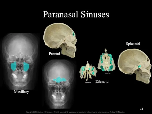
Maxillary
Frontal
Ethmoid
Sphenoid
Paranasal Sinuses
Maxillary
Frontal
Ethmoid
Sphenoid
Paranasal Sinuses
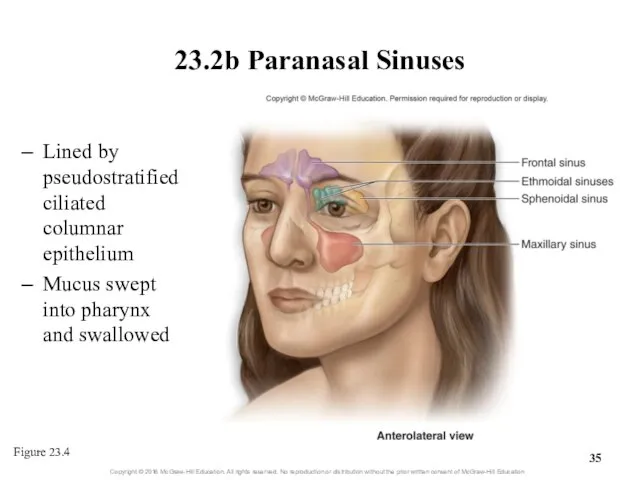
23.2b Paranasal Sinuses
Lined by pseudostratified ciliated columnar epithelium
Mucus swept into
23.2b Paranasal Sinuses
Lined by pseudostratified ciliated columnar epithelium
Mucus swept into
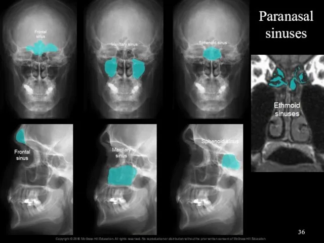
Paranasal sinuses
Paranasal sinuses
Clinical View: Sinus Infections
and Sinus Headaches
Respiratory infection or allergy can cause
Clinical View: Sinus Infections
and Sinus Headaches
Respiratory infection or allergy can cause
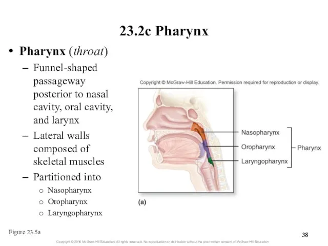
23.2c Pharynx
Pharynx (throat)
Funnel-shaped passageway posterior to nasal cavity, oral cavity, and
23.2c Pharynx
Pharynx (throat)
Funnel-shaped passageway posterior to nasal cavity, oral cavity, and
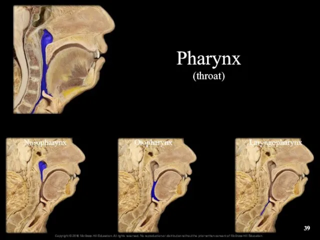
Nasopharynx
Oropharynx
Laryngopharynx
Pharynx
(throat)
Nasopharynx
Oropharynx
Laryngopharynx
Pharynx
(throat)
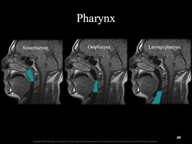
Pharynx
Oropharynx
Nasopharynx
Laryngopharynx
Pharynx
Oropharynx
Nasopharynx
Laryngopharynx

23.2c Pharynx
Nasopharynx: most superior part of pharynx
Posterior to nasal cavity, superior
23.2c Pharynx
Nasopharynx: most superior part of pharynx
Posterior to nasal cavity, superior
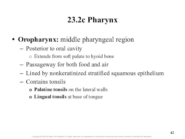
23.2c Pharynx
Oropharynx: middle pharyngeal region
Posterior to oral cavity
Extends from soft palate
23.2c Pharynx
Oropharynx: middle pharyngeal region
Posterior to oral cavity
Extends from soft palate
23.2c Pharynx
Laryngopharynx: inferior, narrow region of pharynx
Posterior to the larynx
From level
23.2c Pharynx
Laryngopharynx: inferior, narrow region of pharynx
Posterior to the larynx
From level
Pharynx
Figure 23.5b
Pharynx
Figure 23.5b
What did you learn?
What are vibrissae?
Between which conchae is the middle
What did you learn?
What are vibrissae?
Between which conchae is the middle
23.3
Lower Respiratory Tract
Describe the general functions and structure of the
23.3
Lower Respiratory Tract
Describe the general functions and structure of the
23.3
Lower Respiratory Tract (continued)
Explain the processes of bronchoconstriction and bronchodilation.
Describe
23.3
Lower Respiratory Tract (continued)
Explain the processes of bronchoconstriction and bronchodilation.
Describe
23.3 Lower Respiratory Tract
Includes conducting pathways from larynx to terminal bronchioles
Includes
23.3 Lower Respiratory Tract
Includes conducting pathways from larynx to terminal bronchioles
Includes
23.3a Larynx
Larynx (voice box)
Cylindrical airway between laryngopharynx and trachea
Several functions
Air passageway
23.3a Larynx
Larynx (voice box)
Cylindrical airway between laryngopharynx and trachea
Several functions
Air passageway
22.3a Larynx
Several functions (continued)
Participates in sneeze and cough reflexes
Help remove
22.3a Larynx
Several functions (continued)
Participates in sneeze and cough reflexes
Help remove
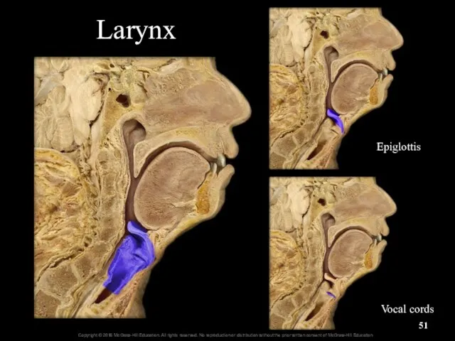
Larynx
Epiglottis
Vocal cords
Larynx
Epiglottis
Vocal cords
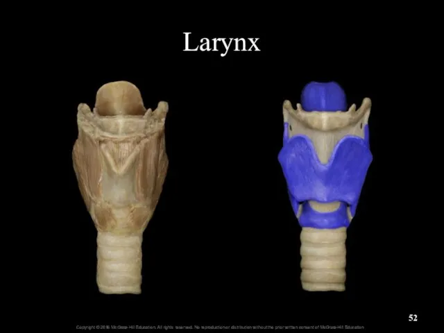
Larynx
Larynx
23.3a Larynx
Larynx anatomy
Laryngeal inlet (laryngeal aperture) connects pharynx and larynx
Larynx
23.3a Larynx
Larynx anatomy
Laryngeal inlet (laryngeal aperture) connects pharynx and larynx
Larynx
23.3a Larynx
Larynx anatomy (continued)
Thyroid cartilage: large, shield-shaped
Forms lateral and anterior walls
23.3a Larynx
Larynx anatomy (continued)
Thyroid cartilage: large, shield-shaped
Forms lateral and anterior walls
23.3a Larynx
Larynx anatomy (continued)
Smaller, paired cartilages located internally
Arytenoid, corniculate, and cuneiform
All
23.3a Larynx
Larynx anatomy (continued)
Smaller, paired cartilages located internally
Arytenoid, corniculate, and cuneiform
All
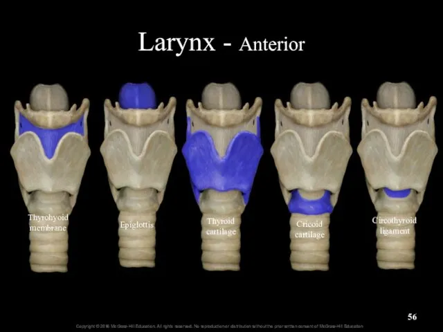
Larynx - Anterior
Thyroid cartilage
Cricoid cartilage
Epiglottis
Circothyroid ligament
Thyrohyoid membrane
Larynx - Anterior
Thyroid cartilage
Cricoid cartilage
Epiglottis
Circothyroid ligament
Thyrohyoid membrane
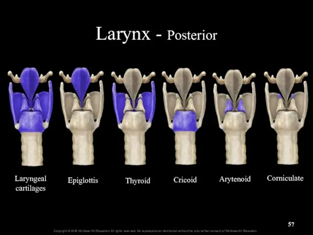
Larynx - Posterior
Laryngeal cartilages
Epiglottis
Thyroid
Cricoid
Arytenoid
Corniculate
Larynx - Posterior
Laryngeal cartilages
Epiglottis
Thyroid
Cricoid
Arytenoid
Corniculate
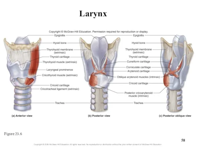
Larynx
Figure 23.6
Larynx
Figure 23.6
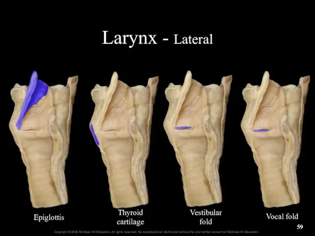
Larynx - Lateral
Epiglottis
Thyroid
cartilage
Vestibular
fold
Vocal fold
Larynx - Lateral
Epiglottis
Thyroid
cartilage
Vestibular
fold
Vocal fold
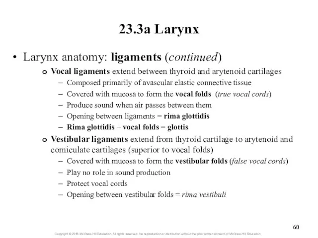
23.3a Larynx
Larynx anatomy: ligaments (continued)
Vocal ligaments extend between thyroid and arytenoid
23.3a Larynx
Larynx anatomy: ligaments (continued)
Vocal ligaments extend between thyroid and arytenoid
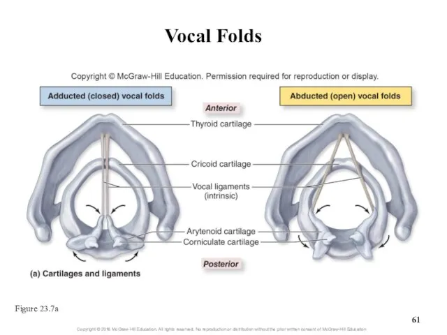
Vocal Folds
Figure 23.7a
Vocal Folds
Figure 23.7a
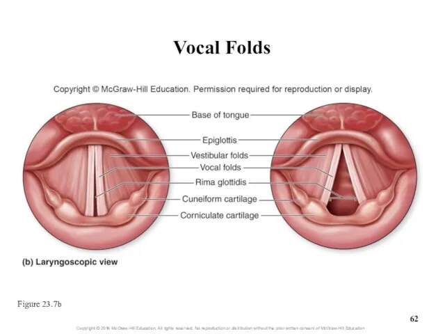
Vocal Folds
Figure 23.7b
Vocal Folds
Figure 23.7b
23.3a Larynx
Larynx anatomy (continued)
Extrinsic skeletal muscles
Stabilize larynx and help it move
23.3a Larynx
Larynx anatomy (continued)
Extrinsic skeletal muscles
Stabilize larynx and help it move
23.3a Larynx
Sound production: vocal cord vibration
Intrinsic laryngeal muscles narrow opening of
23.3a Larynx
Sound production: vocal cord vibration
Intrinsic laryngeal muscles narrow opening of
23.3a Larynx
Sound production (continued)
Other structures are also necessary for speech
Pharynx, nasal
23.3a Larynx
Sound production (continued)
Other structures are also necessary for speech
Pharynx, nasal
Clinical View: Laryngitis
Inflammation of the larynx
Symptoms of hoarse voice, sore throat,
Clinical View: Laryngitis
Inflammation of the larynx
Symptoms of hoarse voice, sore throat,
23.3b Trachea
Gross anatomy of trachea (windpipe)
Flexible, slightly rigid, tubular organ
Goes from
23.3b Trachea
Gross anatomy of trachea (windpipe)
Flexible, slightly rigid, tubular organ
Goes from
23.3b Trachea
Gross anatomy of the trachea (continued)
Carina: internal ridge at inferior
23.3b Trachea
Gross anatomy of the trachea (continued)
Carina: internal ridge at inferior
Trachea
Carina
Trachea
Carina
Trachea
Figure 23.8a-c
Trachea
Figure 23.8a-c
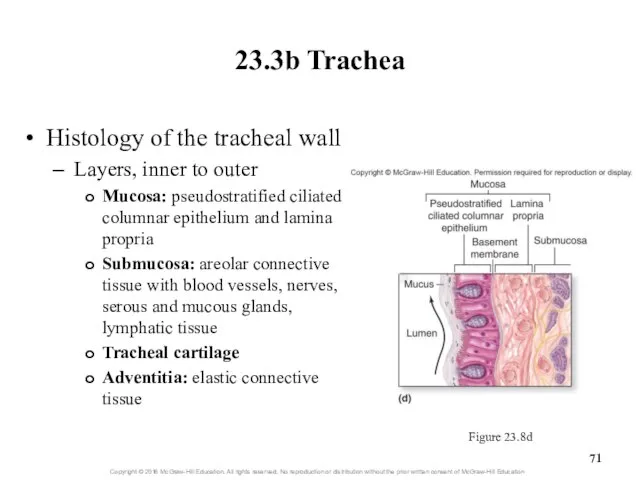
23.3b Trachea
Histology of the tracheal wall
Layers, inner to outer
Mucosa: pseudostratified ciliated
23.3b Trachea
Histology of the tracheal wall
Layers, inner to outer
Mucosa: pseudostratified ciliated
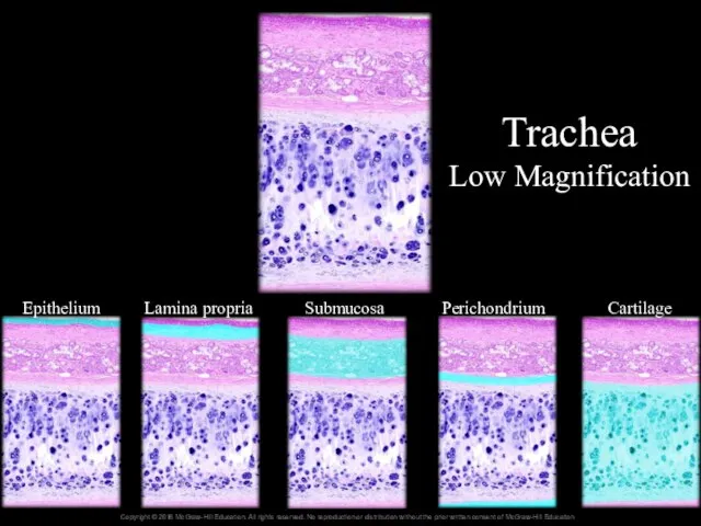
Trachea
Low Magnification
Epithelium
Lamina propria
Submucosa
Perichondrium
Cartilage
Trachea
Low Magnification
Epithelium
Lamina propria
Submucosa
Perichondrium
Cartilage
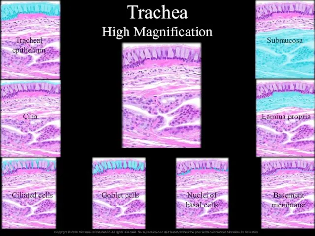
Trachea
High Magnification
Tracheal epithelium
Cilia
Ciliated cells
Goblet cells
Nuclei of basal cells
Basement membrane
Lamina propria
Submucosa
Trachea
High Magnification
Tracheal epithelium
Cilia
Ciliated cells
Goblet cells
Nuclei of basal cells
Basement membrane
Lamina propria
Submucosa
23.3c Bronchial Tree
Bronchial tree: system of highly branched air passages
Originates at
23.3c Bronchial Tree
Bronchial tree: system of highly branched air passages
Originates at
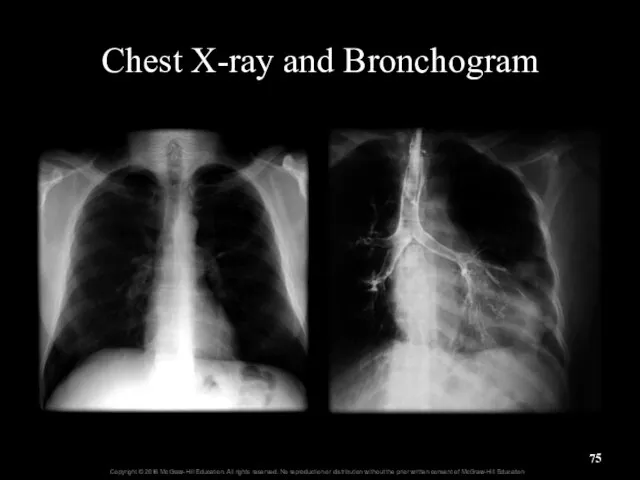
Chest X-ray and Bronchogram
Chest X-ray and Bronchogram
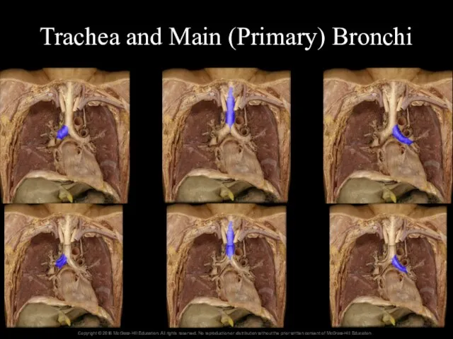
Trachea and Main (Primary) Bronchi
Trachea and Main (Primary) Bronchi
23.3c Bronchial Tree
Gross anatomy of the bronchial tree (continued)
Each main bronchus
23.3c Bronchial Tree
Gross anatomy of the bronchial tree (continued)
Each main bronchus
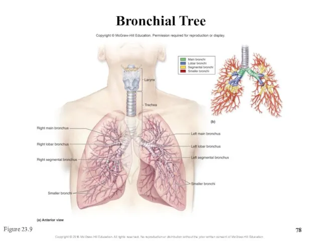
Bronchial Tree
Figure 23.9
Bronchial Tree
Figure 23.9
Clinical View: Bronchitis
Inflammation of the bronchi caused by bacterial or viral
Clinical View: Bronchitis
Inflammation of the bronchi caused by bacterial or viral
23.3c Bronchial Tree
Histology of the bronchial tree
Main bronchi are supported by
23.3c Bronchial Tree
Histology of the bronchial tree
Main bronchi are supported by
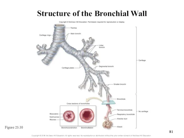
Structure of the Bronchial Wall
Figure 23.10
Structure of the Bronchial Wall
Figure 23.10
Clinical View: Asthma
Episodes of bronchoconstriction, wheezing, coughing, shortness of breath, and
Clinical View: Asthma
Episodes of bronchoconstriction, wheezing, coughing, shortness of breath, and
23.3d Respiratory Zone: Respiratory
Bronchioles, Alveolar Ducts, and Alveoli
Respiratory zone structures are
23.3d Respiratory Zone: Respiratory
Bronchioles, Alveolar Ducts, and Alveoli
Respiratory zone structures are
Bronchioles and Alveoli
Figure 23.11a
Bronchioles and Alveoli
Figure 23.11a
23.3d Respiratory Zone: Respiratory
Bronchioles, Alveolar Ducts, and Alveoli
Alveoli
Each lung contains 300
23.3d Respiratory Zone: Respiratory
Bronchioles, Alveolar Ducts, and Alveoli
Alveoli
Each lung contains 300
23.3d Respiratory Zone: Respiratory
Bronchioles, Alveolar Ducts, and Alveoli
Cell types of alveolar
23.3d Respiratory Zone: Respiratory
Bronchioles, Alveolar Ducts, and Alveoli
Cell types of alveolar
23.3e Respiratory Membrane
The respiratory membrane
Thin barrier between alveoli and pulmonary capillaries
Consists
23.3e Respiratory Membrane
The respiratory membrane
Thin barrier between alveoli and pulmonary capillaries
Consists
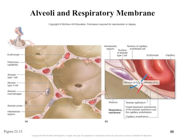
Alveoli and Respiratory Membrane
Figure 23.12
Alveoli and Respiratory Membrane
Figure 23.12
What did you learn?
What makes one speech sound have a higher
What did you learn?
What makes one speech sound have a higher
23.4
Lungs
Describe the location and general structure of the lungs.
Compare and
23.4
Lungs
Describe the location and general structure of the lungs.
Compare and
23.4
Lungs (continued)
Describe the pleural membranes and pleural cavity.
Explain the function
23.4
Lungs (continued)
Describe the pleural membranes and pleural cavity.
Explain the function
23.4a Gross Anatomy of the Lung
Lungs are in thorax on either
23.4a Gross Anatomy of the Lung
Lungs are in thorax on either
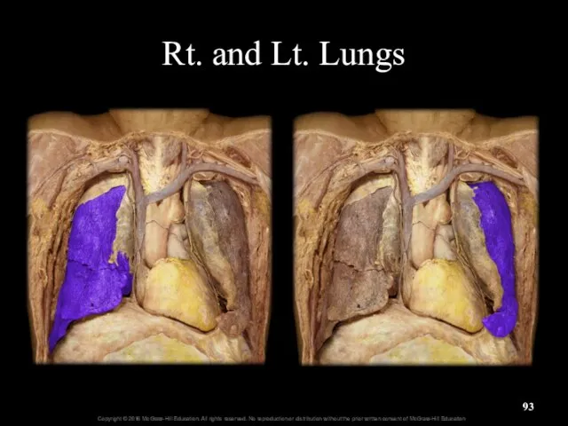
Rt. and Lt. Lungs
Rt. and Lt. Lungs
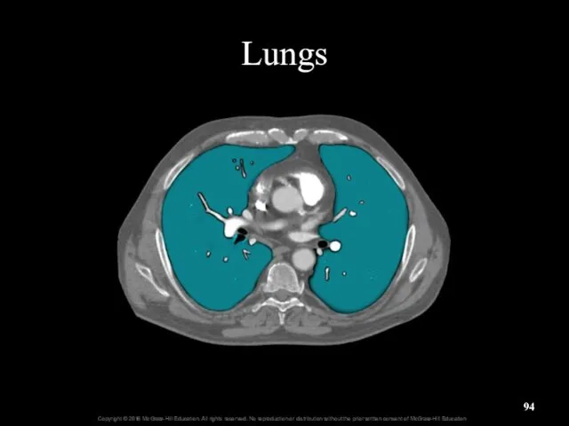
Lungs
Lungs

Chest X-ray and Bronchogram
Chest X-ray and Bronchogram
23.4a Gross Anatomy of the Lung
Lung surfaces
Costal surface adjacent to ribs
Mediastinal
23.4a Gross Anatomy of the Lung
Lung surfaces
Costal surface adjacent to ribs
Mediastinal
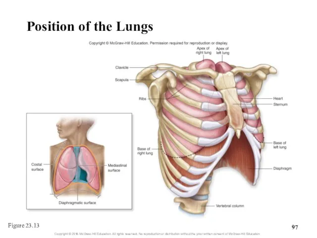
Position of the Lungs
Figure 23.13
Position of the Lungs
Figure 23.13
23.4a Gross Anatomy of the Lung
Right lung is larger and wider
23.4a Gross Anatomy of the Lung
Right lung is larger and wider
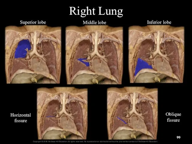
Right Lung
Superior lobe
Middle lobe
Inferior lobe
Horizontal
fissure
Oblique
fissure
Right Lung
Superior lobe
Middle lobe
Inferior lobe
Horizontal
fissure
Oblique
fissure
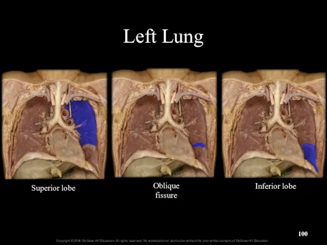
Left Lung
Oblique
fissure
Superior lobe
Inferior lobe
Left Lung
Oblique
fissure
Superior lobe
Inferior lobe
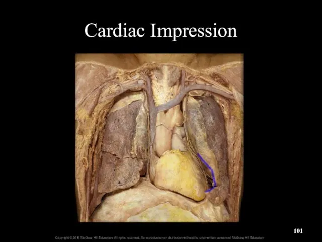
Cardiac Impression
Cardiac Impression
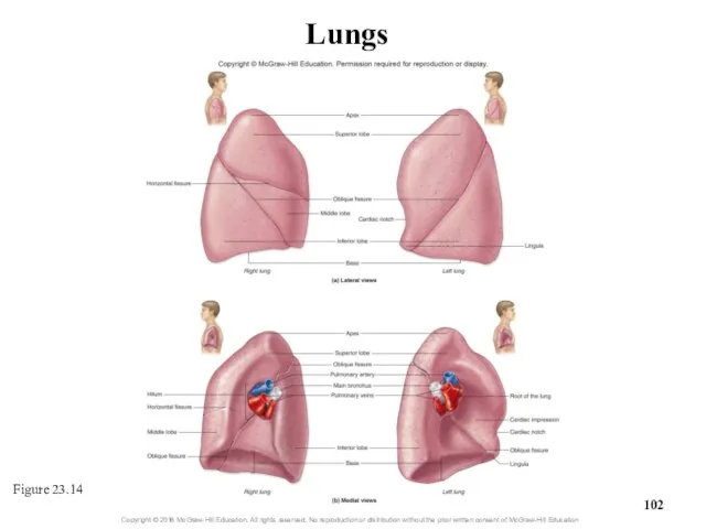
Figure 23.14
Lungs
Figure 23.14
Lungs
23.4a Gross Anatomy of the Lung
Each lung has multiple bronchopulmonary segments
10
23.4a Gross Anatomy of the Lung
Each lung has multiple bronchopulmonary segments
10

Bronchopulmonary Segments
and Lobules of the Lungs
Figure 23.15
Bronchopulmonary Segments
and Lobules of the Lungs
Figure 23.15
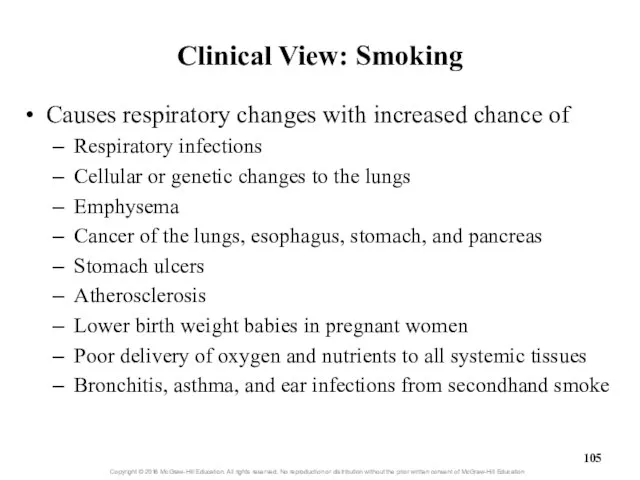
Clinical View: Smoking
Causes respiratory changes with increased chance of
Respiratory infections
Cellular or
Clinical View: Smoking
Causes respiratory changes with increased chance of
Respiratory infections
Cellular or
23.4b Circulation to and Innervation of the Lungs
Blood supply
Two types of
23.4b Circulation to and Innervation of the Lungs
Blood supply
Two types of
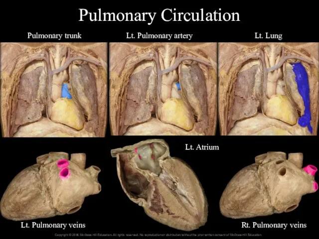
Pulmonary Circulation
Pulmonary trunk
Lt. Pulmonary artery
Lt. Lung
Lt. Pulmonary veins
Rt. Pulmonary veins
Lt. Atrium
Pulmonary Circulation
Pulmonary trunk
Lt. Pulmonary artery
Lt. Lung
Lt. Pulmonary veins
Rt. Pulmonary veins
Lt. Atrium
23.4b Circulation to and Innervation of the Lungs
Blood supply (continued)
Bronchial circulation
23.4b Circulation to and Innervation of the Lungs
Blood supply (continued)
Bronchial circulation
Pulmonary Circulation of the Lungs
Figure 23.16
Pulmonary Circulation of the Lungs
Figure 23.16
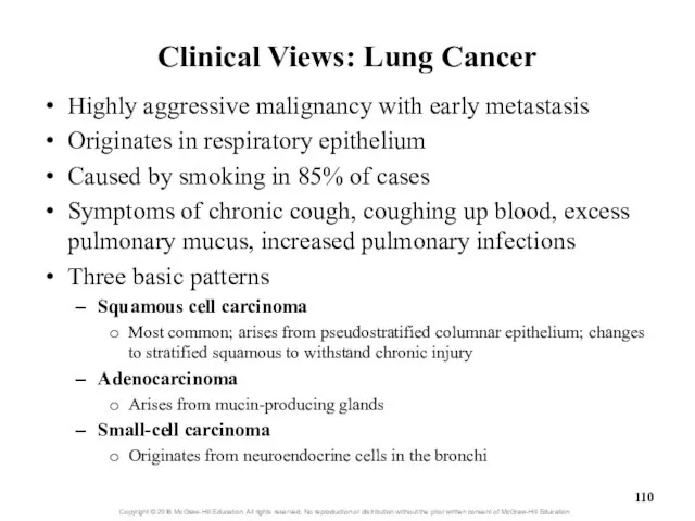
Clinical Views: Lung Cancer
Highly aggressive malignancy with early metastasis
Originates in respiratory
Clinical Views: Lung Cancer
Highly aggressive malignancy with early metastasis
Originates in respiratory
23.4b Circulation to and Innervation of the Lungs
Lymph drainage
Lymph vessels and
23.4b Circulation to and Innervation of the Lungs
Lymph drainage
Lymph vessels and
23.4b Circulation to and Innervation of the Lungs
Innervation of the respiratory
23.4b Circulation to and Innervation of the Lungs
Innervation of the respiratory
23.4c Pleura Membranes
and Pleural Cavity
Pleura: serous membrane
Outer lining of lung
23.4c Pleura Membranes
and Pleural Cavity
Pleura: serous membrane
Outer lining of lung

Pleura Membranes
Visceral pleura
Parietal pleura
Pleural cavity
Pleura Membranes
Visceral pleura
Parietal pleura
Pleural cavity
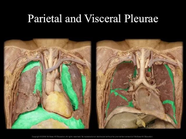
Parietal and Visceral Pleurae
Parietal and Visceral Pleurae
23.4c Pleura Membranes
and Pleural Cavity
Pleural cavity
Located between visceral and parietal
23.4c Pleura Membranes
and Pleural Cavity
Pleural cavity
Located between visceral and parietal
Clinical View: Pleurisy and Pleural Effusion
Pleurisy = inflammation of the pleural
Clinical View: Pleurisy and Pleural Effusion
Pleurisy = inflammation of the pleural
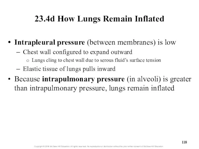
23.4d How Lungs Remain Inflated
Intrapleural pressure (between membranes) is low
Chest wall
23.4d How Lungs Remain Inflated
Intrapleural pressure (between membranes) is low
Chest wall
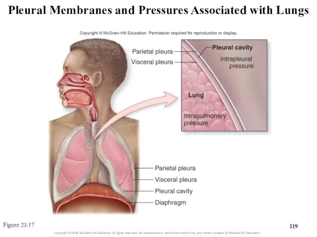
Pleural Membranes and Pressures Associated with Lungs
Figure 23.17
Pleural Membranes and Pressures Associated with Lungs
Figure 23.17
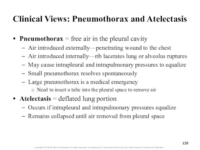
Clinical Views: Pneumothorax and Atelectasis
Pneumothorax = free air in the pleural
Clinical Views: Pneumothorax and Atelectasis
Pneumothorax = free air in the pleural
 Домашнее задание: Самостоятельно придумайте сюжет для анимации. Дайте ему название и подробно опишите планируемую последовател
 Язык SQL. Понятие базы данных
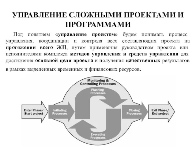 Управление сложными проектами и программами. (Лекция 3)
 Структура книги Excel
 Тема урока: «Знакомство с клавиатурой» «Дорогу осилит идущий, а информатику – мыслящий» Гюстав Гийома
 Администрирование информационных систем Основы сетевого администрирования
  Динамические структуры данных
 Существенные свойства и принятие решения.
 «Списки» Составила: Смирнова Анна, обучающаяся 6 класса. Руководитель: Медведева Елена Валерьевна, учитель музыки и информатики.
 Информационная безопасность
 Измерение и представление информации
 Пульт управления здоровьем в смартфоне
 Базовые понятия языка Си
 Домашнее задание по информатике
 Программное обеспечение
 Сеть Internet
 Администрирование информационных систем Управление контентом веб-узла
  Алгоритмы сжатия данных с потерями и без потерь
 5eb5a808-6762-48a9-a855-a31e57533794
 Программа формирования УУД и ИКТ Программа формирования культуры ЗОЖ Учитель начальных классов Волкова Светлана Николаевна
 Информатика и ИКТ 10-11 класс Системы счисления
  Урок-игра. «Умники и умницы»
 Коллективное дело как инструмент приобретения социального и трудового опыта
 Коммуникационные операции «точка-точка» параллельное программирование
  Основополагающие принципы информационного моделирования в строительстве. ISO 19650-1, 19650-2
 Текстовый процессор Word
 Администрирование информационных систем Установка и начальная настройка SQL Server 2000
 Мультимедиа. Выходной контроль уровня овладения материалами курса (Online - тест)