- Antigen recognition and activation of T-cells

Содержание
- 2. T-cells can be distinguished from other lymphocyte types, such as B cells and NK cells by
- 3. Subsets of T cells: T helper cell (TH cells) assist other white blood cells in immunologic
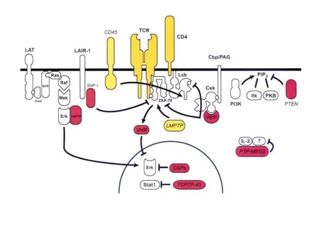
- 4. TCR and CD3 complex ITAM - immunoreceptor tyrosine-based activation motifs
- 5. TCR co-receptors: CD4 – for Th (that is specific for class II MHC). CD8 – for
- 6. CD4 CD4 uses its D1 domain to interact with the β2-domain of MHC class II molecules.
- 7. CD8 The extracellular domain of CD8-α interacts with to the α3 portion of the Class I
- 8. T-cell activation The mechanism by which a T-cell elicits this response upon contact with its unique
- 9. Early signaling steps implicate the following molecules: Lck - Associated with the transmembrane tail of CD4
- 10. Lck (lymphocyte-specific protein tyrosine kinase) and Fyn are members of the Src family of tyrosine kinases.
- 11. Src (pronounced "sarc" as it is short for sarcoma) is a proto-oncogene encoding a tyrosine kinase
- 12. Structure of Src kinase P Binds to proline-rich region of proteins Binds to phosphotyrosines in proteins
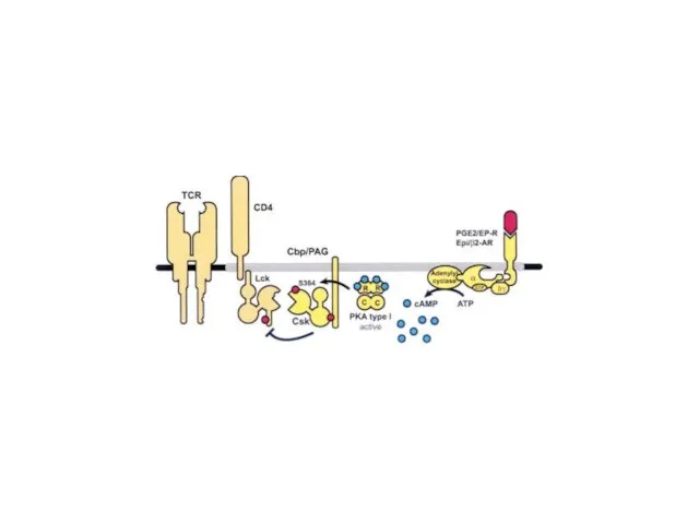
- 15. In resting normal T-cells (a), Csk is present in lipid rafts through interaction with phospho-Tyr-317 in
- 16. SH2 and SH3 domains were found in several other protein families Kinases Phosphatases Phospholipases Adaptor proteins
- 17. ZAP-70 kinase ZAP-70 is an abbreviation for Zeta-chain-associated protein kinase 70 (70 is the molecular weight
- 18. 3D view of Zap-70
- 19. The tandem SH2-domains of ZAP-70 are engaged by the doubly phosphorylated ITAMs of CD3-zeta Y P
- 20. ZAP-70 could phosphorylate the transmembrane protein LAT (linker of activated T cells). LAT localizes to lipid
- 21. Lipid raft The plasma membrane of cells is made of a combination of glycosphingolipids and protein
- 22. Sphingomyelin
- 23. T-cell signaling in lipid rafts
- 24. Human LAT is a 233 amino-acid type III transmembrane protein
- 25. A model of the signaling complexes assembled through LAT in T cells
- 26. “LAT signalosome”
- 27. LAT complexes. Balagopalan L et al. Cold Spring Harb Perspect Biol 2010;2:a005512 ©2010 by Cold Spring
- 28. LAT signaling complexes and microclusters. Balagopalan L et al. Cold Spring Harb Perspect Biol 2010;2:a005512 ©2010
- 29. Lymphoproliferative disease in LAT Y136F KI mice. Balagopalan L et al. Cold Spring Harb Perspect Biol
- 30. PLCγ PI3-K
- 31. PIP2 cleavage to IP3 and DAG initiates intracellular calcium release and PKC activation.
- 32. Protein kinase C also known as PKC activate PKC
- 33. Pleckstrin homology domain (PH domain) is a protein domain of approximately 120 amino acids can bind
- 34. PH domain
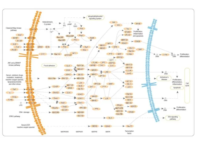
- 35. Mitogen-activated protein (MAP) kinases MAPKKK -> MAPKK -> MAPK -> Transcription factor
- 37. Guanine nucleotide exchange factor (GEF)
- 42. Скачать презентацию
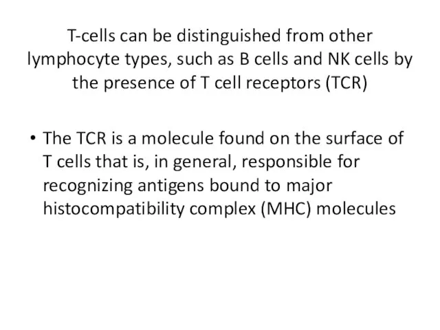
T-cells can be distinguished from other lymphocyte types, such as B
T-cells can be distinguished from other lymphocyte types, such as B
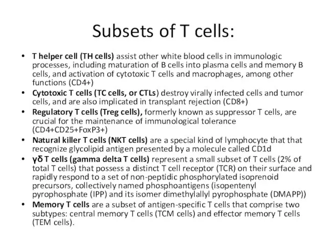
Subsets of T cells:
T helper cell (TH cells) assist other white
Subsets of T cells:
T helper cell (TH cells) assist other white
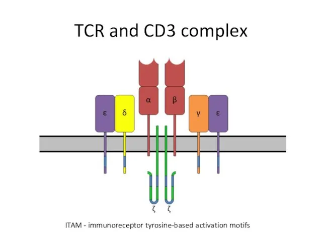
TCR and CD3 complex
ITAM - immunoreceptor tyrosine-based activation motifs
TCR and CD3 complex
ITAM - immunoreceptor tyrosine-based activation motifs
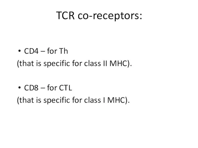
TCR co-receptors:
CD4 – for Th
(that is specific for class II
TCR co-receptors:
CD4 – for Th
(that is specific for class II
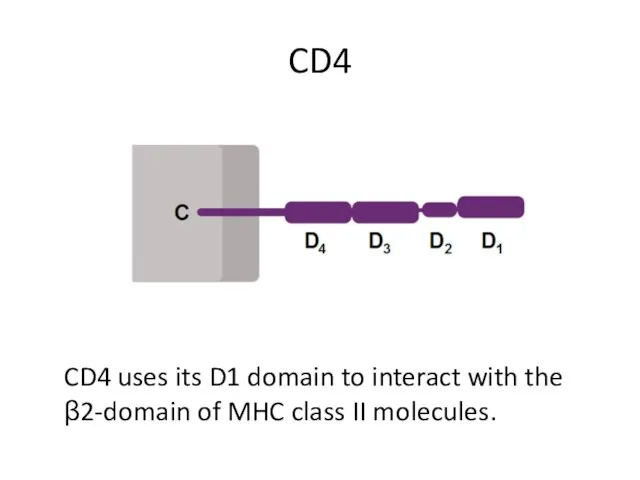
CD4
CD4 uses its D1 domain to interact with the β2-domain of
CD4
CD4 uses its D1 domain to interact with the β2-domain of
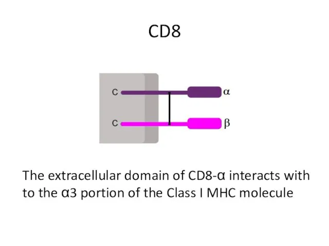
CD8
The extracellular domain of CD8-α interacts with to the α3 portion
CD8
The extracellular domain of CD8-α interacts with to the α3 portion
T-cell activation
The mechanism by which a T-cell elicits this response upon
T-cell activation
The mechanism by which a T-cell elicits this response upon
Early signaling steps implicate the following molecules:
Lck - Associated with
Early signaling steps implicate the following molecules:
Lck - Associated with
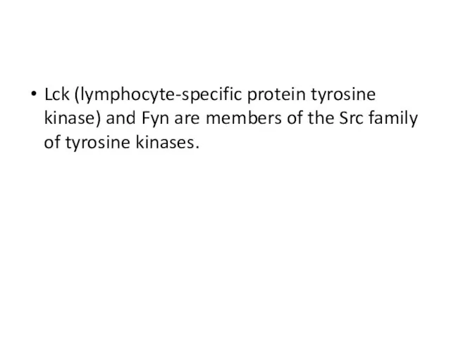
Lck (lymphocyte-specific protein tyrosine kinase) and Fyn are members of the
Lck (lymphocyte-specific protein tyrosine kinase) and Fyn are members of the
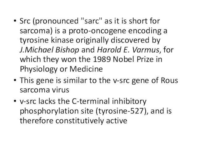
Src (pronounced "sarc" as it is short for sarcoma) is a
Src (pronounced "sarc" as it is short for sarcoma) is a
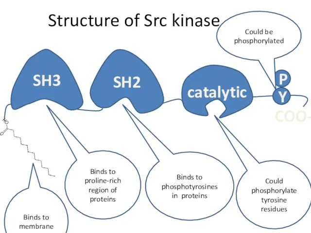
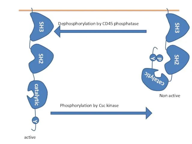
Structure of Src kinase
P
Binds to proline-rich region of proteins
Binds to phosphotyrosines
Structure of Src kinase
P
Binds to proline-rich region of proteins
Binds to phosphotyrosines
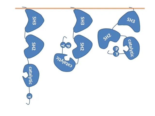
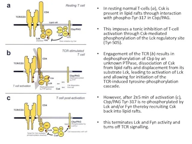
In resting normal T-cells (a), Csk is present in lipid rafts
In resting normal T-cells (a), Csk is present in lipid rafts
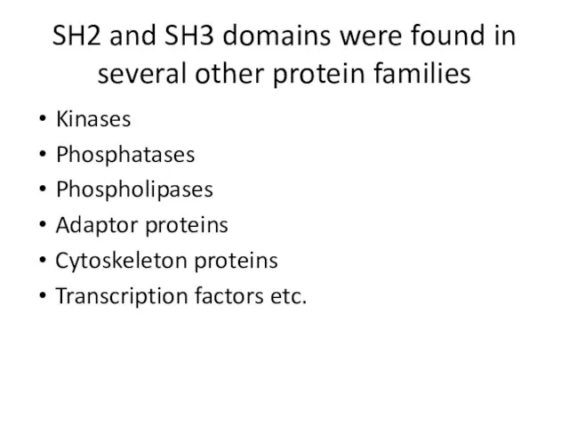
SH2 and SH3 domains were found in several other protein families
Kinases
Phosphatases
Phospholipases
Adaptor
SH2 and SH3 domains were found in several other protein families
Kinases
Phosphatases
Phospholipases
Adaptor
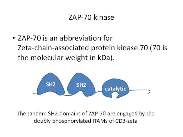
ZAP-70 kinase
ZAP-70 is an abbreviation for Zeta-chain-associated protein kinase 70 (70
ZAP-70 kinase
ZAP-70 is an abbreviation for Zeta-chain-associated protein kinase 70 (70
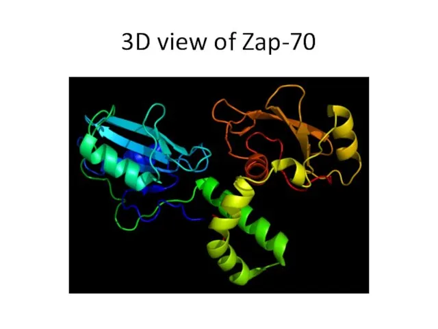
3D view of Zap-70
3D view of Zap-70
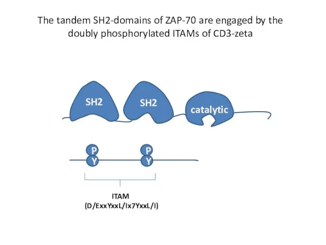
The tandem SH2-domains of ZAP-70 are engaged by the doubly phosphorylated
The tandem SH2-domains of ZAP-70 are engaged by the doubly phosphorylated

ZAP-70 could phosphorylate the transmembrane protein LAT (linker of activated T
ZAP-70 could phosphorylate the transmembrane protein LAT (linker of activated T
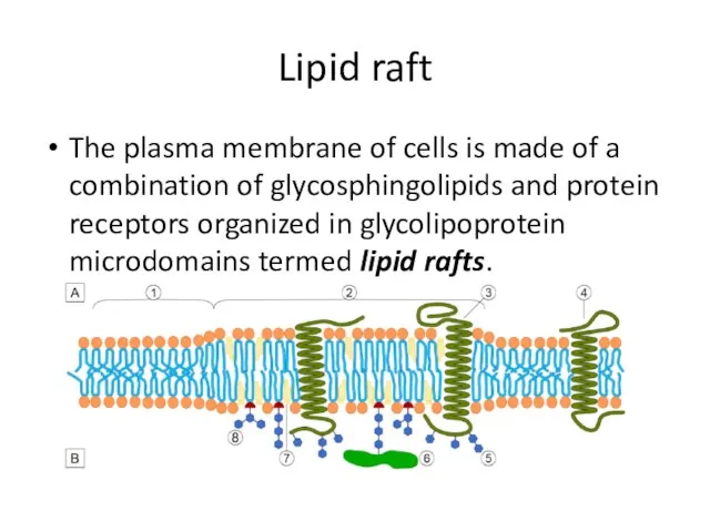
Lipid raft
The plasma membrane of cells is made of a combination
Lipid raft
The plasma membrane of cells is made of a combination
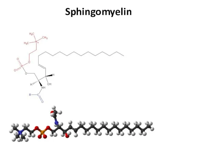
Sphingomyelin
Sphingomyelin
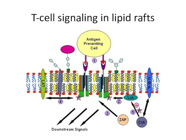
T-cell signaling in lipid rafts
T-cell signaling in lipid rafts
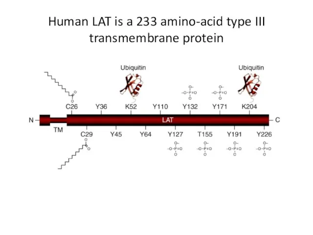
Human LAT is a 233 amino-acid type III transmembrane protein
Human LAT is a 233 amino-acid type III transmembrane protein
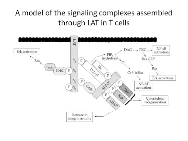
A model of the signaling complexes assembled through LAT in T
A model of the signaling complexes assembled through LAT in T
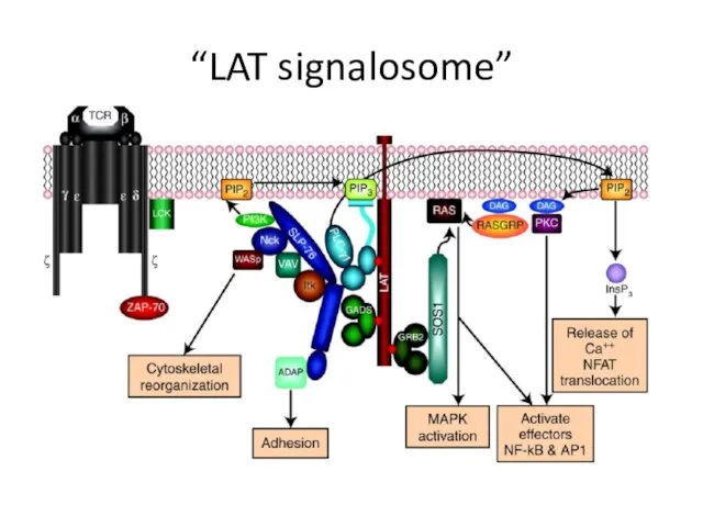
“LAT signalosome”
“LAT signalosome”
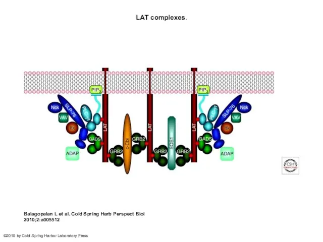
LAT complexes.
Balagopalan L et al. Cold Spring Harb Perspect Biol 2010;2:a005512
©2010
LAT complexes.
Balagopalan L et al. Cold Spring Harb Perspect Biol 2010;2:a005512
©2010
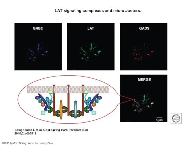
LAT signaling complexes and microclusters.
Balagopalan L et al. Cold Spring Harb
LAT signaling complexes and microclusters.
Balagopalan L et al. Cold Spring Harb
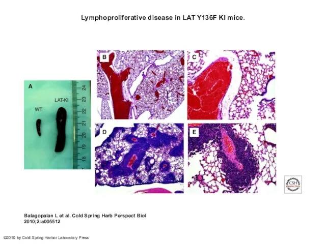
Lymphoproliferative disease in LAT Y136F KI mice.
Balagopalan L et al. Cold
Lymphoproliferative disease in LAT Y136F KI mice.
Balagopalan L et al. Cold
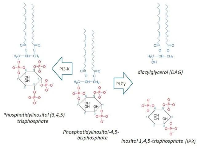
PLCγ
PI3-K
PLCγ
PI3-K
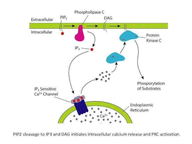
PIP2 cleavage to IP3 and DAG initiates intracellular calcium release and
PIP2 cleavage to IP3 and DAG initiates intracellular calcium release and
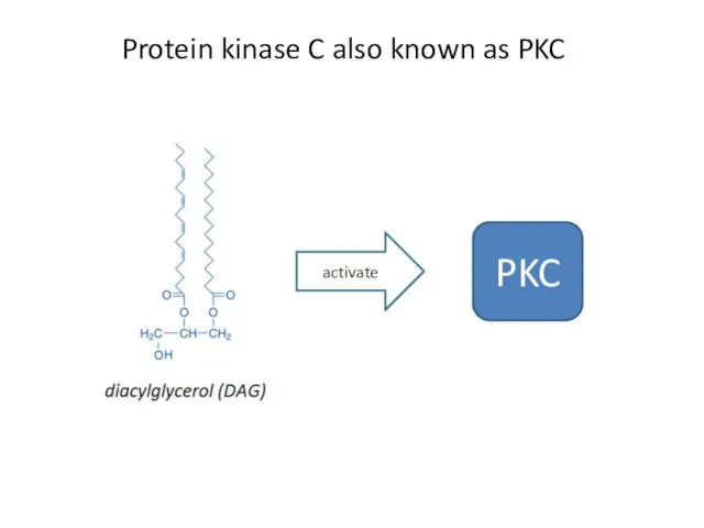
Protein kinase C also known as PKC
activate
PKC
Protein kinase C also known as PKC
activate
PKC
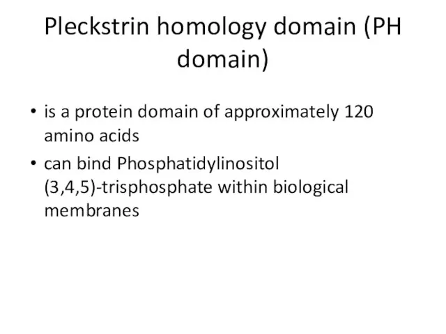
Pleckstrin homology domain (PH domain)
is a protein domain of approximately 120
Pleckstrin homology domain (PH domain)
is a protein domain of approximately 120
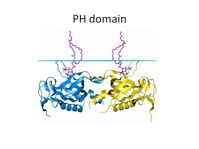
PH domain
PH domain
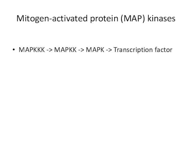
Mitogen-activated protein (MAP) kinases
MAPKKK -> MAPKK -> MAPK -> Transcription factor
Mitogen-activated protein (MAP) kinases
MAPKKK -> MAPKK -> MAPK -> Transcription factor
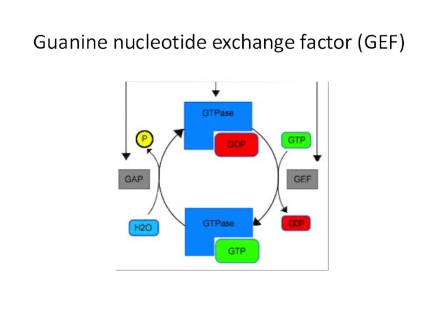
Guanine nucleotide exchange factor (GEF)
Guanine nucleotide exchange factor (GEF)
 Занятие 4. ЛР и ЛРС, содержащие эфирные масла (Apiaceae и Juniperus communis). Сырьевые источники камфоры
 Иммунопатология
 Диффузные заболевания соединительной ткани: дерматологические аспекты
 Клинический разбор пациента
 Развитие потребностей младшего школьника
 Кинопропаганда. Кино как средство информационно-психологической войны
 Учение об инфекции патогенность и вирулентность микробов
 Нарушения функции почек
 Абдоминальный ишемический синдром
 Земские врачи
 Марафон «Энергия жизни». Часть 3 «Путь к мечте». Неделя 2
 Операции на мозговом отделе головы
 Ларингеальная трубка
 Армысыңдар менің бауырларым мен қарындастарым. Болашақ тигрлер ( жолбарыстар)
 Xəstəxanadaxili infeksiyalar və onlarla mübarizənin əsasları, əksepidemik tədbirlər
 Қоршаған орта факторларының салдарлық итж дамуындағы маңызы
 Азбука витаминов плюс детские витамины для поддержки зрения. Строение зрительной системы
 Мой профессиональный выбор
 Особенности эмоционально-волевой и личностной сфер при нарушениях слуха
 СРСП 3 Сайлауов Б.С. 2-067 ОМ
 Омыртқа туберкулезінің салыстырмалы диагностикасы
 Быть хорошим соседом, это значит
 Поражение ЦНС вирусами герпеса
 Себорея. Себорейный дерматит
 Клеточные типичные патологические процессы
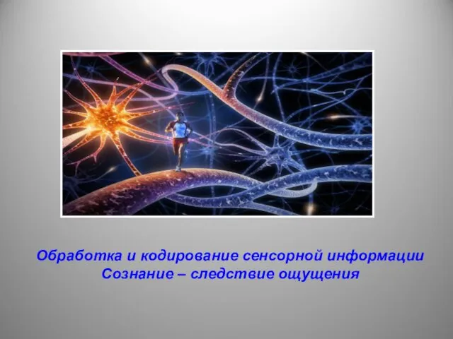 Обработка и кодирование сенсорной информации Сознание – следствие ощущения
 Вирус геномы
 Педагогические конфликты