- Cardiac emergencies and cpr

Содержание
- 2. HISTORICAL REVIEW 5000 - 3000 BC - first artificial mouth to mouth ventilation 1780 – first
- 3. Introduction Basic Life Support needed for patient whose breathing or heart has stopped Ventilations are given
- 4. Review of Circulatory System Circulatory system consists of heart, blood, and blood vessels.
- 5. Cardiovascular System: Primary Functions Transports blood to lungs Delivers carbon dioxide and picks up oxygen Transports
- 6. Anatomy and Physiology of the Heart Ventricles pump blood through two loops or cycles in body
- 7. Heart is composed of a unique type of muscle (myocardium) that contracts to make pumping action.
- 8. Contractions are controlled by electrical signals under nervous system control Heart Muscle
- 9. Arteries Arterial blood is oxygenated, bright red, and under pressure Carotid arteries — major arteries passing
- 10. Pulse When left ventricle contracts, wave of blood is sent through arteries causing pulsing blood pressure
- 11. Pulse continued Palpate femoral pulse in crease between abdomen and thigh Palpate radial pulse on the
- 12. Capillaries Arteries progressively branch into smaller vessels that eventually reach capillaries Capillaries are very small blood
- 13. Veins From capillaries, blood drains back to heart through extensive system of veins Venous blood is
- 14. Heart Rate Heart rate, measured as pulse, is affected by many factors With exercise, fever, or
- 15. Circulatory System: Emergencies Any condition that affects respiration Reduces ability to deliver oxygen Severe bleeding Shock
- 16. Circulatory System: Emergencies continued Heart attack Can lead to cardiac arrest Ventricular fibrillation Heart muscle flutters
- 17. Cardiac Arrest Heart may stop (cardiac arrest) as a result of heart attack Brain damage begins
- 18. Causes of cardiac arrest cardiac extracardiac Primary lesion of cardiac muscle leading to the progressive decline
- 19. Causes of Cardiac Arrest Heart attack Drowning Suffocation Stroke Allergic reaction Diabetic emergency Prolonged seizures Drug
- 20. Causes of circulatory arrest Cardiac Ischemic heart disease (myocardial infarction, stenocardia) Arrhythmias of different origin and
- 21. Cardiac Chain of Survival
- 22. Diagnosis of cardiac arrest Symptoms of cardiac arrest absence of pulse on carotid arteries – a
- 23. Sequence of operations Check responsiveness Call for help Correctly place the victim and ensure the open
- 24. In case of unconsciousness it is necessary to estimate quickly the open airway respiration hemodynamics
- 25. Main stages of resuscitation C (Circulation) – restore the circulation by external cardiac massage A (Airway)
- 26. A (Airway) ensure open airway
- 27. Open the airway using a head tilt lifting of chin. Do not tilt the head too
- 28. B (Breathing) Tilt the head back and listen for. If not breathing normally, pinch nose and
- 29. mouth to mouth or mouth to nose respiration ventilation by a face mask and a self-inflating
- 30. C. Circulation Restore the circulation, that is start external cardiac massage
- 31. 2 mechanisms explaining the restoration of circulation by external cardiac massage Cardiac pump Thoracic pump
- 32. Cardiac pump during the cardiac massage Blood pumping is assured by the compression of heart between
- 33. Thoracic pump at the cardiac massage Blood circulation is restored due to the change in intra
- 34. ALGORITHM of Cardiopulmonary resuscitation 4 cycles: 15 compression and 2 breaths 10 cycles: 5 compression and
- 35. VENTRICULAR FIBRILLATION OR PULSELESS TACHYCARDIA Witnessed Unwitnessed Precordial thump Check pulse, if none: Begin CPR Defibrillate
- 36. Possible arrhythmias after cardiac defibrillation ventricular tachycardia bradyarrythmia including electromechanical dissociation and asystole supraventricular arrhythmia accompanied
- 37. Operations in case of asystole Asystole Start CPR IV line Adrenaline:IV 1 mg, each 3-5 min.
- 38. Call First vs. Call Fast Call First If alone with adult victim Any victim of any
- 39. Cardiopulmonary Resuscitation (CPR) CPR helps keep patient alive by circulating some oxygenated blood to vital organs
- 40. CPR continued Blood circulation resulting from chest compressions not as strong as circulation from heartbeat Can
- 41. CPR continued Often electric shock from AED is needed to restore a heartbeat—and CPR can keep
- 42. CPR Saves Lives CPR and defibrillation within 3-5 minutes can save over 50% of cardiac arrest
- 43. General Technique of CPR If unresponsive, not breathing, and no pulse, start chest compressions Find the
- 44. General Technique of CPR continued Compress chest hard and fast at a rate of 100 compressions/minute
- 45. General Technique of CPR continued If alone, alternate 30 chest compressions and 2 ventilations for any
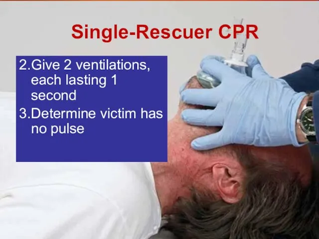
- 46. Single-Rescuer CPR Check patient’s responsiveness, open airway, and determine that patient is not breathing adequately 2.
- 47. Single-Rescuer CPR 2. Give 2 ventilations, each lasting 1 second 3. Determine victim has no pulse
- 48. Put hand(s) in correct position for chest compressions
- 49. Give 30 chest compressions at rate of 100 per minute Then give 2 ventilations
- 50. Continue CPR until: Patient begins to move AED brought to scene and ready to use Professional
- 51. If patient starts moving, check for adequate breathing If patient is breathing adequately, put patient in
- 52. Chest Compressions Alert Be careful with your hand position For adults/children, keep your fingers off patient’s
- 53. Chest Compressions Alert When compressing, keep elbows straight and hands in contact with patient’s chest at
- 54. Chest Compressions Alert Compress chest hard and fast, but let chest recoil completely between compressions. Minimize
- 55. Problems with CPR Technique CPR often ineffective because of poor technique Compressions not delivered steadily and
- 56. Chest Compressions: Bradycardia in Child Infant or child being given rescue breaths or oxygen may have
- 57. Skill: CPR For Adult or Child (Two Rescuers)
- 58. Two-Rescuer CPR for Adults and Children Minimizes time between rescue breaths and compressions CPR becomes more
- 59. Two-Rescuer CPR Performed in cycles of 30:2 for adult (15:2 for infant or child) One rescuer
- 60. Two-Rescuer CPR continued If AED present, one rescuer gives CPR while the other sets up unit
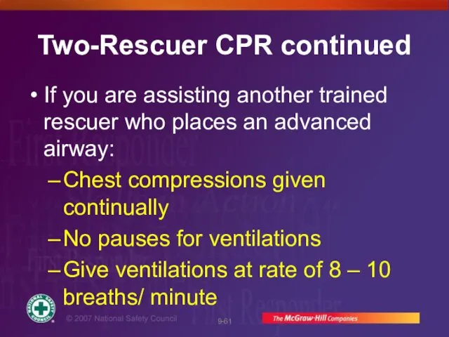
- 61. Two-Rescuer CPR continued If you are assisting another trained rescuer who places an advanced airway: Chest
- 62. Transitioning from One-Rescuer CPR to Two-Rescuer CPR Second rescuer moves into position on other side to
- 63. Transitioning from One-Rescuer CPR to Two-Rescuer CPR When first rescuer says, “No pulse, continue CPR,” second
- 64. Differences in Two-Rescuer Training If First Responder started CPR, arriving second rescuer may have a higher
- 65. Rescuer 1 checks ABCs. Rescuer 2 locates site for chest compressions.
- 66. If no pulse, rescuer 2 gives 30 compressions for adult (15 for child) at rate of
- 67. Rescuer 1 gives 2 breaths.
- 68. Continue cycles of 30:2 for adults (15:2 for child). After 5 cycles (~ 2 minutes) switch
- 69. Adult or Child Two-Rescuer CPR Continued Continue CPR until: Patient moves AED brought to scene and
- 70. Uses different hand position Place thumbs of both hands on sternum while fingers encircle chest Compress
- 71. Skill: CPR: Infants Two Rescuers
- 72. Rescuer 1 checks ABCs. Rescuer 2 locates site for chest compressions.
- 73. If no pulse, rescuer 2 gives 15 chest compressions.
- 74. Rescuer 1 gives 2 breaths.
- 75. Infant Two-Rescuer CPR Continued Continue cycles of 15:2 for ~ 2 minutes then switch roles Continue
- 76. When Not to Perform CPR Presence of a Do-Not-Resuscitate (DNR) order Patient obviously dead (decapitation; incineration;
- 78. Скачать презентацию
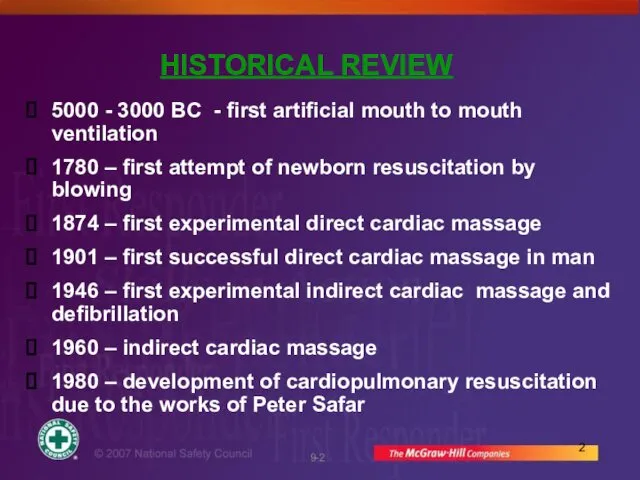
HISTORICAL REVIEW
5000 - 3000 BC - first artificial mouth to
HISTORICAL REVIEW
5000 - 3000 BC - first artificial mouth to
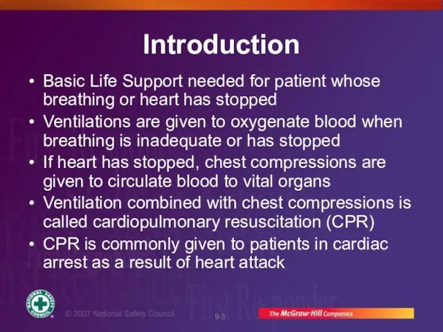
Introduction
Basic Life Support needed for patient whose breathing or heart has
Introduction
Basic Life Support needed for patient whose breathing or heart has

Review of Circulatory System
Circulatory system consists of heart, blood, and blood
Review of Circulatory System
Circulatory system consists of heart, blood, and blood
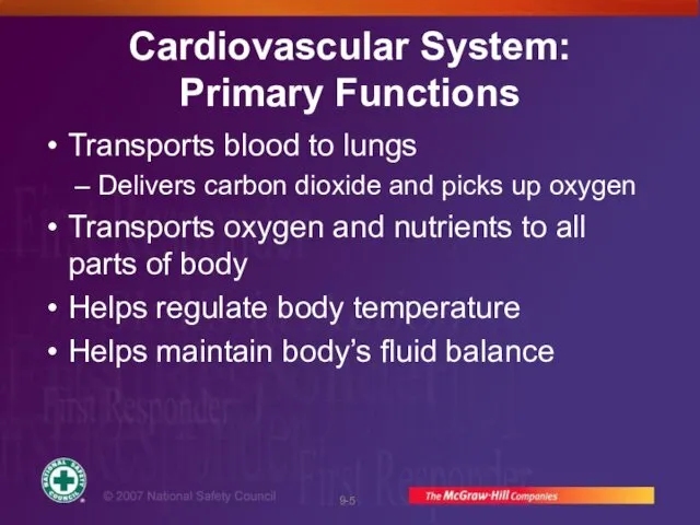
Cardiovascular System:
Primary Functions
Transports blood to lungs
Delivers carbon dioxide and picks
Cardiovascular System:
Primary Functions
Transports blood to lungs
Delivers carbon dioxide and picks

Anatomy and Physiology of the Heart
Ventricles pump blood through two
Anatomy and Physiology of the Heart
Ventricles pump blood through two
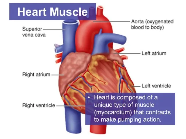
Heart is composed of a unique type of muscle (myocardium) that
Heart is composed of a unique type of muscle (myocardium) that

Contractions are controlled by electrical signals under nervous system control
Heart Muscle
Contractions are controlled by electrical signals under nervous system control
Heart Muscle

Arteries
Arterial blood is oxygenated, bright red, and under pressure
Carotid arteries —
Arteries
Arterial blood is oxygenated, bright red, and under pressure
Carotid arteries —

Pulse
When left ventricle contracts, wave of blood is sent through arteries
Pulse
When left ventricle contracts, wave of blood is sent through arteries

Pulse continued
Palpate femoral pulse in crease between abdomen and thigh
Palpate radial
Pulse continued
Palpate femoral pulse in crease between abdomen and thigh
Palpate radial

Capillaries
Arteries progressively branch into smaller vessels that eventually reach capillaries
Capillaries are
Capillaries
Arteries progressively branch into smaller vessels that eventually reach capillaries
Capillaries are
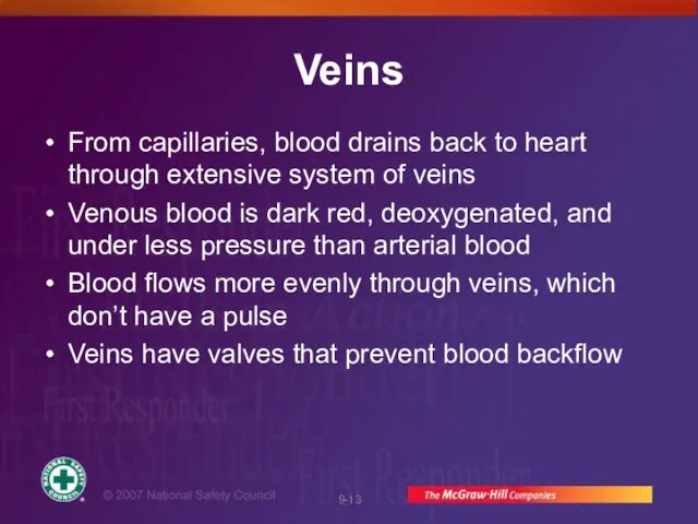
Veins
From capillaries, blood drains back to heart through extensive system of
Veins
From capillaries, blood drains back to heart through extensive system of
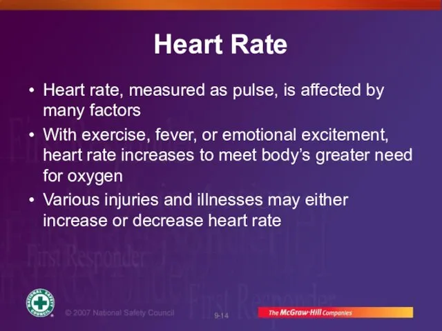
Heart Rate
Heart rate, measured as pulse, is affected by many factors
With
Heart Rate
Heart rate, measured as pulse, is affected by many factors
With

Circulatory System: Emergencies
Any condition that affects respiration
Reduces ability to deliver oxygen
Severe
Circulatory System: Emergencies
Any condition that affects respiration
Reduces ability to deliver oxygen
Severe

Circulatory System: Emergencies continued
Heart attack
Can lead to cardiac arrest
Ventricular fibrillation
Heart muscle
Circulatory System: Emergencies continued
Heart attack
Can lead to cardiac arrest
Ventricular fibrillation
Heart muscle

Cardiac Arrest
Heart may stop (cardiac arrest) as a result of heart
Cardiac Arrest
Heart may stop (cardiac arrest) as a result of heart
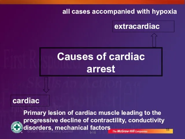
Causes of cardiac arrest
cardiac
extracardiac
Primary lesion of cardiac muscle leading to the
Causes of cardiac arrest
cardiac
extracardiac
Primary lesion of cardiac muscle leading to the

Causes of Cardiac Arrest
Heart attack
Drowning
Suffocation
Stroke
Allergic reaction
Diabetic emergency
Prolonged seizures
Drug overdose
Electric shock
Certain injuries
Causes of Cardiac Arrest
Heart attack
Drowning
Suffocation
Stroke
Allergic reaction
Diabetic emergency
Prolonged seizures
Drug overdose
Electric shock
Certain injuries

Causes of circulatory arrest
Cardiac
Ischemic heart disease (myocardial infarction, stenocardia)
Arrhythmias of different
Causes of circulatory arrest
Cardiac
Ischemic heart disease (myocardial infarction, stenocardia)
Arrhythmias of different

Cardiac Chain of Survival
Cardiac Chain of Survival
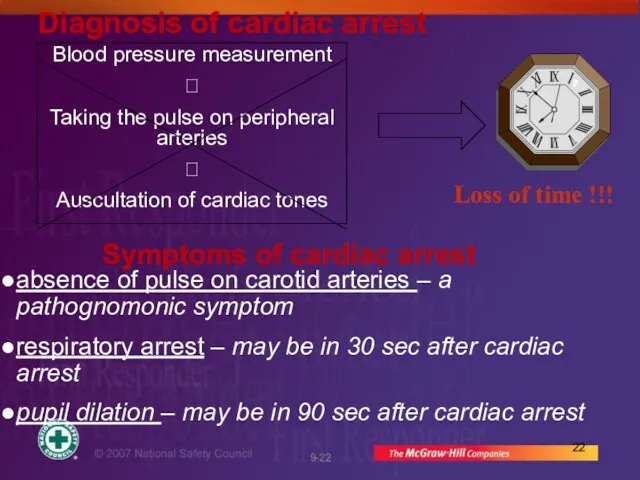
Diagnosis of cardiac arrest
Symptoms of cardiac arrest
absence of pulse on
Diagnosis of cardiac arrest
Symptoms of cardiac arrest
absence of pulse on

Sequence of operations
Check responsiveness
Call for help
Correctly place the
Sequence of operations
Check responsiveness
Call for help
Correctly place the
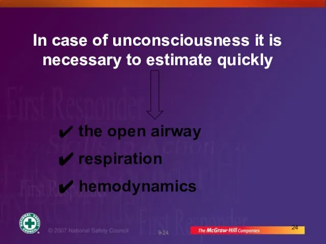
In case of unconsciousness it is necessary to estimate quickly
the
In case of unconsciousness it is necessary to estimate quickly
the

Main stages of resuscitation
C (Circulation) – restore the circulation by external
Main stages of resuscitation
C (Circulation) – restore the circulation by external

A (Airway)
ensure open airway
A (Airway)
ensure open airway

Open the airway using a head tilt lifting of chin. Do
Open the airway using a head tilt lifting of chin. Do

B (Breathing)
Tilt the head back and listen for. If not breathing
B (Breathing)
Tilt the head back and listen for. If not breathing
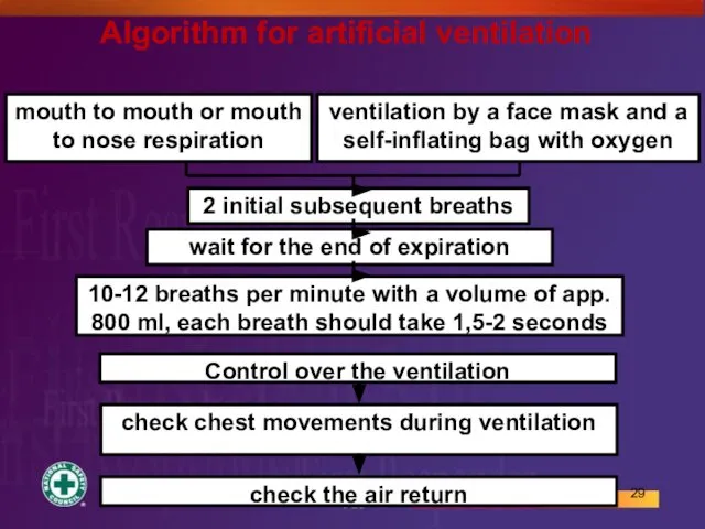
mouth to mouth or mouth to nose respiration
ventilation by a face
mouth to mouth or mouth to nose respiration
ventilation by a face

C. Circulation
Restore the circulation, that is start external cardiac massage
C. Circulation
Restore the circulation, that is start external cardiac massage

2 mechanisms explaining the restoration of circulation by external cardiac massage
Cardiac
2 mechanisms explaining the restoration of circulation by external cardiac massage
Cardiac

Cardiac pump during the cardiac massage
Blood pumping is assured by the
Cardiac pump during the cardiac massage
Blood pumping is assured by the
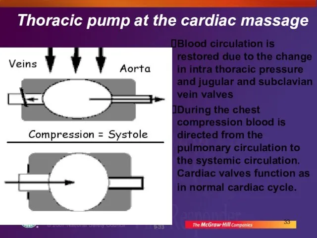
Thoracic pump at the cardiac massage
Blood circulation is restored due to
Thoracic pump at the cardiac massage
Blood circulation is restored due to

ALGORITHM of Cardiopulmonary resuscitation
4 cycles: 15 compression and 2 breaths
10 cycles:
ALGORITHM of Cardiopulmonary resuscitation
4 cycles: 15 compression and 2 breaths
10 cycles:
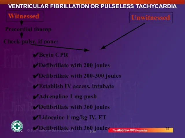
VENTRICULAR FIBRILLATION OR PULSELESS TACHYCARDIA
Witnessed
Unwitnessed
Precordial thump
Check pulse, if none:
Begin CPR
Defibrillate with
VENTRICULAR FIBRILLATION OR PULSELESS TACHYCARDIA
Witnessed
Unwitnessed
Precordial thump
Check pulse, if none:
Begin CPR
Defibrillate with
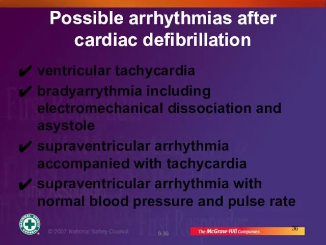
Possible arrhythmias after cardiac defibrillation
ventricular tachycardia
bradyarrythmia including electromechanical dissociation and asystole
supraventricular
Possible arrhythmias after cardiac defibrillation
ventricular tachycardia
bradyarrythmia including electromechanical dissociation and asystole
supraventricular

Operations in case of asystole
Asystole
Start CPR
IV line
Adrenaline:IV 1
Operations in case of asystole
Asystole
Start CPR
IV line
Adrenaline:IV 1
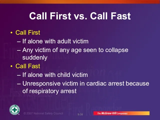
Call First vs. Call Fast
Call First
If alone with adult victim
Any victim
Call First vs. Call Fast
Call First
If alone with adult victim
Any victim
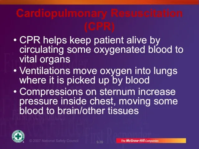
Cardiopulmonary Resuscitation (CPR)
CPR helps keep patient alive by circulating some oxygenated
Cardiopulmonary Resuscitation (CPR)
CPR helps keep patient alive by circulating some oxygenated

CPR continued
Blood circulation resulting from chest compressions not as strong as
CPR continued
Blood circulation resulting from chest compressions not as strong as

CPR continued
Often electric shock from AED is needed to restore a
CPR continued
Often electric shock from AED is needed to restore a

CPR Saves Lives
CPR and defibrillation within 3-5 minutes can save over
CPR Saves Lives
CPR and defibrillation within 3-5 minutes can save over

General Technique of CPR
If unresponsive, not breathing, and no pulse,
General Technique of CPR
If unresponsive, not breathing, and no pulse,

General Technique of CPR continued
Compress chest hard and fast at a
General Technique of CPR continued
Compress chest hard and fast at a
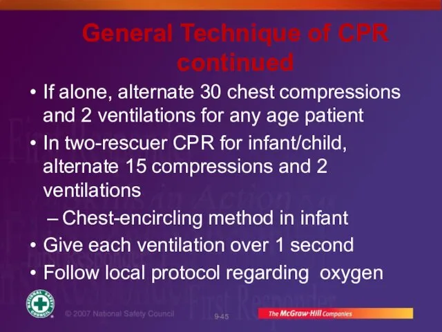
General Technique of CPR continued
If alone, alternate 30 chest compressions and
General Technique of CPR continued
If alone, alternate 30 chest compressions and

Single-Rescuer CPR
Check patient’s responsiveness,
open airway,
and determine that patient is
Single-Rescuer CPR
Check patient’s responsiveness,
open airway,
and determine that patient is
Single-Rescuer CPR
2. Give 2 ventilations, each lasting 1 second
3. Determine victim
Single-Rescuer CPR
2. Give 2 ventilations, each lasting 1 second
3. Determine victim
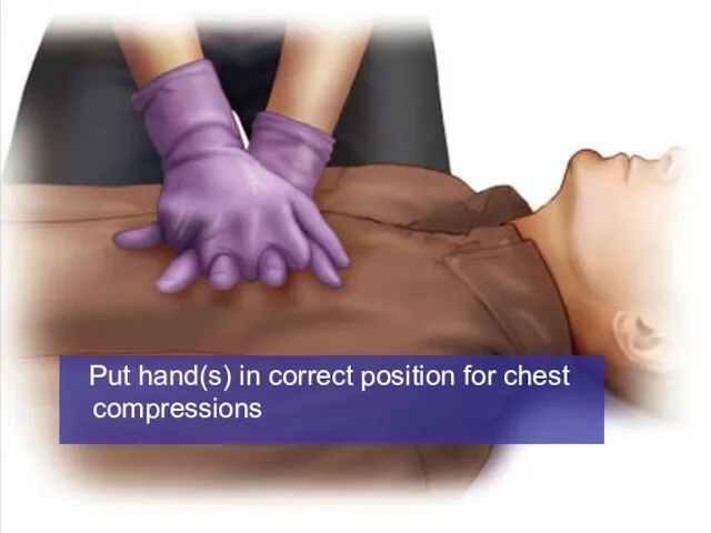
Put hand(s) in correct position for chest compressions
Put hand(s) in correct position for chest compressions

Give 30 chest compressions at rate of 100 per minute
Give 30 chest compressions at rate of 100 per minute

Continue CPR until:
Patient begins to move
AED brought to scene and ready
Continue CPR until:
Patient begins to move
AED brought to scene and ready
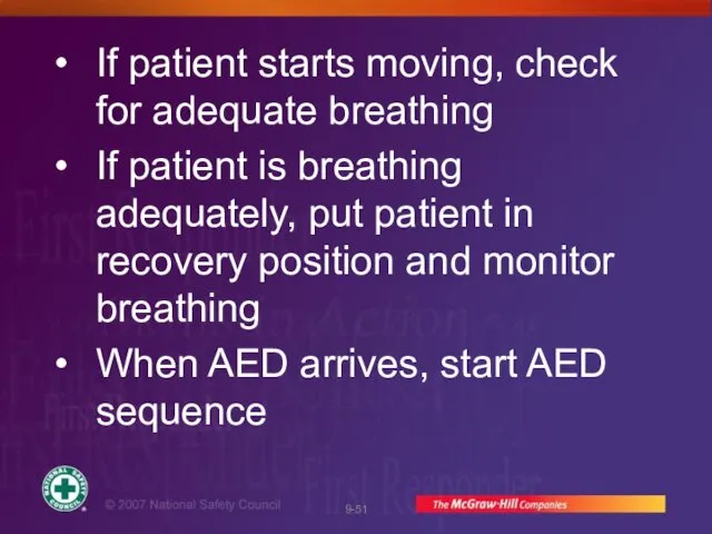
If patient starts moving, check for adequate breathing
If patient is breathing
If patient starts moving, check for adequate breathing
If patient is breathing

Chest Compressions Alert
Be careful with your hand position
For adults/children, keep your
Chest Compressions Alert
Be careful with your hand position
For adults/children, keep your

Chest Compressions Alert
When compressing, keep elbows straight and hands in contact
Chest Compressions Alert
When compressing, keep elbows straight and hands in contact

Chest Compressions Alert
Compress chest hard and fast, but let chest recoil
Chest Compressions Alert
Compress chest hard and fast, but let chest recoil

Problems with CPR Technique
CPR often ineffective because of poor technique
Compressions not
Problems with CPR Technique
CPR often ineffective because of poor technique
Compressions not

Chest Compressions: Bradycardia in Child
Infant or child being given rescue breaths
Chest Compressions: Bradycardia in Child
Infant or child being given rescue breaths

Skill:
CPR For Adult
or Child
(Two Rescuers)
Skill:
CPR For Adult
or Child
(Two Rescuers)

Two-Rescuer CPR for Adults and Children
Minimizes time between rescue breaths and
Two-Rescuer CPR for Adults and Children
Minimizes time between rescue breaths and

Two-Rescuer CPR
Performed in cycles of 30:2 for adult (15:2 for infant
Two-Rescuer CPR
Performed in cycles of 30:2 for adult (15:2 for infant
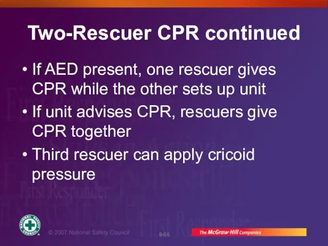
Two-Rescuer CPR continued
If AED present, one rescuer gives CPR while the
Two-Rescuer CPR continued
If AED present, one rescuer gives CPR while the
Two-Rescuer CPR continued
If you are assisting another trained rescuer who places
Two-Rescuer CPR continued
If you are assisting another trained rescuer who places
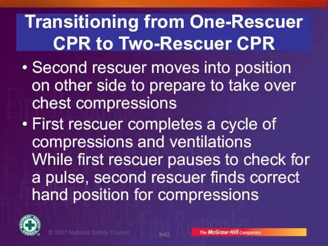
Transitioning from One-Rescuer CPR to Two-Rescuer CPR
Second rescuer moves into position
Transitioning from One-Rescuer CPR to Two-Rescuer CPR
Second rescuer moves into position

Transitioning from One-Rescuer CPR to Two-Rescuer CPR
When first rescuer says, “No
Transitioning from One-Rescuer CPR to Two-Rescuer CPR
When first rescuer says, “No

Differences in Two-Rescuer Training
If First Responder started CPR, arriving second rescuer
Differences in Two-Rescuer Training
If First Responder started CPR, arriving second rescuer

Rescuer 1 checks ABCs. Rescuer 2 locates site for chest
Rescuer 1 checks ABCs. Rescuer 2 locates site for chest

If no pulse, rescuer 2 gives 30 compressions for adult
If no pulse, rescuer 2 gives 30 compressions for adult

Rescuer 1 gives 2 breaths.
Rescuer 1 gives 2 breaths.

Continue cycles of 30:2 for adults (15:2 for child). After
Continue cycles of 30:2 for adults (15:2 for child). After

Adult or Child Two-Rescuer CPR Continued
Continue CPR until:
Patient moves
AED brought to
Adult or Child Two-Rescuer CPR Continued
Continue CPR until:
Patient moves
AED brought to
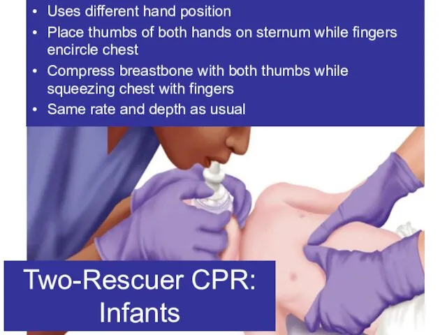
Uses different hand position
Place thumbs of both hands on sternum while
Uses different hand position
Place thumbs of both hands on sternum while
Skill:
CPR: Infants
Two Rescuers
Skill:
CPR: Infants
Two Rescuers

Rescuer 1 checks ABCs. Rescuer 2 locates site for chest
Rescuer 1 checks ABCs. Rescuer 2 locates site for chest

If no pulse, rescuer 2 gives 15 chest compressions.
If no pulse, rescuer 2 gives 15 chest compressions.

Rescuer 1 gives 2 breaths.
Rescuer 1 gives 2 breaths.
Infant Two-Rescuer CPR Continued
Continue cycles of 15:2 for ~ 2
Infant Two-Rescuer CPR Continued
Continue cycles of 15:2 for ~ 2

When Not to Perform CPR
Presence of a Do-Not-Resuscitate (DNR) order
Patient
When Not to Perform CPR
Presence of a Do-Not-Resuscitate (DNR) order
Patient
 Паллиативные операции на толстой кишке
 Perm State Medical University named after Academician E.A. Wagner. Medical education in Germany
 Переживання захворювання онкологічно хворими
 Анализ статей и их критическая оценка. Специальность: психиатрия и наркология
 Влияние детского массажа на новорожденных с неонатальной желтухой, получающих фототерапию
 Жатыр қатерлі ісігі
 Иммунопатология
 Патология пищеварительной системы
 Неврит и невралгия тройничного нерва
 Учет медицинской помощи по профилю химиотерапии
 Методы обследования стоматологического больного
 Инвазивный мониторинг в нейрохирургии - дорогие игрушки или путь к спасению жизни человека
 Медицинское страхование в Российской Федерации и за рубежом
 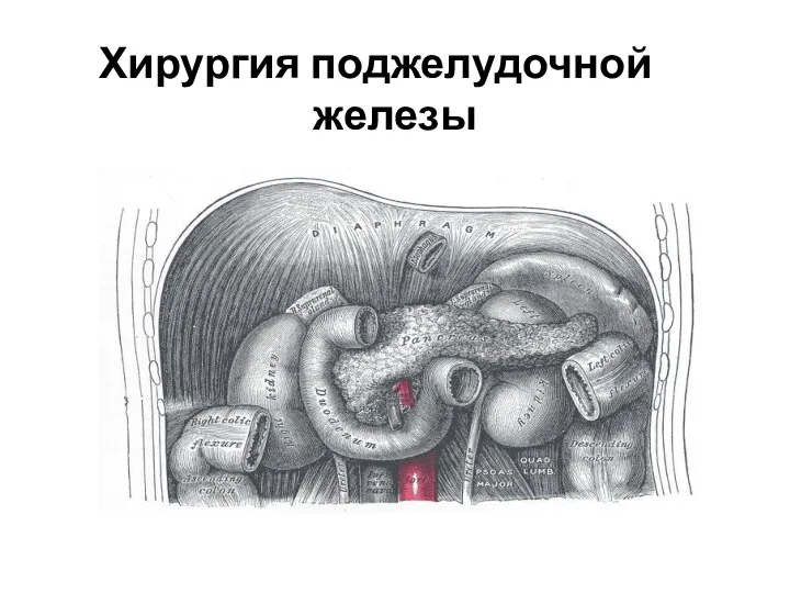 Хирургия поджелудочной железы
 Психологическая помощь в Калуге
 Планирование семьи. Регулирование рождаемости. Бесплодный брак
 Лекарства и яды в древности
 Обмен отдельных классов органических соединений
 Кистовидные отёки
 Вирусный гепатит В
 Инсулинома. Разбор клинического случая
 Общение как взаимодействие
 Неотложные состояния в аллергологии
 Пороки развития уха
 Киста гартнерова хода
 Преимущества грудного вскармливания
 Научные статьи в электронных базах о длительности грудного вскармливания и заболевании детей
 Clinica stomatologică srl „omni dent” teza de licență aspecte ale tratamentului chirurgical în afecțiunile parodonțiului marginal