- Electromyography

Содержание
- 2. What is electromyography? Electromyography (EMG) is a diagnostic procedure that evaluates the health condition of muscles
- 3. Why is electromyography performed? Your doctor may perform an EMG if you’re experiencing symptoms that may
- 4. What happens during an electromyography? There are two components to an EMG test: the nerve conduction
- 5. What do my electromyography results mean? If your EMG shows any electrical activity in a resting
- 7. Скачать презентацию
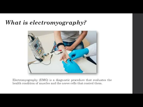
What is electromyography?
Electromyography (EMG) is a diagnostic procedure that evaluates the
What is electromyography?
Electromyography (EMG) is a diagnostic procedure that evaluates the

Why is electromyography performed?
Your doctor may perform an EMG if you’re
Why is electromyography performed?
Your doctor may perform an EMG if you’re
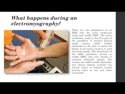
What happens during an electromyography?
There are two components to an EMG
What happens during an electromyography?
There are two components to an EMG
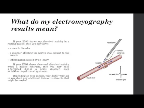
What do my electromyography results mean?
If your EMG shows any electrical
What do my electromyography results mean?
If your EMG shows any electrical
 Трансплантология. Тері, бұлшық ет, сіңір, жүйке, сүйек тінді қуысты ағзалардың пластикасы
 Синдром Мэллори-Вейса
 Роль сестринского персонала при оперативных вмешательствах
 Гарантия качества планов IMRT в РНЦРР
 Профилактика коронавирусной инфекции (covid-19)
 Кесарево сечение в современном акушерстве
 Лептоспіроз
 YogaSiddhi_Sceleton System
 Диализ. Принцип действия диализа
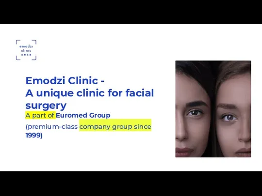 Emodzi Clinic A unique clinic for facial surgery
 Акушерлік стационардағы санитарлық – гигиеналық режимнің ерекшеліктері
 Роль прививок в поддержании здоровья населения
 Фармакология наружных лекарственных препаратов
 Сердечно-легочная реанимация
 Тромбоз. Эмболия
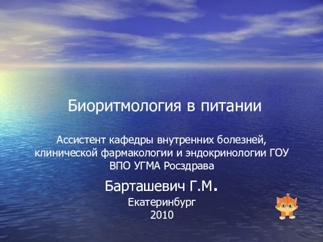 Биоритмология в питании
 Рак желудка
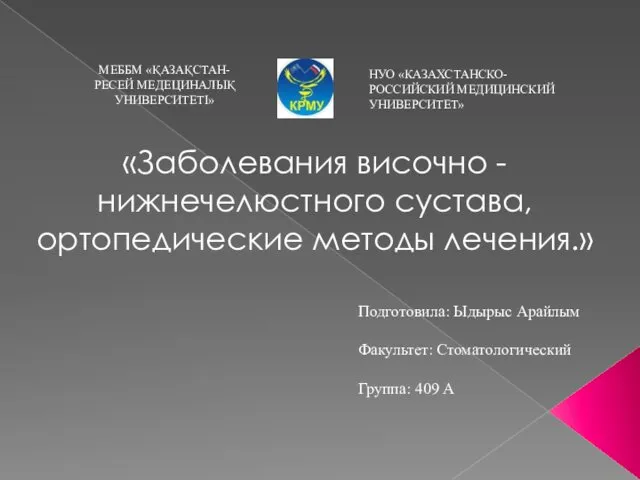 Заболевания височно-нижнечелюстного сустава, ортопедические методы лечения
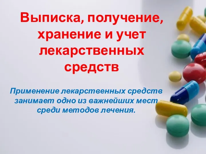 Выписка, получение, хранение и учет лекарственных средств
 Онкологические заболевания женских половых органов
 Хирургическая анатомия фасций и клетчаточных пространств конечностей
 Отчет о работе акушерского дистанционного центра
 Роль православия в развитии педиатрии: от истоков к современности
 Injections. Punctures
 Жыныстық жетілудің болмауы
 Балалардың ақпаратты қабылдау қабілетін бағалаудың психофизиологиялық негіздері
 Здоровая пища
 Методы исследования в психологии