- Features of childrens` blood. The semiotics of major hematological syndromes

Содержание
- 2. The blood creations or hemogenesis is a process of origin and subsequent ripening of blood cells
- 3. The single cell that gives life to all blood cells is the polypotentic trunk cell of
- 4. About the blood condition we can judge on the base of peripheral blood composition making blood
- 5. Functions of blood and its formed elements. The functions of blood and its formed elements are
- 6. Using blood circulation the nutrients and oxygen are delivered to the tissues and the final metabolites
- 7. The blood cells also execute the great number of functions. The long-living (to 100 days) red
- 8. The leucocytes disintegrating feed intensively proliferative tissues by the products contained in their kernels. The platelet
- 9. The concept of embryonic hemogenesis. In the period of prenatal life of fetus 3 periods of
- 10. The first stage of hemogenesis is revealed in a 19-th daily aged embryo in the bloody
- 11. The second (hepatic-splenic) period begins after 6 weeks of intrauteral development and achieves its maximum at
- 12. The third or bone marrow period of hemogenesis which gradually becomes determinative in the production of
- 13. According to the different periods of hemogenesis (extraembryonic, hepatic and bone marrow) there are three different
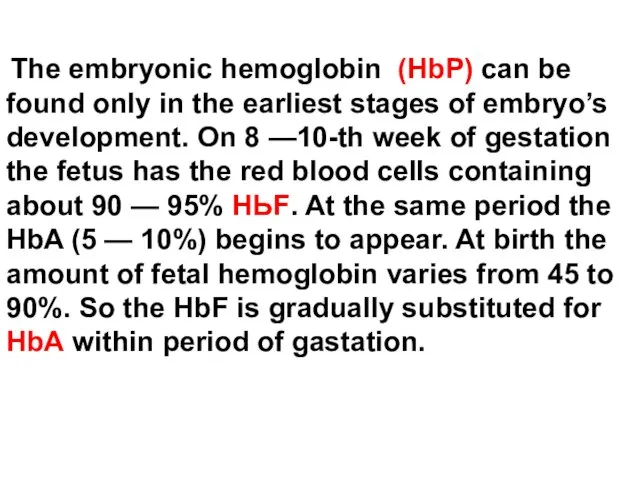
- 14. The embryonic hemoglobin (HbP) can be found only in the earliest stages of embryo’s development. On
- 15. In children aged one year the common hemoglobin level remains about 15% of НЬF and in
- 16. Hemogenesis after the birth. Newborns’ marrow is the general source of all blood cells production. At
- 17. The rapidly exhausted marrow is incident to the neonatal period of life. After an unfavorable influences
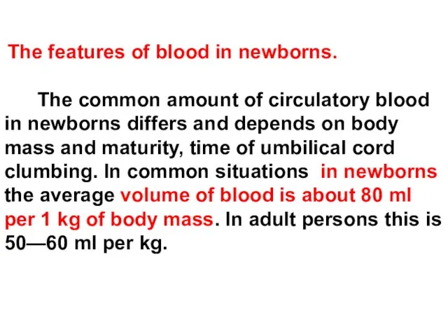
- 18. The features of blood in newborns. The common amount of circulatory blood in newborns differs and
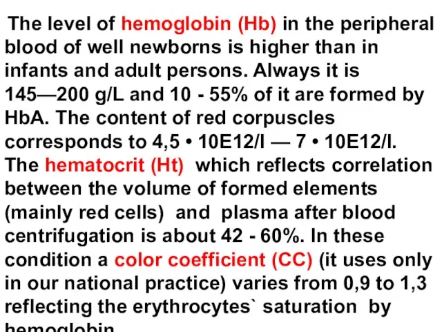
- 19. The level of hemoglobin (Hb) in the peripheral blood of well newborns is higher than in
- 20. From the first hours after birth the disintegration of red cells begins. This process clinically causes
- 21. The range of normal variations of general leucocytes count (WBC) at birth is wide enough and
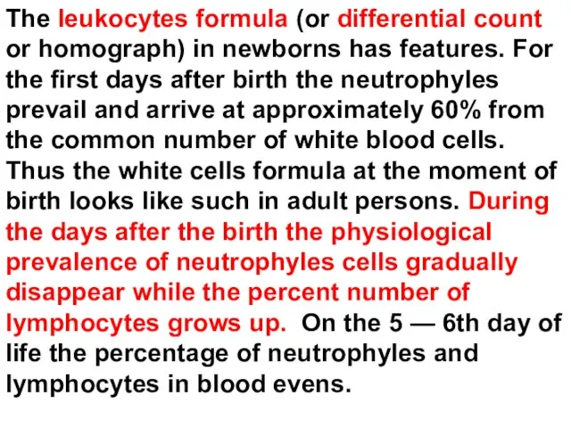
- 22. The leukocytes formula (or differential count or homograph) in newborns has features. For the first days
- 23. The physiological prevalence of lymphocytes in comparison with other white cells is being the normal phenomenon
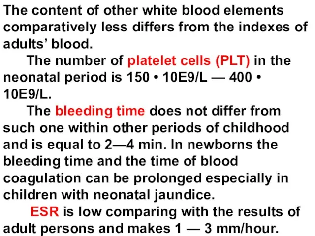
- 24. The content of other white blood elements comparatively less differs from the indexes of adults’ blood.
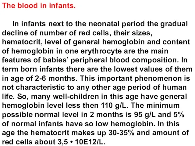
- 25. The blood in infants. In infants next to the neonatal period the gradual decline of number
- 26. The average volume of one erythrocyte in children in this age is equal to 75 -
- 27. The amount of leucocytes in healthy infants vary in limits since 5 • 10E9/L until 15•
- 28. Second and subsequent years of child’s life Since the beginning of the second year of life
- 29. In a leukocytic formula in children aged 3-4 years there is the tendency to gradual growth
- 30. Semyotics of blood changes. The modern blood test includes: - Research of blood cellular composition. -
- 31. red blood cells
- 32. The state of red blood cells is characterized by next clinical and laboratory parameters. 1. The
- 33. There are some exceptions. This value is necessary to increase up to 135 g/L in newborns
- 34. If the average content of Hb (Mean corpuscular hemoglobin concentration MCH) in one red cell is
- 35. With high accuracy the hematocryt (Ht, %) reflects a volume which is occupied by red cells
- 36. The amount of red cells (RBC) can be also calculated. The content of red cells less
- 37. The average volume of one red cell (Mean corpuscular volume – MCV) can be also measured
- 38. There is the extra - systematic value of erythrocytes system called color coefficient. In proper measure
- 39. Morphologic abnormalities of the red blood cell. A.(left above) Normal. B.(right above etc.) Macrocytes (folic acid
- 40. The anemic syndromes. The anemic syndrome is defined as a deficit of hemoglobin and/or red cells
- 41. According laboratory data all cases of anemic syndrome or anemia can be divided into a several
- 42. Iron - deficiency anemia and anemia, caused by deficit of other components necessary for the Hb
- 43. For iron-deficient anemia it is typical of: The general hemoglobin is below than age dependant normal
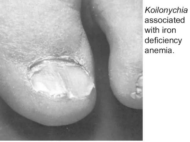
- 44. Koilonychia associated with iron deficiency anemia.
- 45. The symptoms of promoted fatigueability in children can look like as diminished motor activity. The pallor
- 46. The cause of anemia can be a deficit in the child’s organism of vitamin В12 and
- 47. The anemia due to bleeding first of all is attributed to them. The fact of the
- 48. As a result of red cells haemolysis also the normochromic, normocytal anemias develop. A jaundice (yellowish
- 49. Finally, the normochromic and normocytal anemias develop as a result of red cells making lack in
- 50. The semyotics of red cells number increase 1. Typically the abnormally high numbers of red cells
- 51. Leucocytes
- 52. The characteristics of leukocytes are: 1) The value of leucocytes count (WBC) in unit of blood
- 53. Leukocytosis The leukocytes count (WBC) is defined as a content of leucocytes of all types. Except
- 54. The leukocytes count (WBC) exceeding 20 х 10E9/L is named hyperleukocytosis or leukemoid reaction because this
- 55. Neutrophylic leukocytosis (ANC) and leukemoid reaction of neutrophylic type appear in festering-inflammatory processes. The appearance in
- 56. Eozynophilia (over 5% from numbers of leukocytes formula or AEOSC 0,5 х 10E9 cells in 1
- 57. Lymphocytosis. The amount of lymphocytes considerably increases in viral diseases like the infectious lymphocytosis and whooping
- 58. Monocytosis is characteristic for infectious mononucleosis caused by the Ebstain-Barr viral disease. MON
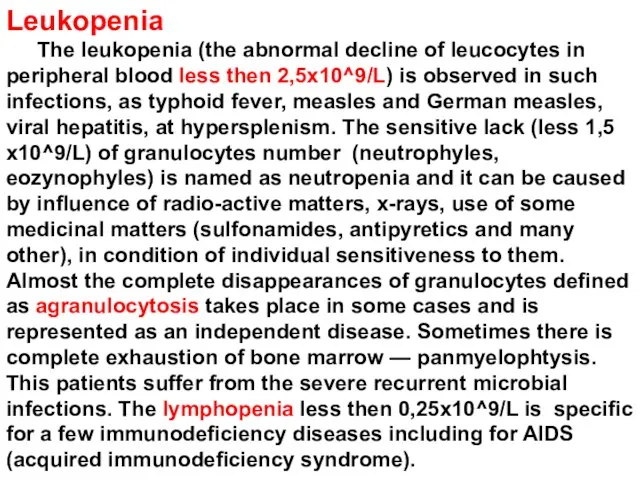
- 59. Leukopenia The leukopenia (the abnormal decline of leucocytes in peripheral blood less then 2,5х10^9/L) is observed
- 60. coagulative properties of blood
- 61. The adequate to the trauma early bleeding is beginning right away after the damage of vessel
- 62. The components of haemostasis are: - vascular wall - thrombocytes (platelet cells) - plasma factors of
- 63. For the hemorrhagic syndrome the inadequate easy bleeding and also late, deferred from a moment of
- 64. The damage of any component of hemostasis causes haemorrhagic syndromes which subdivide on: - vasopathy (vessel
- 65. In accordance of represented factors of hemorrhagic disease 3 clinical types of pathologic bleeding are: 1.
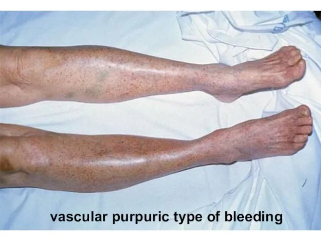
- 66. The vascular purpuric type of bleeding shows up as the small-spotted hemorrhagic rash (skin eruptions) sometimes
- 67. vascular purpuric type of bleeding
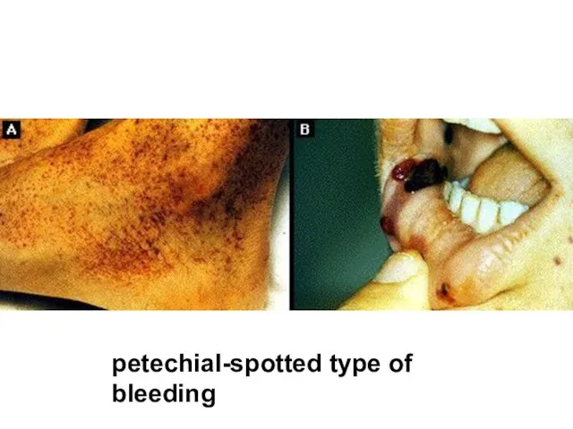
- 68. The petechial-spotted type of bleeding is also wide spread in pediatric practice. On a skin spontaneously
- 69. petechial-spotted type of bleeding
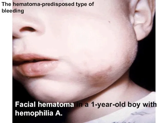
- 70. The hematoma-predisposed type. There are large, deep, very painful intermuscular haemorrhage and bleeding in to the
- 71. Facial hematoma in a 1-year-old boy with hemophilia A. The hematoma-predisposed type of bleeding
- 72. biophysical properties of blood (ESR)
- 73. ESR (erythrocytes sedimentation reaction). The spontaneous (not to mix up with hematocrit) concretion of red cells
- 74. ESR increases due to raise of serum fybrinogen, immunoglobulines, gaptoglobin, cholesterol concentrations. It elevates also in
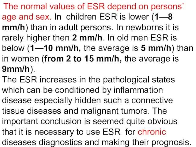
- 75. The normal values of ESR depend on persons` age and sex. In children ESR is lower
- 76. At what age in childhood the hematocrit has the lowest normal level? A. 1 hour B.
- 77. At what age in childhood the hematocrit has the lowest normal level? A. 1 hour B.
- 78. Tiny, flat, round, red or purple spot on skin caused by minute submucosal or intradermal hemorrhage
- 79. Tiny, flat, round, red or purple spot on skin caused by minute submucosal or intradermal hemorrhage
- 80. The microcytic anemia is associated with: hemolytic disorders folic acid deficiency due to bleeding chronic inflammation
- 81. The microcytic anemia is associated with: hemolytic disorders folic acid deficiency due to bleeding chronic inflammation
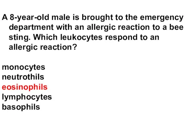
- 82. A 8-year-old male is brought to the emergency department with an allergic reaction to a bee
- 83. A 8-year-old male is brought to the emergency department with an allergic reaction to a bee
- 84. Normal hemoglobin (Hb) values to neonate is: 70 to 110 g/l 110 to 130 g/l 140
- 86. Скачать презентацию
The blood creations or hemogenesis is a process of origin and
The blood creations or hemogenesis is a process of origin and
The single cell that gives life to all blood cells is
About the blood condition we can judge on the base
About the blood condition we can judge on the base
Functions of blood and its formed elements.
The functions of blood
Functions of blood and its formed elements.
The functions of blood
Using blood circulation the nutrients and oxygen are delivered to the
Using blood circulation the nutrients and oxygen are delivered to the
The blood cells also execute the great number of functions. The
The blood cells also execute the great number of functions. The
The leucocytes disintegrating feed intensively proliferative tissues by the products contained
The leucocytes disintegrating feed intensively proliferative tissues by the products contained
The concept of embryonic hemogenesis.
In the period of prenatal life
The concept of embryonic hemogenesis.
In the period of prenatal life
The first stage of hemogenesis is revealed in a 19-th daily
The second (hepatic-splenic) period begins after 6 weeks of intrauteral development
The second (hepatic-splenic) period begins after 6 weeks of intrauteral development
The third or bone marrow period of hemogenesis which gradually
The third or bone marrow period of hemogenesis which gradually
According to the different periods of hemogenesis (extraembryonic, hepatic and bone
The embryonic hemoglobin (HbP) can be found only in the
The embryonic hemoglobin (HbP) can be found only in the
In children aged one year the common hemoglobin level remains about
In children aged one year the common hemoglobin level remains about
Hemogenesis after the birth.
Newborns’ marrow is the general source of
Hemogenesis after the birth.
Newborns’ marrow is the general source of
The rapidly exhausted marrow is incident to the neonatal period of
The rapidly exhausted marrow is incident to the neonatal period of
The features of blood in newborns.
The common amount of
The features of blood in newborns.
The common amount of
The level of hemoglobin (Hb) in the peripheral blood of
The level of hemoglobin (Hb) in the peripheral blood of
From the first hours after birth the disintegration of red cells
The range of normal variations of general leucocytes count (WBC)
The range of normal variations of general leucocytes count (WBC)
The leukocytes formula (or differential count or homograph) in newborns has
The leukocytes formula (or differential count or homograph) in newborns has
The physiological prevalence of lymphocytes in comparison with other white cells
The physiological prevalence of lymphocytes in comparison with other white cells
The content of other white blood elements comparatively less differs from
The content of other white blood elements comparatively less differs from
The blood in infants.
In infants next to the neonatal
The blood in infants.
In infants next to the neonatal
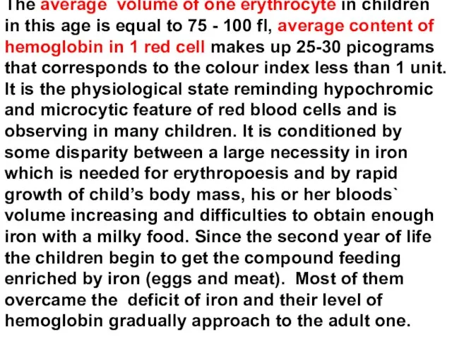
The average volume of one erythrocyte in children in this age
The average volume of one erythrocyte in children in this age
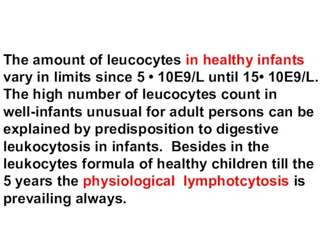
The amount of leucocytes in healthy infants vary in limits since
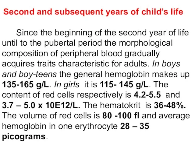
Second and subsequent years of child’s life
Since the beginning of
Second and subsequent years of child’s life
Since the beginning of
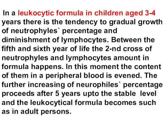
In a leukocytic formula in children aged 3-4 years there
In a leukocytic formula in children aged 3-4 years there
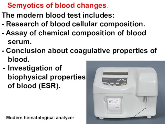
Semyotics of blood changes.
The modern blood test includes:
- Research of
Semyotics of blood changes.
The modern blood test includes:
- Research of
red blood cells
red blood cells
The state of red blood cells is characterized by next clinical
The state of red blood cells is characterized by next clinical
There are some exceptions. This value is necessary to increase up
There are some exceptions. This value is necessary to increase up
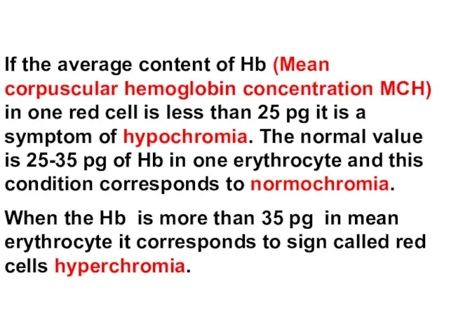
If the average content of Hb (Mean corpuscular hemoglobin concentration MCH)
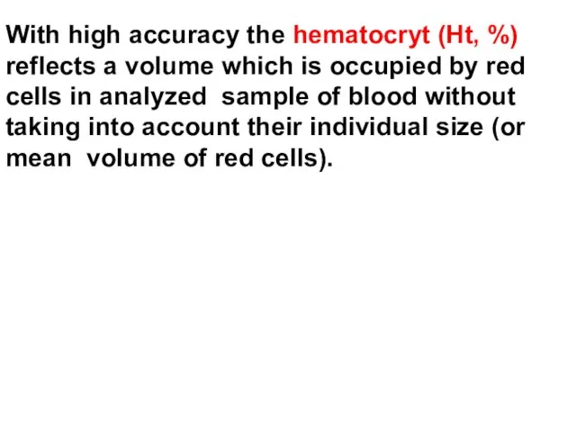
With high accuracy the hematocryt (Ht, %) reflects a volume which
With high accuracy the hematocryt (Ht, %) reflects a volume which
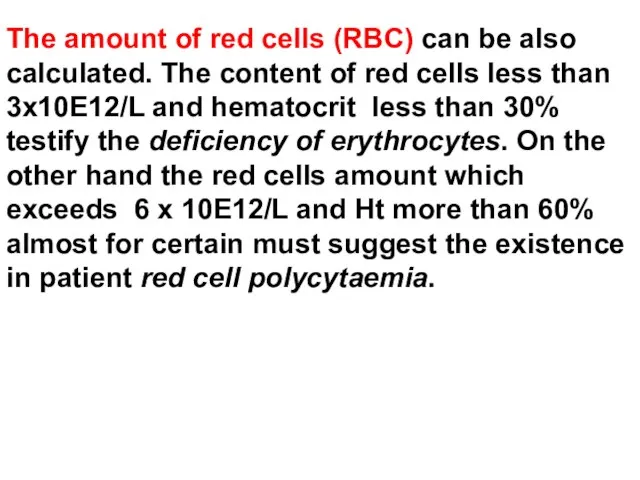
The amount of red cells (RBC) can be also calculated. The
The amount of red cells (RBC) can be also calculated. The
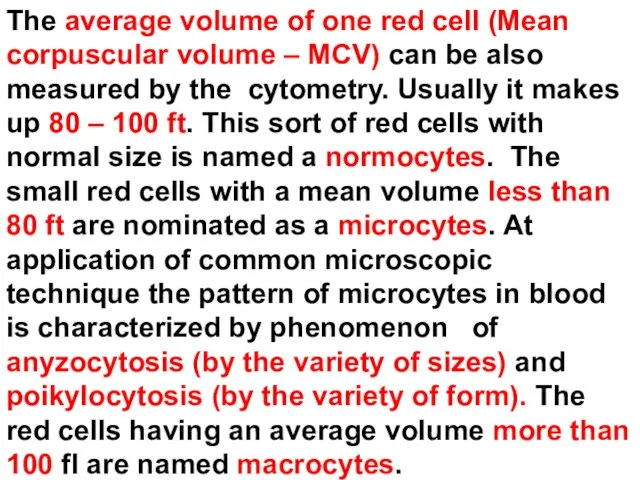
The average volume of one red cell (Mean corpuscular volume –
The average volume of one red cell (Mean corpuscular volume –
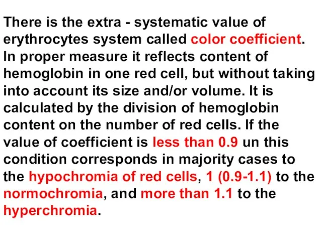
There is the extra - systematic value of erythrocytes system called
There is the extra - systematic value of erythrocytes system called
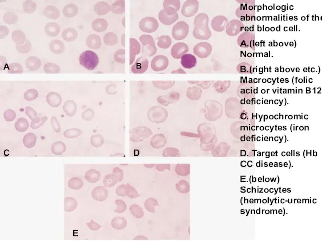
Morphologic abnormalities of the red blood cell.
A.(left above) Normal.
B.(right
Morphologic abnormalities of the red blood cell.
A.(left above) Normal.
B.(right
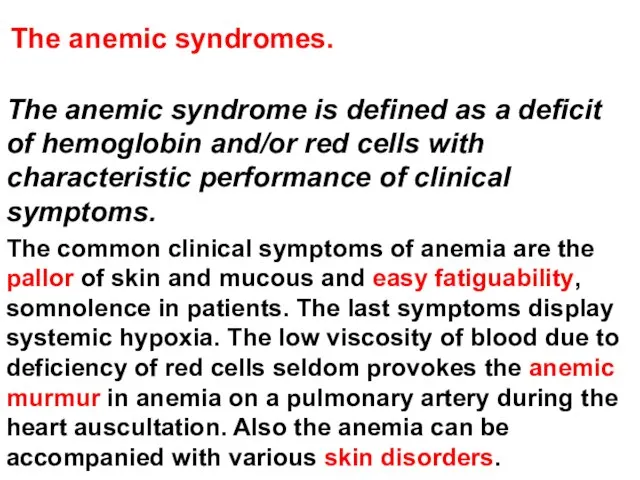
The anemic syndromes.
The anemic syndrome is defined as a deficit
The anemic syndromes.
The anemic syndrome is defined as a deficit
According laboratory data all cases of anemic syndrome or anemia
According laboratory data all cases of anemic syndrome or anemia
Iron - deficiency anemia and anemia, caused by deficit of other
Iron - deficiency anemia and anemia, caused by deficit of other
For iron-deficient anemia it is typical of:
The general hemoglobin is below
For iron-deficient anemia it is typical of:
The general hemoglobin is below
Koilonychia associated with iron deficiency anemia.
Koilonychia associated with iron deficiency anemia.
The symptoms of promoted fatigueability in children can look like as
The symptoms of promoted fatigueability in children can look like as
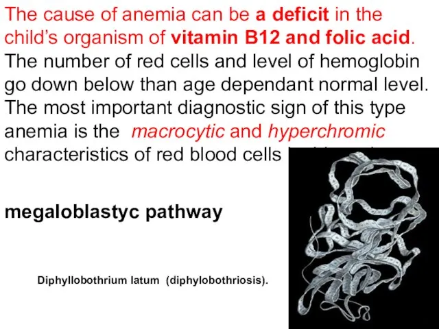
The cause of anemia can be a deficit in the child’s
The cause of anemia can be a deficit in the child’s
The anemia due to bleeding first of all is attributed to
The anemia due to bleeding first of all is attributed to
As a result of red cells haemolysis also the normochromic,
As a result of red cells haemolysis also the normochromic,
Finally, the normochromic and normocytal anemias develop as a result
Finally, the normochromic and normocytal anemias develop as a result
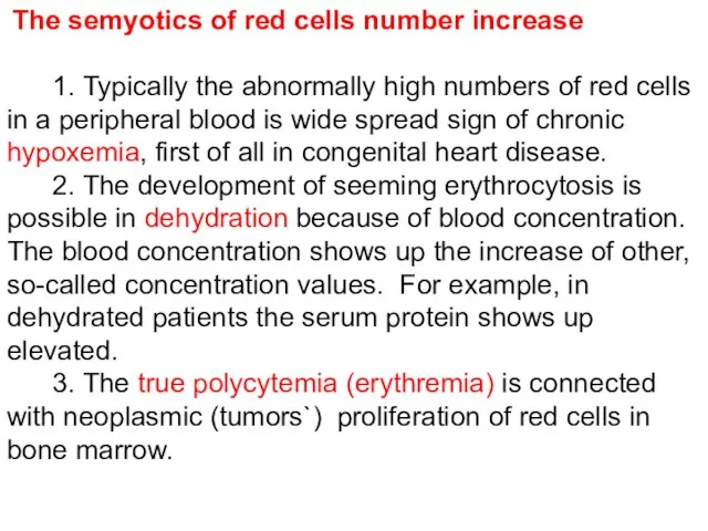
The semyotics of red cells number increase
1. Typically
The semyotics of red cells number increase
1. Typically

Leucocytes
Leucocytes
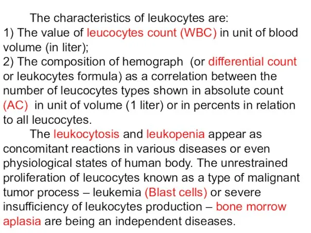
The characteristics of leukocytes are:
1) The value of
The characteristics of leukocytes are:
1) The value of
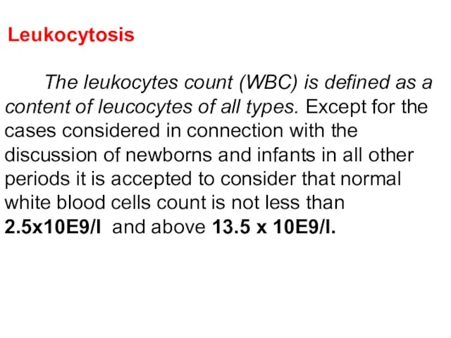
Leukocytosis
The leukocytes count (WBC) is defined as a content
Leukocytosis
The leukocytes count (WBC) is defined as a content
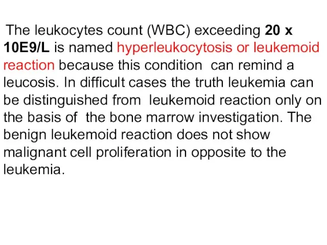
The leukocytes count (WBC) exceeding 20 х 10E9/L is named
The leukocytes count (WBC) exceeding 20 х 10E9/L is named
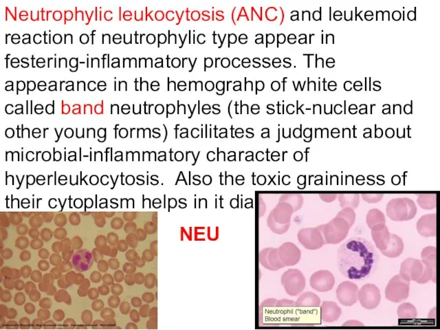
Neutrophylic leukocytosis (ANC) and leukemoid reaction of neutrophylic type appear in
Neutrophylic leukocytosis (ANC) and leukemoid reaction of neutrophylic type appear in
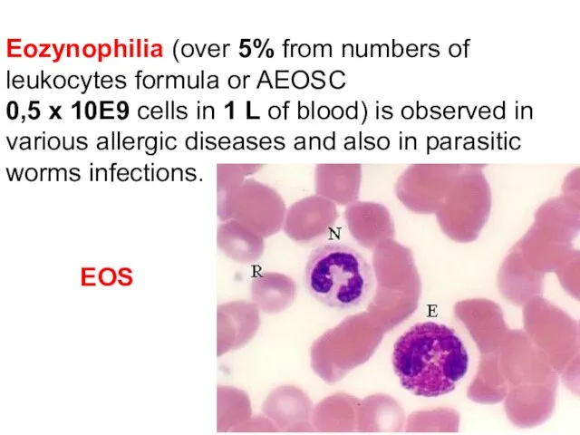
Eozynophilia (over 5% from numbers of leukocytes formula or AEOSC
0,5 х
Eozynophilia (over 5% from numbers of leukocytes formula or AEOSC
0,5 х
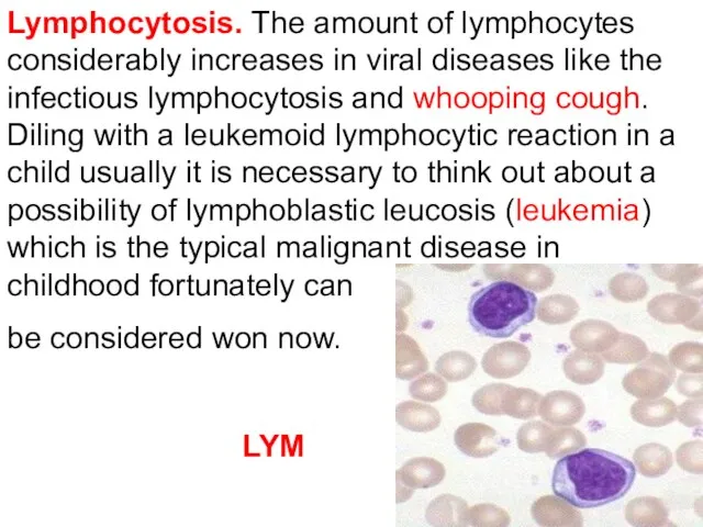
Lymphocytosis. The amount of lymphocytes considerably increases in viral diseases like
Lymphocytosis. The amount of lymphocytes considerably increases in viral diseases like
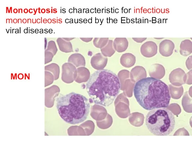
Monocytosis is characteristic for infectious mononucleosis caused by the Ebstain-Barr viral
Monocytosis is characteristic for infectious mononucleosis caused by the Ebstain-Barr viral
Leukopenia
The leukopenia (the abnormal decline of leucocytes in peripheral blood
Leukopenia
The leukopenia (the abnormal decline of leucocytes in peripheral blood
coagulative properties of blood
The adequate to the trauma early bleeding is beginning right away
The adequate to the trauma early bleeding is beginning right away
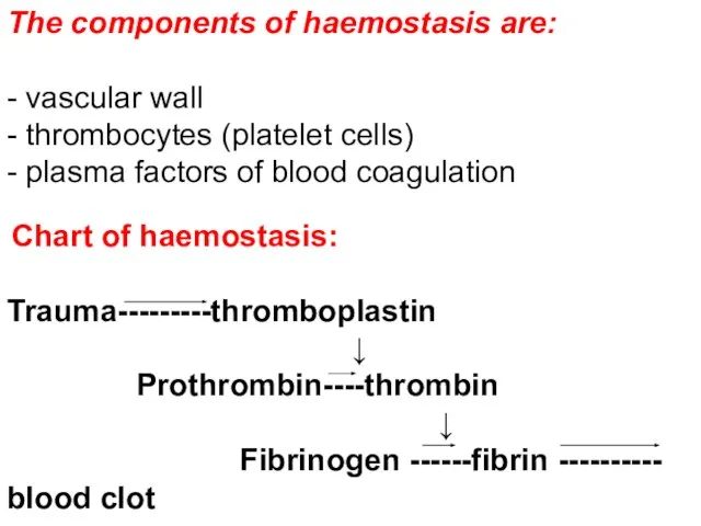
The components of haemostasis are:
- vascular wall
- thrombocytes (platelet cells)
- plasma
The components of haemostasis are:
- vascular wall
- thrombocytes (platelet cells)
- plasma
For the hemorrhagic syndrome the inadequate easy bleeding and also late,
The damage of any component of hemostasis causes haemorrhagic syndromes which
In accordance of represented factors of hemorrhagic disease 3 clinical types
In accordance of represented factors of hemorrhagic disease 3 clinical types
The vascular purpuric type of bleeding shows up as the
The vascular purpuric type of bleeding shows up as the
vascular purpuric type of bleeding
vascular purpuric type of bleeding
The petechial-spotted type of bleeding is also wide spread in pediatric
The petechial-spotted type of bleeding is also wide spread in pediatric
petechial-spotted type of bleeding
petechial-spotted type of bleeding
The hematoma-predisposed type. There are large, deep, very painful intermuscular haemorrhage
The hematoma-predisposed type. There are large, deep, very painful intermuscular haemorrhage
Facial hematoma in a 1-year-old boy with hemophilia A.
The hematoma-predisposed
Facial hematoma in a 1-year-old boy with hemophilia A.
The hematoma-predisposed
biophysical properties of blood
(ESR)
biophysical properties of blood
(ESR)
ESR (erythrocytes sedimentation reaction).
The spontaneous (not to mix up with
ESR (erythrocytes sedimentation reaction).
The spontaneous (not to mix up with
ESR increases due to raise of serum fybrinogen, immunoglobulines, gaptoglobin, cholesterol
The normal values of ESR depend on persons` age and
The normal values of ESR depend on persons` age and
At what age in childhood the hematocrit has the lowest normal
At what age in childhood the hematocrit has the lowest normal
At what age in childhood the hematocrit has the lowest normal
At what age in childhood the hematocrit has the lowest normal
Tiny, flat, round, red or purple spot on skin caused by
Tiny, flat, round, red or purple spot on skin caused by
Tiny, flat, round, red or purple spot on skin caused by
Tiny, flat, round, red or purple spot on skin caused by
The microcytic anemia is associated with:
hemolytic disorders
folic acid
The microcytic anemia is associated with:
hemolytic disorders
folic acid
The microcytic anemia is associated with:
hemolytic disorders
folic acid
The microcytic anemia is associated with:
hemolytic disorders
folic acid
A 8-year-old male is brought to the emergency department with an
A 8-year-old male is brought to the emergency department with an
A 8-year-old male is brought to the emergency department with an
A 8-year-old male is brought to the emergency department with an
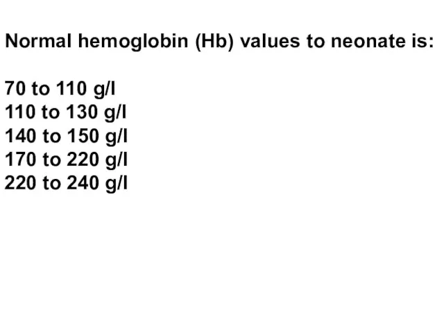
Normal hemoglobin (Hb) values to neonate is:
70 to 110 g/l
110 to
Normal hemoglobin (Hb) values to neonate is:
70 to 110 g/l
110 to
 Сестринская помощь при повреждениях мягких тканей, костей конечностей
 Сообщение печальных новостей
 Угревая болезнь
 Пять эффективных способов решения конфликтов
 Эпилепсия. Клинические проявления
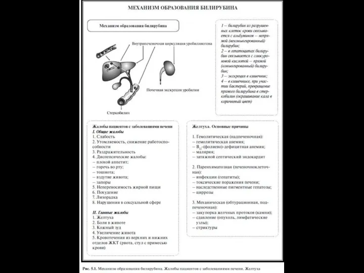 Механизм образования билирубина
 Блокаторы калиевых каналов. Амиодарон
 Паркинсон ауруы
 Зейін және ес бұзылыстарының клиникалық сипаттамасы
 Сестринский персонал в программах профилактики ВИЧ. Лекция 1
 Бронх демікпесі
 Выделительная система
 Балалардағы қант диабетінің алдын алу
 Дәрігер-науқас-мейірбеке қарым-қатнасы
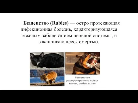 Бешенство (Rabies)
 Пункция заднего свода влагалища (кульдоцентез)
 ЭКГ при нарушениях ритма и проводимости
 Предпрофильная подготовка. Тип темперамента
 Формирование благоприятного социально-психологического климата в классном коллективе учащихся и родителей
 Трахеостомия
 Влагалищные пессарии: виды, показания, правила подбора, плюсы и минусы использования
 Школа Флоренс Найнтингейл
 Алғашқы медико-санитарлық көмекті ұйымдастыру
 Артериальная гипертензия и гипотензия во время анестезии и ближайшем послеоперационном периоде
 Осанка. Комплекс упражнений для формирования правильной осанки
 Балалардың асқазан - ішек аурулары туралы түсінік беру
 Гипреактивные дети
 Дифференциальная диагностика. Миастения