- Skin cancer. Melanoma

Содержание
- 2. UV radiation increase risk of skin cancer on 70%
- 3. Common Premalignant and Malignant Neoplasms of the Skin
- 4. Obligate precancerous Pigment kseroderma, Bowen disease, Eritroplasia Keyra
- 5. Facultative precancerous Keratoacantoma of nose, Diskeratosis in old man, seborrhea keratosis, skin horn
- 6. Basal cell carcinoma is a malignant tumor that rarely metastasizes. It is composed of cells that
- 7. Basal cell carcinomas account for more than 75% of keratinocytic skin cancers diagnosed in the United
- 8. Basal cell carcinoma
- 9. Basal cell carcinoma (pigment form)
- 10. Ulcerous type (ulcus rоdеns) (Basalioma terebrans)
- 11. Perforating basalioma
- 12. Cryosurgery
- 13. Basal cell carcinoma before and after cryosurgery
- 14. Squamous cell carcinoma is a malignant tumor arising from epidermal or appendageal keratinocytes or from the
- 15. Ways of lymphatic spread
- 16. Cancer of skin
- 17. Cancer of skin (exophyt form)
- 18. Cancer of skin. Advanced form
- 19. Malignant melanoma of skin accounts for 160,000 new cases annually, with slightly more occurring in women
- 20. Melanoma
- 21. Epidemiology
- 22. Dangerous nevus Nevus of Settona, Intradermal nevus, Blue nevus
- 23. Dangerous nevus gigantic nevus, Oto’s nevus, Ito’s nevus
- 24. Dangerous nevus Melanoz of Dubrea
- 26. Melanoma
- 27. Differential Clinical Features of the Common Types of Melanoma
- 29. Clark’s method of microstaging defines five levels of penetration through the dermis to the subcutaneous fat
- 31. Скачать презентацию
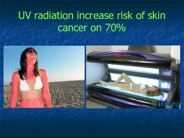
UV radiation increase risk of skin cancer on 70%
UV radiation increase risk of skin cancer on 70%
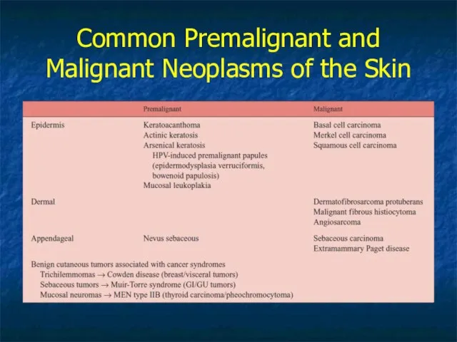
Common Premalignant and Malignant Neoplasms of the Skin
Common Premalignant and Malignant Neoplasms of the Skin
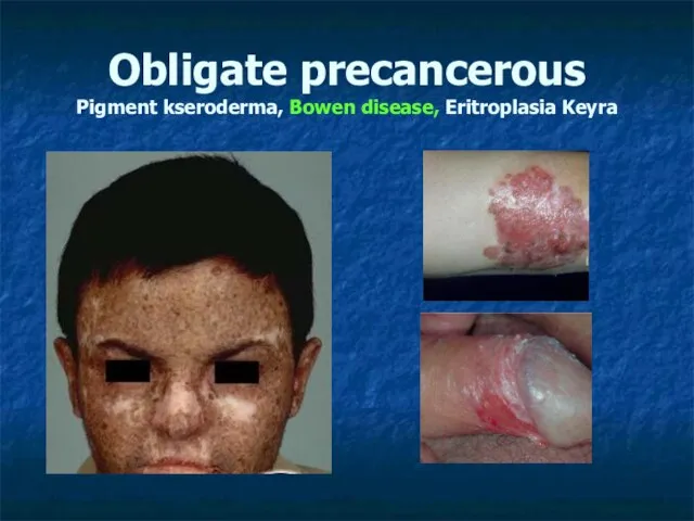
Obligate precancerous
Pigment kseroderma, Bowen disease, Eritroplasia Keyra
Obligate precancerous
Pigment kseroderma, Bowen disease, Eritroplasia Keyra
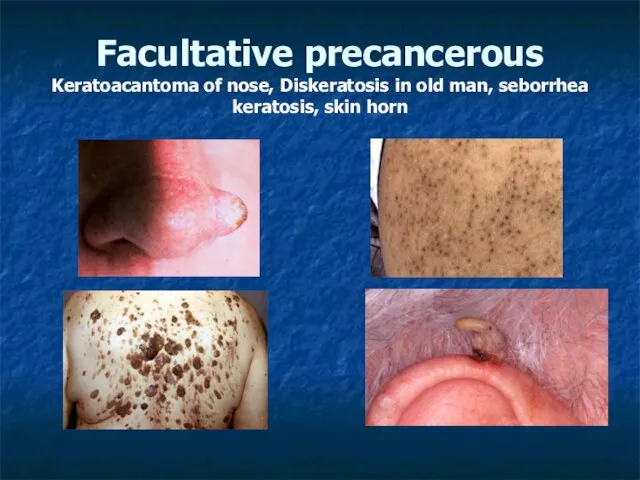
Facultative precancerous Keratoacantoma of nose, Diskeratosis in old man, seborrhea keratosis,
Facultative precancerous Keratoacantoma of nose, Diskeratosis in old man, seborrhea keratosis,
Basal
cell carcinoma is a malignant tumor that rarely
metastasizes. It is composed
Basal cell carcinoma is a malignant tumor that rarely metastasizes. It is composed
Basal cell carcinomas
account for more than 75% of keratinocytic skin
cancers diagnosed
Basal cell carcinomas account for more than 75% of keratinocytic skin cancers diagnosed

Basal cell carcinoma
Basal cell carcinoma

Basal cell carcinoma
(pigment form)
Basal cell carcinoma
(pigment form)

Ulcerous type
(ulcus rоdеns)
(Basalioma terebrans)
Ulcerous type
(ulcus rоdеns)
(Basalioma terebrans)

Perforating basalioma
Perforating basalioma

Cryosurgery
Cryosurgery

Basal cell carcinoma before and after cryosurgery
Basal cell carcinoma before and after cryosurgery

Squamous cell carcinoma is a malignant tumor
arising from epidermal or appendageal
Squamous cell carcinoma is a malignant tumor arising from epidermal or appendageal
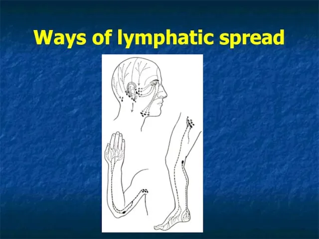
Ways of lymphatic spread
Ways of lymphatic spread

Cancer of skin
Cancer of skin
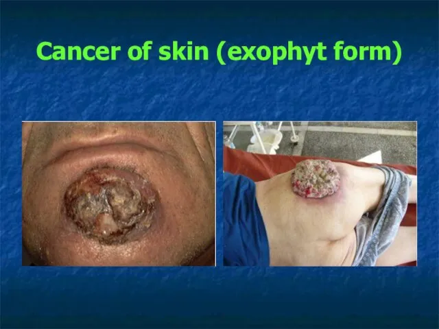
Cancer of skin (exophyt form)
Cancer of skin (exophyt form)

Cancer of skin. Advanced form
Cancer of skin. Advanced form

Malignant melanoma of skin accounts for
160,000 new cases annually, with slightly
Malignant melanoma of skin accounts for 160,000 new cases annually, with slightly

Melanoma
Melanoma

Epidemiology
Epidemiology

Dangerous nevus
Nevus of Settona, Intradermal nevus, Blue nevus
Dangerous nevus
Nevus of Settona, Intradermal nevus, Blue nevus
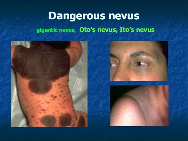
Dangerous nevus gigantic nevus, Oto’s nevus, Ito’s nevus
Dangerous nevus gigantic nevus, Oto’s nevus, Ito’s nevus
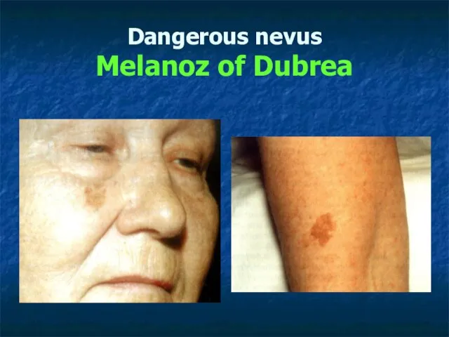
Dangerous nevus
Melanoz of Dubrea
Dangerous nevus
Melanoz of Dubrea

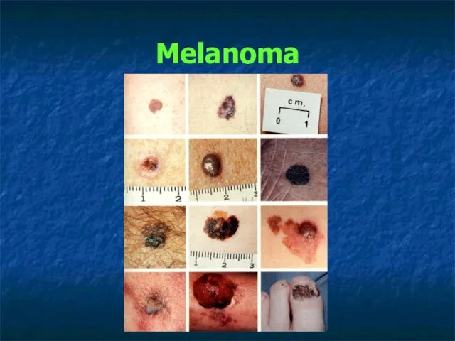
Melanoma
Melanoma
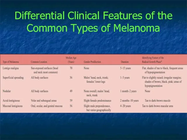
Differential Clinical Features of the Common Types of Melanoma
Differential Clinical Features of the Common Types of Melanoma

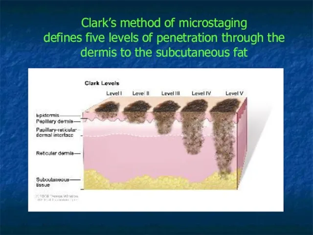
Clark’s method of microstaging
defines five levels of penetration through the
dermis to
Clark’s method of microstaging defines five levels of penetration through the dermis to
 Дәрігер тәжірибесіндегі дәлелдемелі медицина. (Курс 3)
 Биохимические механизмы нарушения обмена углеводов и липидов
 Оказание первой помощи при несчастных случаях на производстве
 Аневризмы сосудов головного мозга
 Материнский и детский травматизм в родах. Лекция 6
 Шықшыт буын дисфункциясының емі
 Как вести себя в конфликтной ситуации: способы решения конфликтов
 Эмоциональное напряжение и его степени. Невроз как функциональное нарушение ВНД
 Обследование ВНЧС
 ООО НПО Биосинтез. Лекарственное средство салсоколлин и салсоколлин-микрон. Лечение болезней печени и желчного пузыря
 Гиперфункция щитовидной железы: причины, патогенез основных проявлений
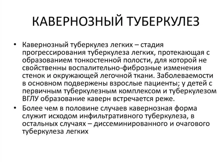 Кавернозный туберкулёз
 Патология носа и околоносовых пазух у детей
 Балалар мен жасөспірімдердің денсаулық жағдайы. Дене дамуы денсаулықтың негізгі көрсеткіштерінің бірі ретінде
 Хронічні розлади харчування (діференційний діагноз, лікування)
 Rhesus (rh) isoimmunization
 Иммунопрофилактика инфекционных заболеваний
 Безопасность использования в медицинских целях листьев подорожника, произрастающего в антропогенно нарушенных местообитаниях
 Viêm gan. Học viện YDHCT Việt nam Bộ môn: Truyền nhiễm
 Клінічна фармакологія бета-лактамних антибіотиків
 Применение лекарственных средств, используеммых для лечения сердечно-сосудистых заболеваний у лиц пожилого возраста
 Туа біткен даму ақаулары
 Пиелонефрит
 Нежелательные лекарственные реакции
 Гематурия. Классификация
 Психодрама
 Физиология гемостаза
 Период взрослости