- Proteins. Functions, structure, classification

Содержание
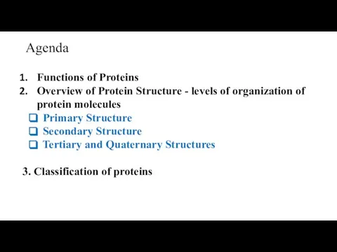
- 2. Agenda Functions of Proteins Overview of Protein Structure - levels of organization of protein molecules Primary
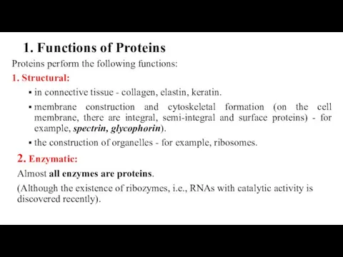
- 3. 1. Functions of Proteins Proteins perform the following functions: 1. Structural: in connective tissue - collagen,
- 4. 3. Signaling (Hormonal) function (Peptide hormones or protein hormones): Regulation and coordination of metabolism in different
- 5. 8. Regulatory Proteins - regulate genes expression; 9. Proteins-toxins: pseudomonas exotoxin (PE), diphtheria toxin (DT), etc.
- 6. Resilin provides soft rubber-elasticity to mechanically active organs and tissue. It helps insects to flap the
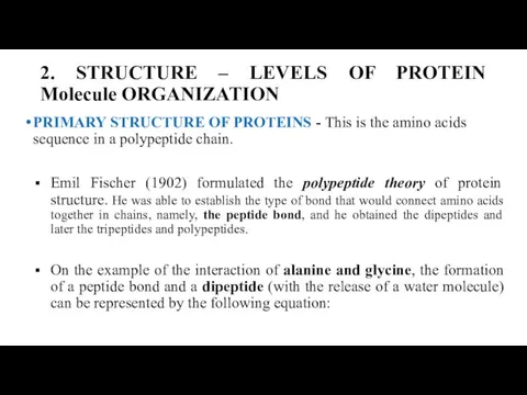
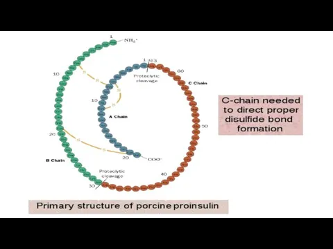
- 8. 2. STRUCTURE – LEVELS OF PROTEIN Molecule ORGANIZATION PRIMARY STRUCTURE OF PROTEINS - This is the
- 9. Alanine Glycine Alanilglycine The sequence and ratio of amino acids in the primary structure determines the
- 11. The primary structure of RNase – ribonuclease
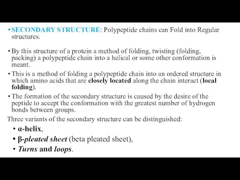
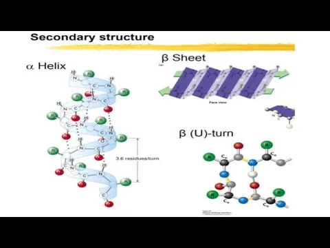
- 12. SECONDARY STRUCTURE: Polypeptide chains can Fold into Regular structures. By this structure of a protein a
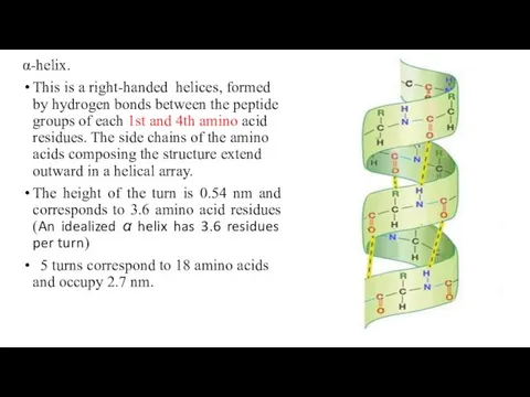
- 14. α-helix. This is a right-handed helices, formed by hydrogen bonds between the peptide groups of each
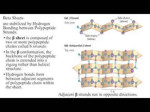
- 16. Beta Sheets are stabilized by Hydrogen Bonding between Polypeptide Strands. the β sheet is composed of
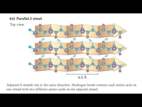
- 17. Adjacent b strands run in the same direction. Hydrogen bonds connect each amino acid on one
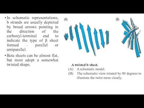
- 18. In schematic representations, b strands are usually depicted by broad arrows pointing in the direction of
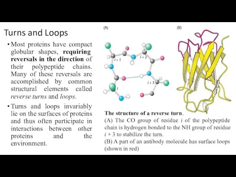
- 19. Turns and Loops Most proteins have compact globular shapes, requiring reversals in the direction of their
- 20. Tertiary Structure: water-soluble Proteins fold into Compact structures The tertiary structure, refers to the spatial arrangement
- 21. Some polypeptide chains fold into two or more compact regions that may be connected by a
- 22. Bonds involved in the formation of the tertiary structure Various bonds are involved in the formation
- 23. The main driving force in the emergence of a three-dimensional structure is the interaction of amino
- 24. Polypeptide chain Types of non-covalent bonds stabilizing the tertiary structure: a – electrostatic interaction; b –
- 25. 1 – Ionic (electrostatic attraction); 2 – hydrogen bond; 3 – hydrophobic interactions of nonpolar groups;
- 26. 1 – Ionic (electrostatic attraction); 2 – hydrogen bond; 3 – disulfide bond 4 – hydrophobic
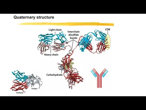
- 27. Quaternary Structure: Multiple Polypeptide Chains Can Assemble into a Single Protein Proteins consisting of more than
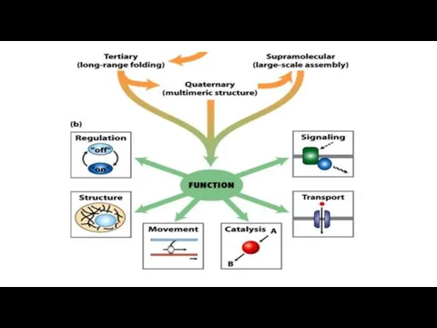
- 29. SUMMARY on Protein Tertiary and Quaternary Structures Tertiary structure is the complete three-dimensional structure of a
- 30. 3. Protein classification Proteins are classified: A) By function (see above - “protein functions”). B) By
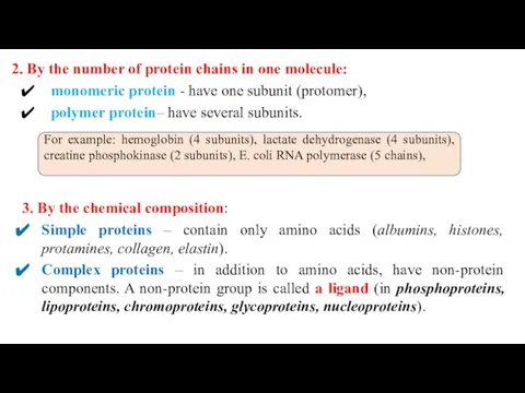
- 31. 2. By the number of protein chains in one molecule: monomeric protein - have one subunit
- 33. Скачать презентацию
Agenda
Functions of Proteins
Overview of Protein Structure - levels of organization
Agenda
Functions of Proteins
Overview of Protein Structure - levels of organization
1. Functions of Proteins
Proteins perform the following functions:
1. Structural:
in connective tissue
1. Functions of Proteins
Proteins perform the following functions:
1. Structural:
in connective tissue
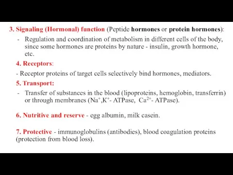
3. Signaling (Hormonal) function (Peptide hormones or protein hormones):
Regulation and coordination of metabolism in
3. Signaling (Hormonal) function (Peptide hormones or protein hormones):
Regulation and coordination of metabolism in

8. Regulatory Proteins - regulate genes expression;
9. Proteins-toxins: pseudomonas exotoxin (PE),
8. Regulatory Proteins - regulate genes expression;
9. Proteins-toxins: pseudomonas exotoxin (PE),

Resilin provides soft rubber-elasticity to mechanically active organs and tissue.
It helps
Resilin provides soft rubber-elasticity to mechanically active organs and tissue.
It helps
2. STRUCTURE – LEVELS OF PROTEIN Molecule ORGANIZATION
PRIMARY STRUCTURE OF PROTEINS
2. STRUCTURE – LEVELS OF PROTEIN Molecule ORGANIZATION
PRIMARY STRUCTURE OF PROTEINS
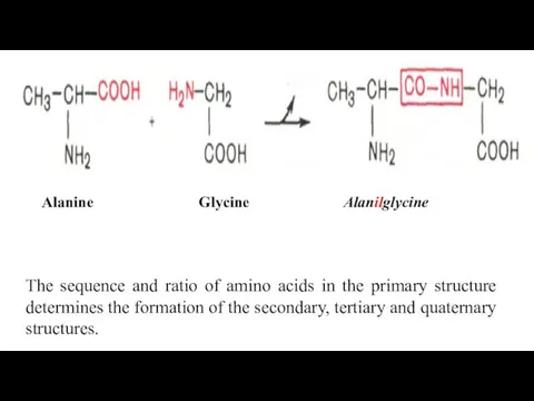
Alanine Glycine Alanilglycine
The sequence and ratio of amino acids in the
Alanine Glycine Alanilglycine
The sequence and ratio of amino acids in the
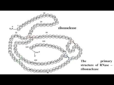
The primary structure of RNase – ribonuclease
The primary structure of RNase – ribonuclease
SECONDARY STRUCTURE: Polypeptide chains can Fold into Regular structures.
By this structure
SECONDARY STRUCTURE: Polypeptide chains can Fold into Regular structures.
By this structure
α-helix.
This is a right-handed helices, formed by hydrogen bonds between the
α-helix.
This is a right-handed helices, formed by hydrogen bonds between the
Beta Sheets
are stabilized by Hydrogen Bonding between Polypeptide Strands.
the β
Beta Sheets
are stabilized by Hydrogen Bonding between Polypeptide Strands.
the β
Adjacent b strands run in the same direction. Hydrogen bonds connect
Adjacent b strands run in the same direction. Hydrogen bonds connect
In schematic representations, b strands are usually depicted by broad arrows
In schematic representations, b strands are usually depicted by broad arrows
Turns and Loops
Most proteins have compact globular shapes, requiring reversals in
Turns and Loops
Most proteins have compact globular shapes, requiring reversals in

Tertiary Structure: water-soluble Proteins fold into Compact structures
The tertiary structure, refers
Tertiary Structure: water-soluble Proteins fold into Compact structures
The tertiary structure, refers

Some polypeptide chains fold into two or more compact regions that
Some polypeptide chains fold into two or more compact regions that
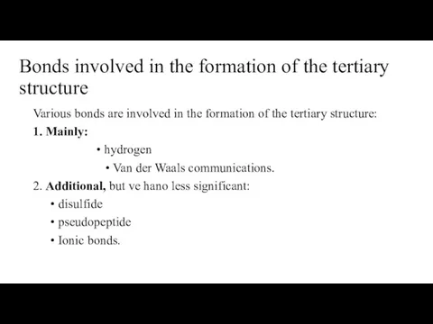
Bonds involved in the formation of the tertiary structure
Various bonds are
Bonds involved in the formation of the tertiary structure
Various bonds are
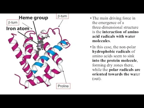
The main driving force in the emergence of a three-dimensional structure
The main driving force in the emergence of a three-dimensional structure
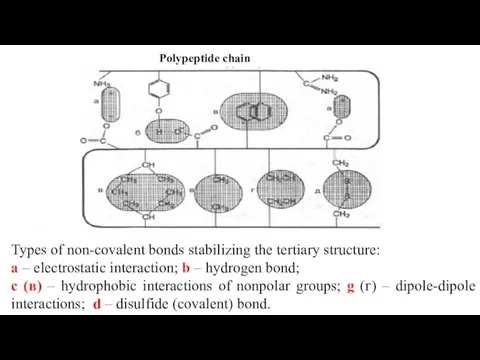
Polypeptide chain
Types of non-covalent bonds stabilizing the tertiary structure:
a –
Polypeptide chain
Types of non-covalent bonds stabilizing the tertiary structure:
a –
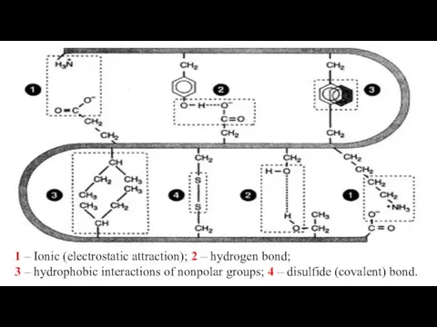
1 – Ionic (electrostatic attraction); 2 – hydrogen bond;
3 –
1 – Ionic (electrostatic attraction); 2 – hydrogen bond;
3 –
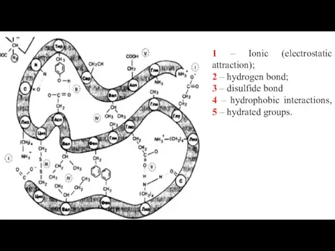
1 – Ionic (electrostatic attraction);
2 – hydrogen bond;
3 –
1 – Ionic (electrostatic attraction);
2 – hydrogen bond;
3 –

Quaternary Structure: Multiple Polypeptide Chains Can Assemble into a Single Protein
Proteins
Quaternary Structure: Multiple Polypeptide Chains Can Assemble into a Single Protein
Proteins
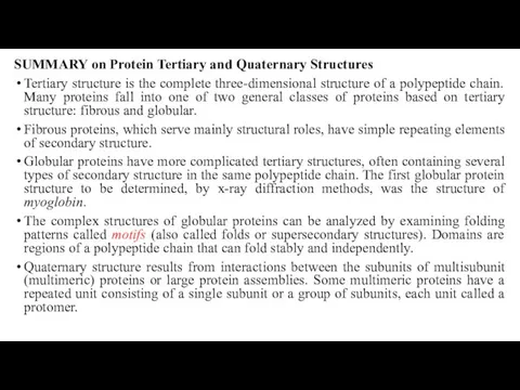
SUMMARY on Protein Tertiary and Quaternary Structures
Tertiary structure is the complete
SUMMARY on Protein Tertiary and Quaternary Structures
Tertiary structure is the complete
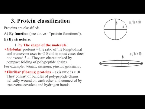
3. Protein classification
Proteins are classified:
A) By function (see above - “protein
3. Protein classification
Proteins are classified:
A) By function (see above - “protein
2. By the number of protein chains in one molecule:
monomeric
2. By the number of protein chains in one molecule:
monomeric
 Природные и попутные нефтяные газы
 Майлардың анықтамасы
 Общая радиохимия. Свойства радиоколлоидов
 Строение атома углерода
 Да здравствует мыло душистое
 Химические и физико-химические методы анализа. Сущность и методы качественного анализа
 Фосфорні добрива. Технології збагачення фосфоровмісної сировини
 Уравнения химических реакций
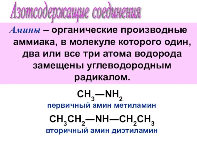 Азотсодержащие соединения. Амины
 Оборудование для дистилляции мисцеллы
 Гидролиз. Сущность процесса гидролиза
 Электронные эффекты заместителей. Типы химических реакций
 Исследовательский проект «Кристаллы»
 Минералы. Классы минералов
 Спирты
 Гидролиз. Виды гидролиза
 Пищевые добавки
 Основания и кислоты. Тема 2
 Зміна ліпідів за технологічної обробки
 Презентация по Химии "Степень окисления" - скачать смотреть бесплатно_
 Презентация по Химии "Высшие природные полимеры - Белки и Нуклеиновые кислоты" - скачать смотреть
  Области применения спиртов
 Применение синтетических полимеров в вооружении
 Пластик, пластмасса
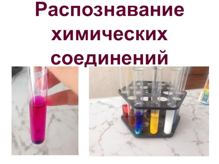 Распознавание химических соединений
 Основы безопасности при уничтожении химического оружия
 Сера и ее аллотропные модификации. (9 класс)
 Виды химической связи