- Abhishek

Содержание
- 3. The scheme of normal pressure in hearts chambers, mm Hg
- 4. c Transthoracic visualization of pulmonary artery is very pure
- 5. RA and RV enlargement Sign of Pulmonary Hypertension #1
- 6. Right ventricle size in during systole and diastole
- 7. Right ventricular diameter measured in apical 4-chamber view at level of left ventricular papillary muscles
- 8. Transesophageal echocardiographic measurements of right ventricular (RV) diameters from mid-esophageal 4-chamber view
- 9. Reference limits and partition values of right ventricular and pulmonary artery size
- 10. Reference limits and partition values of right ventricular size and function as measured in the apical
- 11. Measurement of right ventricular outflow tract diameter at sub-pulmonary region (RVOT1) and pulmonic valve annulus (RVOT2)
- 12. Measure RV wall in M-regiment- long parasternal position and in B-regiment –subcostal position Sign of pulmonary
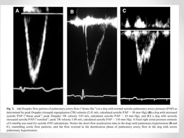
- 13. Variants of Pulmonary arteries pressure calculating AT/ET connection= middle pulmonary pressure Equation of Kitabatake Calculation by
- 14. Acceleration time (AT) of Right ventricle Ejection time (ET) of Right ventricle Normal AT/ET 0,4-0,45
- 15. Decrease of AT/ET time is the Pulmonary Hypertension
- 17. Equation of Kitabatake P middle PA =(0,0068хАТ)+2,1=90-(0,62хАТ)
- 18. The diameter of the Inferior vena cava (IVC) and the percent decrease in the diameter during
- 19. Inferior Vena Cava View
- 20. Assessment of pulmonary artery hypertension degree
- 22. Скачать презентацию
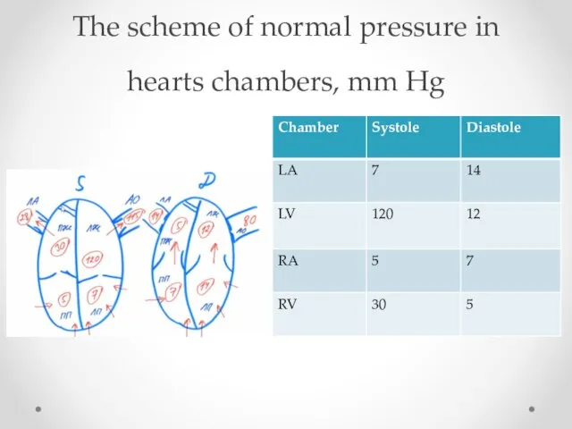
The scheme of normal pressure in hearts chambers, mm Hg
The scheme of normal pressure in hearts chambers, mm Hg
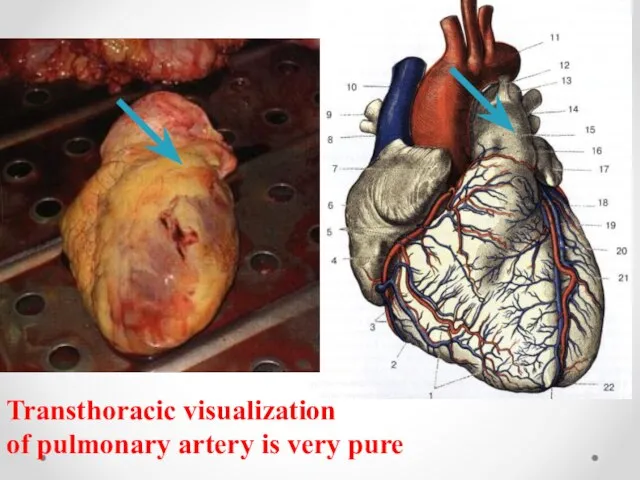
c
Transthoracic visualization
of pulmonary artery is very pure
c
Transthoracic visualization
of pulmonary artery is very pure
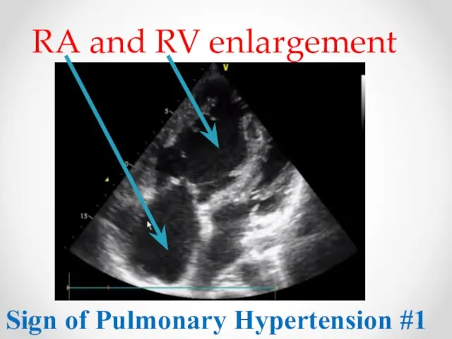
RA and RV enlargement
Sign of Pulmonary Hypertension #1
RA and RV enlargement
Sign of Pulmonary Hypertension #1
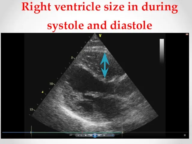
Right ventricle size in during systole and diastole
Right ventricle size in during systole and diastole
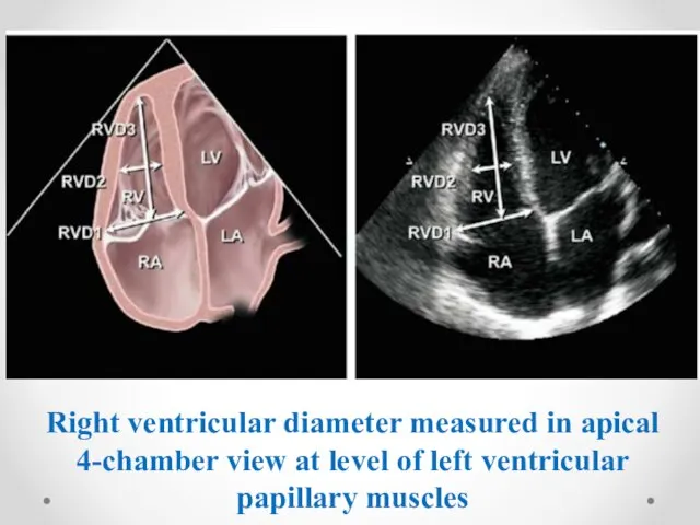
Right ventricular diameter measured in apical 4-chamber view at level of
Right ventricular diameter measured in apical 4-chamber view at level of
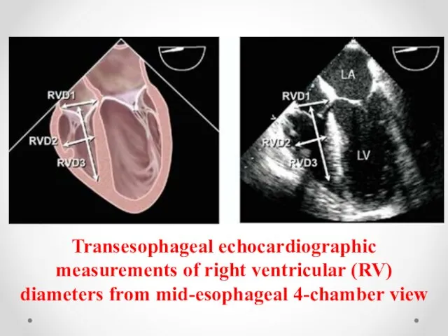
Transesophageal echocardiographic measurements of right ventricular (RV) diameters from mid-esophageal 4-chamber
Transesophageal echocardiographic measurements of right ventricular (RV) diameters from mid-esophageal 4-chamber
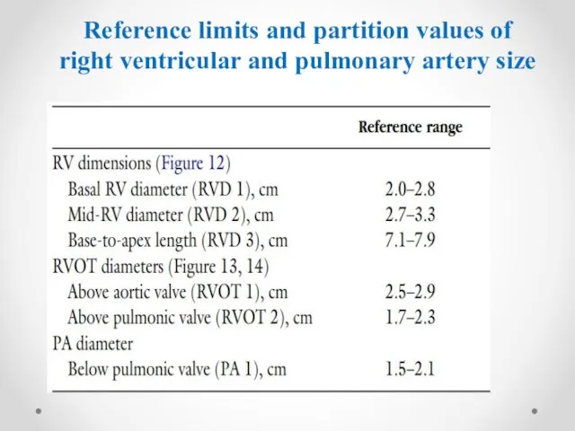
Reference limits and partition values of right ventricular and pulmonary artery
Reference limits and partition values of right ventricular and pulmonary artery

Reference limits and partition values of right ventricular size and function
Reference limits and partition values of right ventricular size and function
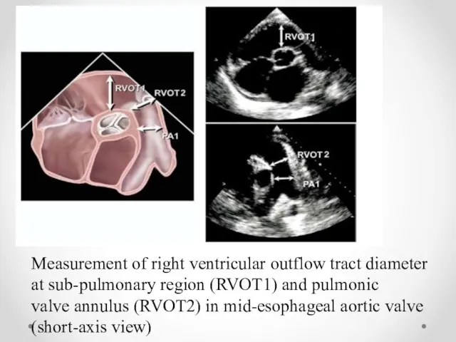
Measurement of right ventricular outflow tract diameter at sub-pulmonary region (RVOT1)
Measurement of right ventricular outflow tract diameter at sub-pulmonary region (RVOT1)
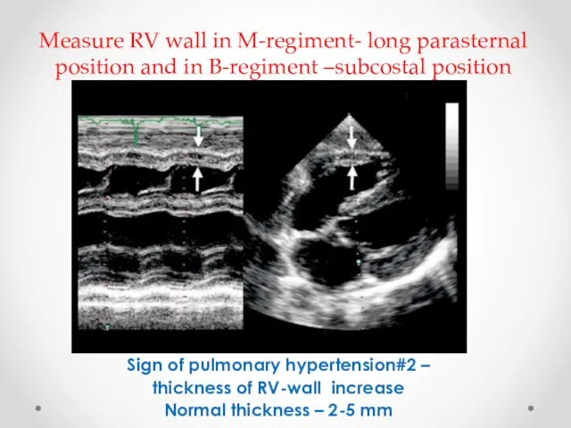
Measure RV wall in M-regiment- long parasternal position and in B-regiment
Measure RV wall in M-regiment- long parasternal position and in B-regiment
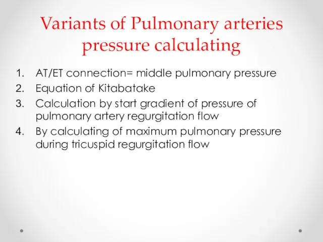
Variants of Pulmonary arteries pressure calculating
AT/ET connection= middle pulmonary pressure
Equation
Variants of Pulmonary arteries pressure calculating
AT/ET connection= middle pulmonary pressure
Equation
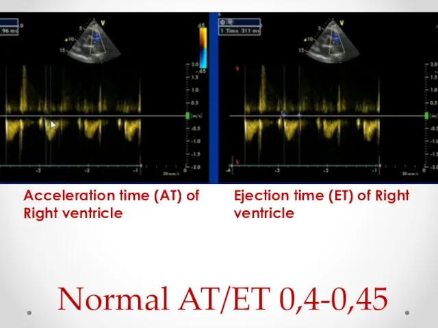
Acceleration time (AT) of Right ventricle
Ejection time (ET) of Right ventricle
Normal
Acceleration time (AT) of Right ventricle
Ejection time (ET) of Right ventricle
Normal
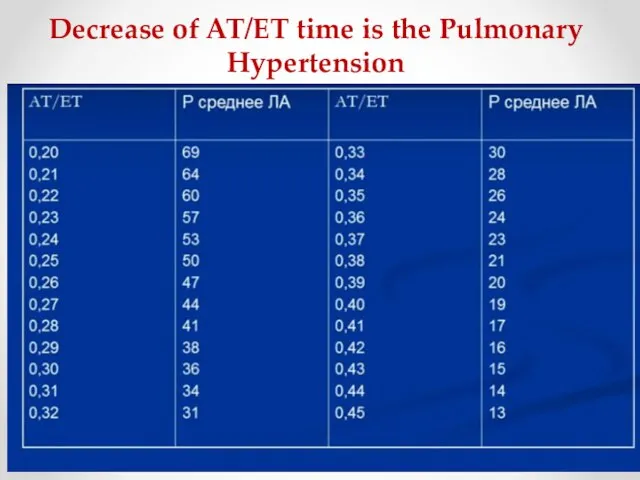
Decrease of AT/ET time is the Pulmonary Hypertension
Decrease of AT/ET time is the Pulmonary Hypertension
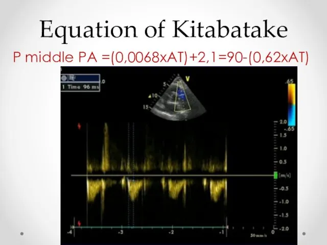
Equation of Kitabatake
P middle PA =(0,0068хАТ)+2,1=90-(0,62хАТ)
Equation of Kitabatake
P middle PA =(0,0068хАТ)+2,1=90-(0,62хАТ)
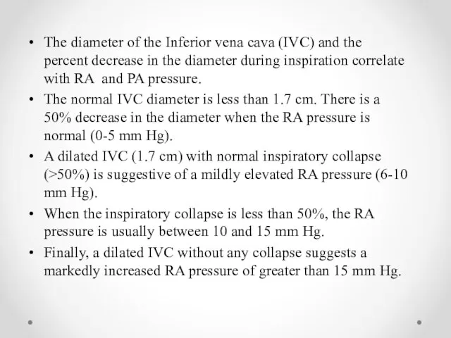
The diameter of the Inferior vena cava (IVC) and the percent
The diameter of the Inferior vena cava (IVC) and the percent
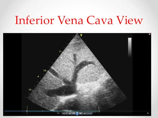
Inferior Vena Cava View
Inferior Vena Cava View
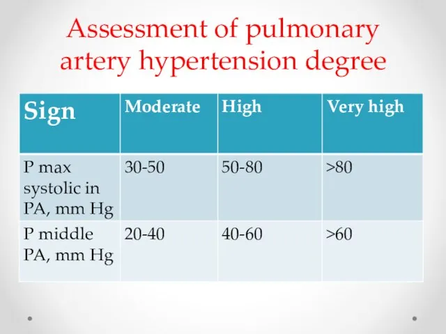
Assessment of pulmonary artery hypertension degree
Assessment of pulmonary artery hypertension degree
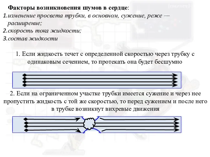 Факторы возникновения шумов в сердце
 Инфекции, передающиеся парентеральным путем (вирусные гепатиты В,С,Д, ВИЧ-инфекция)
 Амбулаторлы хирургия
 Углеводы. Классификация углеводов
 Пищеварение у детей
 Общая распространенность нарушений функционирования, жизнедеятельности и здоровья у реконвалисцентов COVID-19: результаты
 Понятие заикание
 Сестринский уход за детьми при рахите, спазмофилии
 Тифлит как осложнение острого аппендицита
 Өндірістік улардың әсерінің жалпы заңдылықтары
 Наставничество как ответственная функция организатора сестринского дела
 Оказание доврачебной помощи пострадавшим
 Наблюдение беременной в женской консультации при физиологической беременности
 Санитарное содержание предприятий ОП
 Вариативная анатомия сосудов головы и шеи
 Санаторно-курортная индустрия
 Хирургиялық аурулардың су-электірлік бұзылыстары және инфузионды терапияның принціпі
 Микробиология острых кишечных вирусных инфекций
 Новые коллекционные КФС Жемчужина Сербии, Варварин Ключ
 Задержка психического развития
 Методы простейшей физиотерапии
 Чума. Этиология
 Виды дренажей
 Медицина мен денсаулық сақтаудағы дисперсиялық анализ. Дисперсиялық анализдің негізгі ұғымдары мен əдістемесі
 Рахит (от греч.rhahis — спинной хребет)
 lektsiya_11_svezhie_formy_vtorichnogo_tuberkuleza_ochagovaya_infiltrativnaya_kazeoznaya_pnevmoniya_tuberkuleznyiy_plevrit
 Диагностика определения уровня готовности ребенка к школе
 Трансплантация лица