- Anatomy of orbit

Содержание
- 2. Development of orbit Develops from mesenchyme by ossification 6 th to 7 th week laying down
- 3. Developmental Anomalies : Craniosynosotosis: Brachycephaly Oxycephaly Scophocephaly Trigonocephaly
- 4. Craniosfacial dysostois / Crouzon’ syndrome Proptosis – shallow orbits Hypertelorim - wide separation of orbits V
- 5. Oxycephaly-syndactlye / Apert’ syndrome: Flattened occiput , steep forehead , supra orbital ridge Midfacial hypoplasia ,
- 6. Bones of Orbit Frontal Ethmoid Sphenoid Lacrimal Palatine Maxillary Zygomatic
- 7. Dimensions - orbit 30 ml –volume 35 mm vertically , 40 mm horizontally 45 degree between
- 8. Boundaries of Orbit Roof Floor Side walls Orbital apex
- 9. Roof of orbit Frontal bone [Orbital plate] & lesser wing of sphenoid Separated from frontal sinus
- 10. Orbital roof anomaly / fracture CSF pulsation pulsatile exophthalmos Orbital meningocele / encephalocele
- 11. Medial wall Body of sphenoid Ethmoid Lacrimal Maxilla[frontal process]
- 12. Orbital cellulitis Extremely thin wall Prone for damage & sinusitis spread Infection across Orbital cellulitis
- 13. Floor of orbit Maxilla Zygomatic Palatine Triangular segment -- thinnest Inferior orbital groove
- 14. Blow out fractures Fragile barrier to maxillary sinus Due to trauma eyeball collapse into Maxillary sinus
- 15. Le fort’s fracture Type 2 - Pyramidal Type 3 - Craniofacial dissociation
- 16. Lateral wall Greater wing –sphenoid Orbital surface – Frontal process of zygomatic Inferiorly – inf orbital
- 17. Behind Zygomatic sphenoidal suture lateral orbitotomy of greater wing ( thin wall ) cancellous bone middle
- 18. At frontal sphenoidal suture -- meningeal foramen Site of anastomosis of Lacrimal artery and meningeal artery
- 19. Orbital apex
- 20. Orbital apex syndrome / Tolosa - hunt syndrome : Damage to structures at apex 2 nd,
- 21. Other causes: Inflammatory Infectious Neoplastic Iatrogenic / traumatic Vascular
- 22. Superior orbital fissure syndrome / Rochon – Duvigneaud syndrome : Lesion anterior to orbital apex excluding
- 23. Contents of orbit Eye ball Orbital fat Connective tissue system Blood vessels Nerves Extraocular muscles
- 24. Eyeball - Applied anatomy: Proptosis : Dystopia Enophthalmosis Ophthalmoplegia
- 25. Connective tissue system Periorbita Orbital septum Tenon’s capsule
- 26. Periorbita: Loosely attached to orbital bone Attached firmly to Arcus marginalis Trochlea Lateral orbital tubercle Optic
- 27. Orbital septum: Interconnecting / circumferential radial webs of fascial system support and transmit forces in trauma
- 28. Anterior fascial system Formed by condensation of fibrous septa Lockwood lig, whitnall sup susp lig Lacrimal
- 29. Tenon’s capsule Dense elastic , vascular Extent : from perilimbal sclera to optic nerve meninges with
- 31. Surgical spaces in orbit : Sub periosteal space Peripheral space Central space Tenon’s space
- 32. Extra ocular muscles 4 rectal muscles 2 oblique muscles Two lid retractors To serve in eyeball
- 33. Arterial supply
- 34. Venous drainage
- 35. Optic nerve Intra orbital part = 25 mm out of 4 cm Enclosed in three meningeal
- 36. Oculomotor nerve Divides at anterior part of cavernous sinus before Entering sup orbital fissure Sup division
- 37. Trochlear nerve Runs medially from lateral wall of cavernous sinus Above Levator palpebral sup Then supplies
- 38. Abducent nerve Running inferior lateral to 3 rd nerve then supplies ocular surface of lateral Rectus
- 39. Trigeminal nerve Three terminal branches of ophthalmic division: Frontal nerve supratrochlear supraorbital Lacrimal nerve Sensory and
- 40. Nasociliary nerve: Communicating branch to sensory root of ciliary ganglion Long ciliary nerves - dilator pupillae
- 42. Скачать презентацию
Development of orbit
Develops from mesenchyme by ossification
6 th to 7 th
Development of orbit
Develops from mesenchyme by ossification
6 th to 7 th
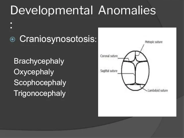
Developmental Anomalies :
Craniosynosotosis:
Brachycephaly
Oxycephaly
Scophocephaly
Trigonocephaly
Developmental Anomalies :
Craniosynosotosis:
Brachycephaly
Oxycephaly
Scophocephaly
Trigonocephaly
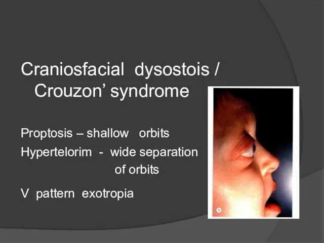
Craniosfacial dysostois / Crouzon’ syndrome
Proptosis – shallow orbits
Hypertelorim - wide separation
Craniosfacial dysostois / Crouzon’ syndrome
Proptosis – shallow orbits
Hypertelorim - wide separation
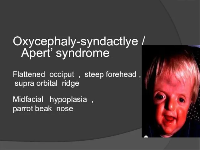
Oxycephaly-syndactlye / Apert’ syndrome:
Flattened occiput , steep forehead ,
supra orbital
Oxycephaly-syndactlye / Apert’ syndrome:
Flattened occiput , steep forehead ,
supra orbital
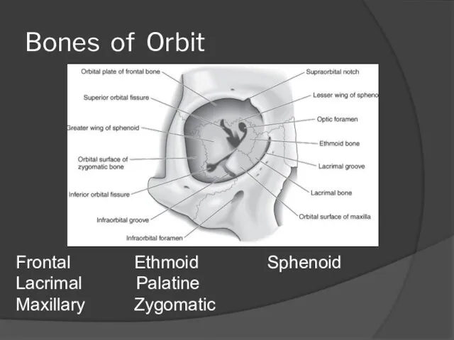
Bones of Orbit
Frontal Ethmoid Sphenoid
Lacrimal Palatine
Maxillary Zygomatic
Bones of Orbit
Frontal Ethmoid Sphenoid
Lacrimal Palatine
Maxillary Zygomatic
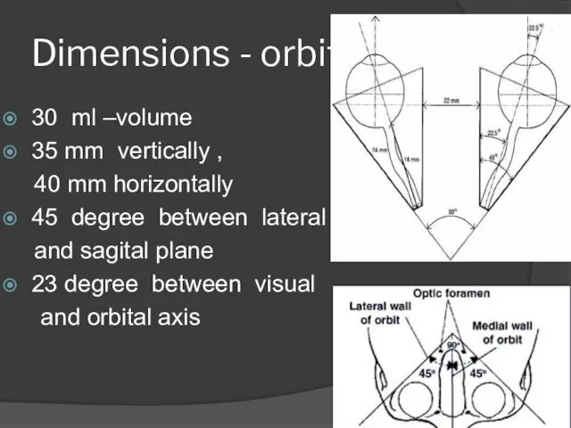
Dimensions - orbit
30 ml –volume
35 mm vertically ,
40 mm horizontally
45
Dimensions - orbit
30 ml –volume
35 mm vertically ,
40 mm horizontally
45
Boundaries of Orbit
Roof
Floor
Side walls
Orbital apex
Boundaries of Orbit
Roof
Floor
Side walls
Orbital apex
![Roof of orbit Frontal bone [Orbital plate] & lesser wing of](/_ipx/f_webp&q_80&fit_contain&s_1440x1080/imagesDir/jpg/462640/slide-8.jpg)
Roof of orbit
Frontal bone [Orbital plate] & lesser wing of sphenoid
Separated
Roof of orbit
Frontal bone [Orbital plate] & lesser wing of sphenoid
Separated
Orbital roof anomaly / fracture
CSF pulsation pulsatile
exophthalmos
Orbital meningocele
Orbital roof anomaly / fracture
CSF pulsation pulsatile
exophthalmos
Orbital meningocele
![Medial wall Body of sphenoid Ethmoid Lacrimal Maxilla[frontal process]](/_ipx/f_webp&q_80&fit_contain&s_1440x1080/imagesDir/jpg/462640/slide-10.jpg)
Medial wall
Body of sphenoid
Ethmoid
Lacrimal
Maxilla[frontal
process]
Medial wall
Body of sphenoid
Ethmoid
Lacrimal
Maxilla[frontal
process]
Orbital cellulitis
Extremely thin wall
Prone for damage & sinusitis spread
Infection across
Orbital cellulitis
Extremely thin wall
Prone for damage & sinusitis spread
Infection across
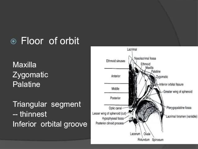
Floor of orbit
Maxilla
Zygomatic
Palatine
Triangular segment
-- thinnest
Inferior orbital groove
Floor of orbit
Maxilla
Zygomatic
Palatine
Triangular segment
-- thinnest
Inferior orbital groove
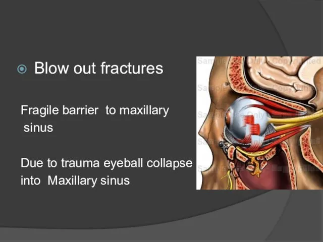
Blow out fractures
Fragile barrier to maxillary
sinus
Due to trauma eyeball collapse
Blow out fractures
Fragile barrier to maxillary
sinus
Due to trauma eyeball collapse
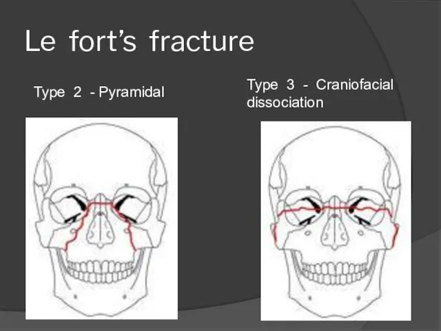
Le fort’s fracture
Type 2 - Pyramidal
Type 3 - Craniofacial dissociation
Le fort’s fracture
Type 2 - Pyramidal
Type 3 - Craniofacial dissociation
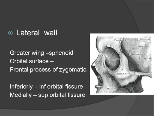
Lateral wall
Greater wing –sphenoid
Orbital surface –
Frontal process of zygomatic
Inferiorly – inf
Lateral wall
Greater wing –sphenoid
Orbital surface –
Frontal process of zygomatic
Inferiorly – inf
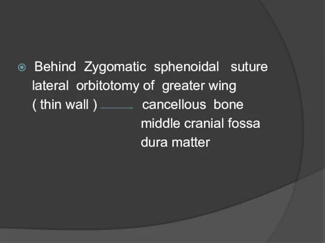
Behind Zygomatic sphenoidal suture
lateral orbitotomy of greater wing
(
Behind Zygomatic sphenoidal suture
lateral orbitotomy of greater wing
(
At frontal sphenoidal suture
-- meningeal foramen
Site of anastomosis
At frontal sphenoidal suture
-- meningeal foramen
Site of anastomosis
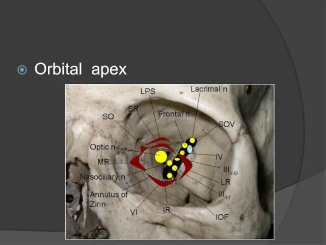
Orbital apex
Orbital apex
Orbital apex syndrome
/ Tolosa - hunt syndrome :
Damage to structures
Orbital apex syndrome
/ Tolosa - hunt syndrome :
Damage to structures
Other causes:
Inflammatory
Infectious
Neoplastic
Iatrogenic / traumatic
Vascular
Other causes:
Inflammatory
Infectious
Neoplastic
Iatrogenic / traumatic
Vascular
Superior orbital fissure syndrome
/ Rochon – Duvigneaud syndrome
Superior orbital fissure syndrome
/ Rochon – Duvigneaud syndrome
Contents of orbit
Eye ball
Orbital fat
Connective tissue system
Blood vessels
Nerves
Extraocular muscles
Contents of orbit
Eye ball
Orbital fat
Connective tissue system
Blood vessels
Nerves
Extraocular muscles
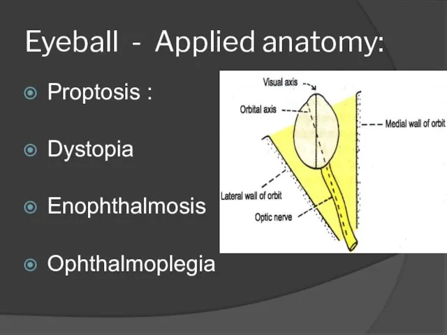
Eyeball - Applied anatomy:
Proptosis :
Dystopia
Enophthalmosis
Ophthalmoplegia
Eyeball - Applied anatomy:
Proptosis :
Dystopia
Enophthalmosis
Ophthalmoplegia
Connective tissue system
Periorbita
Orbital septum
Tenon’s capsule
Connective tissue system
Periorbita
Orbital septum
Tenon’s capsule
Periorbita:
Loosely attached to orbital bone
Attached firmly to
Arcus marginalis
Trochlea
Lateral orbital
Periorbita:
Loosely attached to orbital bone
Attached firmly to
Arcus marginalis
Trochlea
Lateral orbital
Orbital septum:
Interconnecting / circumferential radial webs of fascial system
support and
Orbital septum:
Interconnecting / circumferential radial webs of fascial system
support and
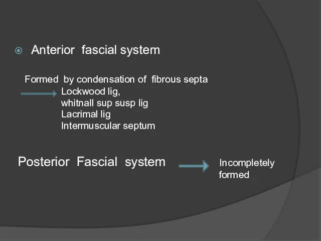
Anterior fascial system
Formed by condensation of fibrous septa
Lockwood
Anterior fascial system
Formed by condensation of fibrous septa
Lockwood
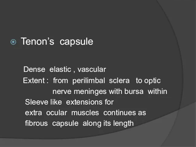
Tenon’s capsule
Dense elastic , vascular
Extent : from perilimbal sclera to
Tenon’s capsule
Dense elastic , vascular
Extent : from perilimbal sclera to
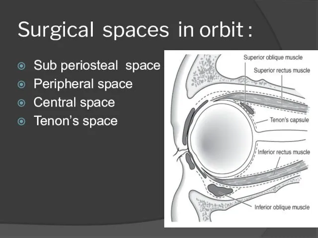
Surgical spaces in orbit :
Sub periosteal space
Peripheral space
Central space
Tenon’s
Surgical spaces in orbit :
Sub periosteal space
Peripheral space
Central space
Tenon’s
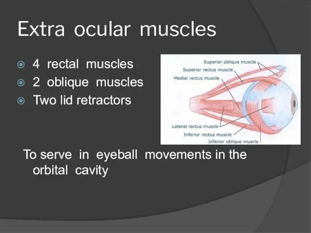
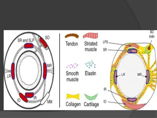
Extra ocular muscles
4 rectal muscles
2 oblique muscles
Two lid retractors
To serve in
Extra ocular muscles
4 rectal muscles
2 oblique muscles
Two lid retractors
To serve in
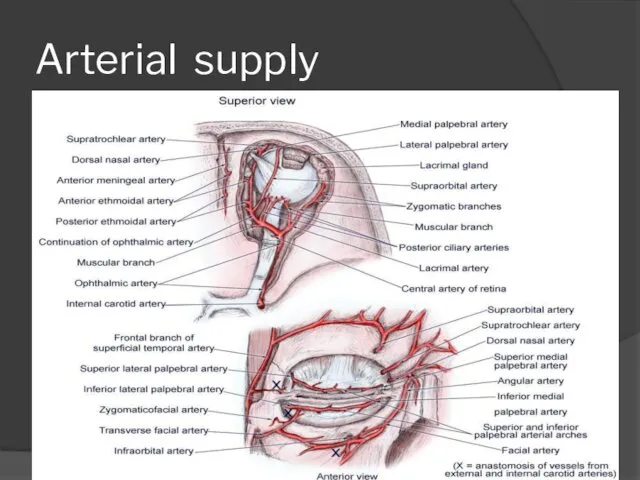
Arterial supply
Arterial supply
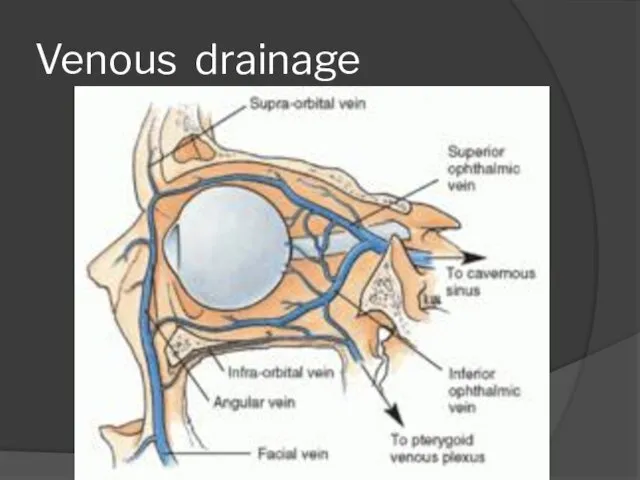
Venous drainage
Venous drainage
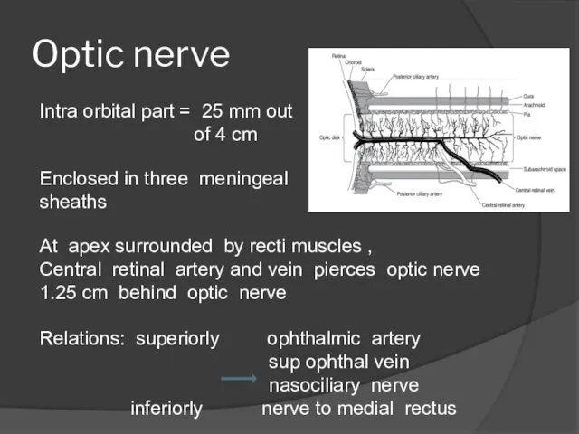
Optic nerve
Intra orbital part = 25 mm out
of 4
Optic nerve
Intra orbital part = 25 mm out
of 4
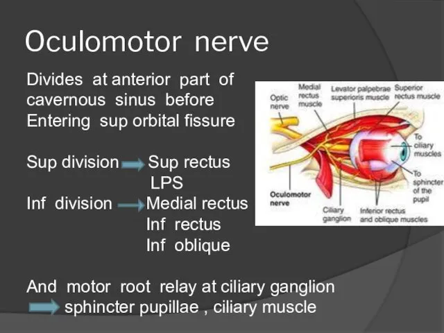
Oculomotor nerve
Divides at anterior part of cavernous sinus before
Entering sup
Oculomotor nerve
Divides at anterior part of cavernous sinus before
Entering sup
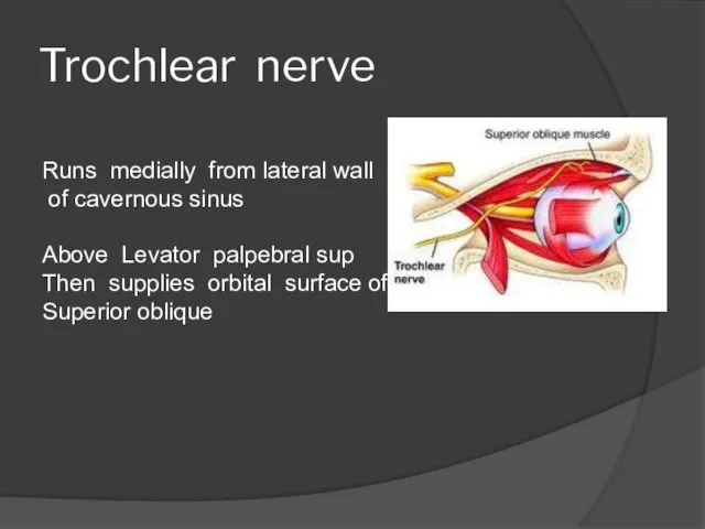
Trochlear nerve
Runs medially from lateral wall
of cavernous sinus
Above Levator
Trochlear nerve
Runs medially from lateral wall
of cavernous sinus
Above Levator
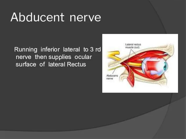
Abducent nerve
Running inferior lateral to 3 rd
nerve then supplies ocular
Abducent nerve
Running inferior lateral to 3 rd
nerve then supplies ocular
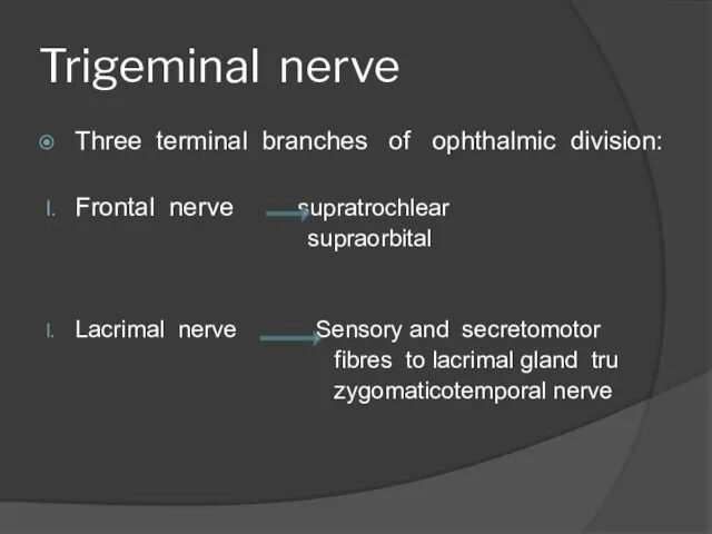
Trigeminal nerve
Three terminal branches of ophthalmic division:
Frontal nerve supratrochlear
supraorbital
Lacrimal
Trigeminal nerve
Three terminal branches of ophthalmic division:
Frontal nerve supratrochlear
supraorbital
Lacrimal
Nasociliary nerve:
Communicating branch to sensory root of ciliary ganglion
Long ciliary nerves
Nasociliary nerve:
Communicating branch to sensory root of ciliary ganglion
Long ciliary nerves
 Роль акушерки в профилактике родовых травм новорожденных
 Обструктивная уропатия
 Паразитические простейшие (протозоология)
 Алгоритм диагностики и неотложной помощи при судорожном синдроме у детей
 Особенности сестринского ухода за пациентом с хламидийным конъюнктивитом
 Кафедра патологической анатомии в Запорожском медицинском институте
 Ожиріння визнане ВООЗ новою неінфекційною епідемією нашого часу
 The weaknesses of the international business in Ukraine during pandemic COVID-19
 Студенческий научный кружок кафедры фармакологии
 Загальна будова та функції органів дихання
 Физиология крови
 Основы патологии. Понятие о болезни. Диагностика. (Лекция 2)
 Исследование раненых животных. Диагностика и лечение свежих ран
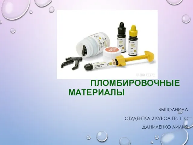 Пломбировочные материалы
 Беттің туа біткен патологиясын алдын алу. Беттің, жақсүйектерінің және ауыз қуысының ағзаларының ақаулары болған кездегі
 Некроз. Апоптоз. Атрофия
 Мутациялық өзгергіштік
 Базовые реанимационные мероприятия
 Почему мы ссоримся?
 Пиелонефрит у беременных
 М’язи спини та живота
 Нарушения регуляции клеточного цикла в неопластических клетках: роль онкогенов и опухолевых супрессоров. Лекция 2
 Виды антигенных детерминант иммуноглобулинов
 Остеохондроз позвоночника
 Лечение детей с заболеваниями органов мочевыделения. Острый пиелонефрит. Острый гломерулонефрит. Острый цистит. ОПН. ХПН
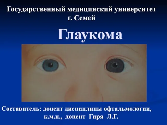 Глаукома. Методы исследования
 Осложнения после удаления зуба
 Тактическая медицина. Современные средства остановки кровотечения