- Efferent peripheral NS: the autonomic votor divisions

Содержание
- 2. Autonomic nervous system: A part of the nervous system that regulates key involuntary functions of the
- 3. Review (again)
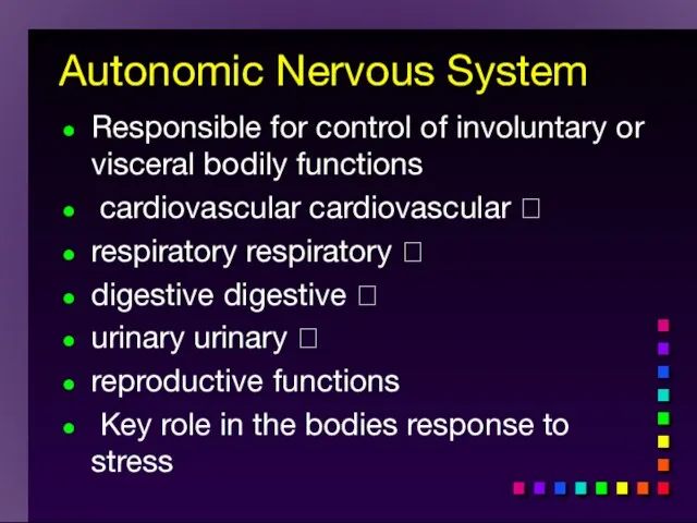
- 4. Autonomic Nervous System Responsible for control of involuntary or visceral bodily functions cardiovascular cardiovascular respiratory
- 6. Overview: The Parts of a Reflex
- 7. Autonomic Targets Smooth Muscle Cardiac Muscle Exocrine Glands Some Endocrine glands Lymphoid Tissue Adipose
- 8. Divisions of ANS Sympathetic Parasympathetic Metasympathetic
- 9. Sympathetic and parasympathetic divisions typically function in opposition to each other. But this opposition is better
- 10. ANS 2 divisions: Sympathetic “Fight or flight” “E” division Exercise, excitement, emergency, and embarrassment Parasympathetic “Rest
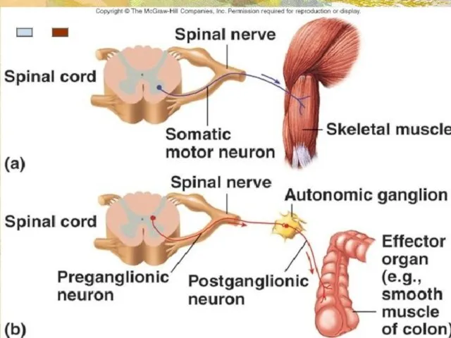
- 11. 1. The autonomic nervous system (ANS) is an involuntary motor (efferent) system. 2. Autonomic nerves are
- 12. Autonomic pathway: Two Efferent Neurons in Series Preganglionic neuron cell body in CNS Synapse in autonomic
- 14. 3. Although “involuntary”, the autonomic nervous system is regulated by higher centers. The best known of
- 15. 4. The autonomic nervous system consists of two divisions: a) the sympathetic (or thoracolumbar) division in
- 16. Sympathetic “Fight or flight” “E” division Exercise, excitement, emergency, and embarrassment
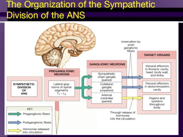
- 17. = Thoracolumbar division (T1 to L2) Preganglionic neurons (N1)from thoracolumbar region of spinal cord Pre and
- 18. Sympathetic (preganglionic ): 1. The cell bodies giving rise to preganglionic neurons (N1) are located in
- 19. Sympathetic (postganglionic ): 1. The cell bodies giving rise to postganglionic neurons (N2) are located in
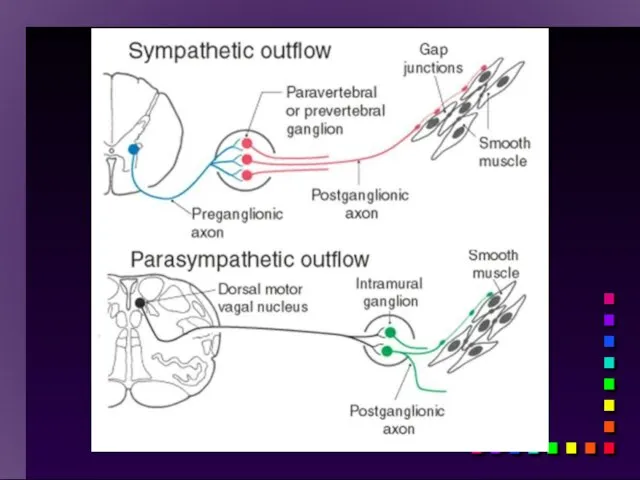
- 20. Sympathetic ganglia Sympathetic chain ganglia (paravertebral ganglia) – preganglionic fibers of the sympathetic NS that carry
- 21. Sympathetic Trunk Ganglia Located on both sides of the vertebral column Linked by short nerves into
- 22. Prevertebral Ganglia Unpaired, not segmentally arranged Occur only in abdomen and pelvis Lie anterior to the
- 23. The Organization of the Sympathetic Division of the ANS
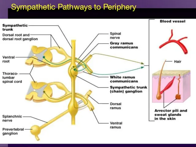
- 24. Copyright Sympathetic Pathways to Periphery Figure 15.9
- 25. Rejoin spinal nerves and reach their destination by way of the dorsal and ventral rami Those
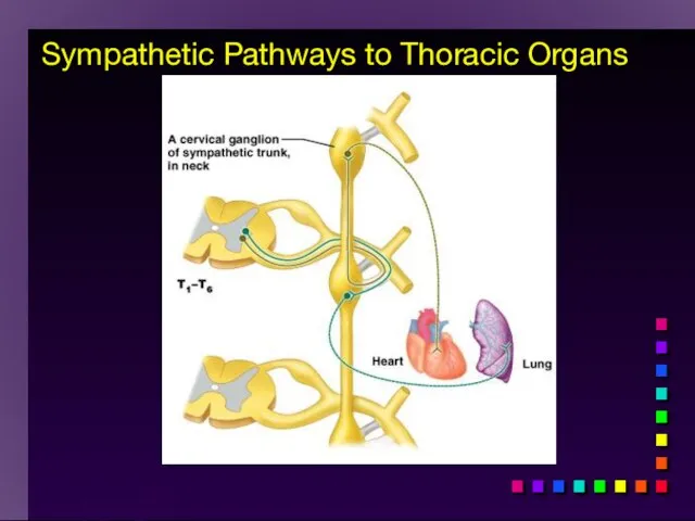
- 26. Copyright Sympathetic Pathways to Thoracic Organs
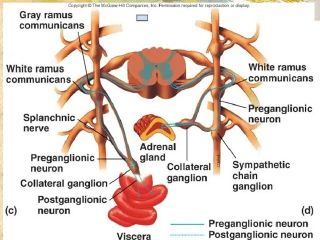
- 27. Sympathetic innervation via preganglionic fibers that synapse within collateral ganglia Splanchic nerves – carry fibers that
- 28. Celiac ganglion Innervates stomach, liver, gall bladder, pancreas, spleen Superior mesenteric ganglion Innervates small intestine and
- 29. Copyright Sympathetic Pathways to the Abdominal Organs
- 30. Copyright Sympathetic Pathways to the Pelvic Organs
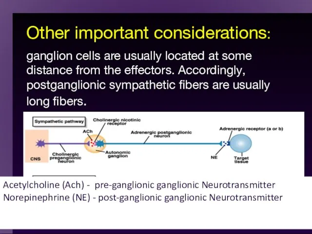
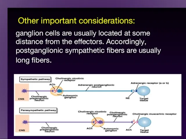
- 31. Other important considerations: ganglion cells are usually located at some distance from the effectors. Accordingly, postganglionic
- 32. Sympathetic Division A single sympathetic preganglionic fiber has many axon collaterals and may synapse with 20
- 33. Sympathetic Variosities
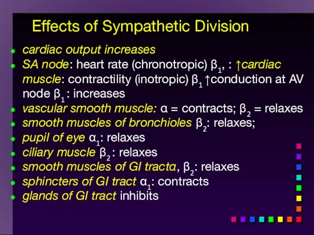
- 34. Effects of Sympathetic Division cardiac output increases SA node: heart rate (chronotropic) β1, : ↑cardiac muscle:
- 35. THE STRESS REACTION A stressful situation activates three major communication systems in the brain that regulate
- 36. THE STRESS REACTION When stress occurs, the sympathetic nervous system is triggered. Norepinephrine is released by
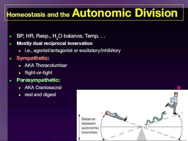
- 38. The two divisions of the autonomic nervous system are not infrequently said to be antagonists in
- 39. Homeostasis and the Autonomic Division BP, HR, Resp., H2O balance, Temp. . . Mostly dual reciprocal
- 40. Other important considerations: ganglion cells are usually located at some distance from the effectors. Accordingly, postganglionic
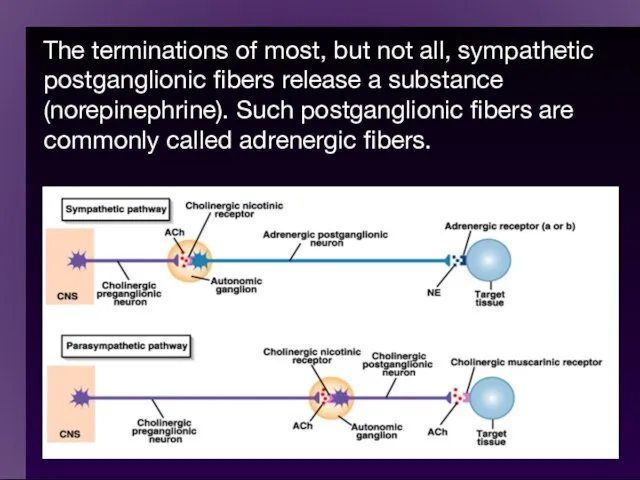
- 41. The terminations of most, but not all, sympathetic postganglionic fibers release a substance (norepinephrine). Such postganglionic
- 43. The effects elicited by the action of the sympathetic division of the ANS are typically effects
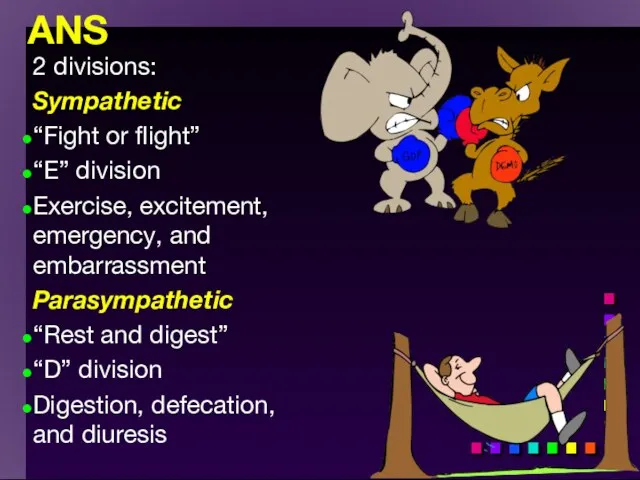
- 44. Parasympathetic “Rest and digest” “D” division Digestion, defecation, and diuresis
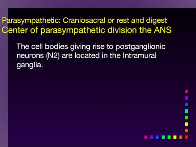
- 45. Parasympathetic: Craniosacral or rest and digest Center of parasympathetic division the ANS Has preganglionic cell bodies
- 46. The Organization of the Parasympathetic Division of the ANS
- 47. The Distribution of Parasympathetic Innervation
- 49. = Craniosacral Division Long preganglionic axons from brain & S2- S4 Intramural ganglia Postganglionic (nonmyelinated) neurons
- 50. Parasympathetic: Craniosacral or rest and digest Center of parasympathetic division the ANS The cell bodies giving
- 51. The ganglion cells of the parasympathetic system are located in or on the wall of the
- 53. Most postganglionic parasympathetic fibers release acetylcholine at their terminations. These fibers are, hence, often called cholinergic
- 54. Summary: Pre- & Postganglionic Parasympathetic Neurons Release ACh muscarinic nicotinic Receptors N1 N2
- 55. All parasympathetic fibers release ACh Short-lived response as ACH is broken down by AChE and tissue
- 56. Parasympathetic (muscarinic) cardiac output M2: decreases SA nodeSA node: heart rate (chronotropic) M2: decreases cardiac musclecardiac
- 57. Effects produced by the parasympathetic division relaxation food processing energy absorption Parasympathetic activation
- 58. The parasympathetic division controls body process during ordinary situations. Generally, it conserves and restores. It slows
- 59. Most Common Autonomic NTs: Acetylcholine (ACh) ACh neurons & ACh receptors are called cholinergic (nicotinic or
- 60. NTs of Autonomic NS Compare to Fig 11-7 α and β N1 N1 N2 N2
- 61. Neuroeffector Junction = Synapse between postganglionic cell and target Most are different from model synapse (compare
- 62. Summary: Pre- & Postganglionic Parasympathetic Neurons Release ACh muscarinic nicotinic Receptors N1 N2
- 63. Two Types of Cholinergic Receptors: Nicotinic and Muscarinic Nicotine = agonist In autonomic ganglia & somatic
- 64. When the neurotransmitter, acetylcholine, attaches to the portion of the nicotinic receptor outside of the cell
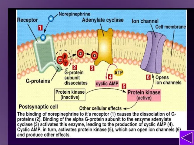
- 65. 2) Muscarinic cholinergic receptor Muscarine = agonist Found in neuro-effector junctions of parasympathetic branch G-protein coupled
- 66. Muscarinic ACh are G-protein Mediated Receptor Mechanism of Sweat Glands: Also some 2nd messenger mechanisms
- 67. Note on G-Proteins: Many functions of the nervous system (e.g., memory) require prolonged changes in neurons
- 68. Adrenergic Receptors Found in neuroeffector junctions of sympathetic branch G protein linked, with various 2nd mess.
- 69. NE Action
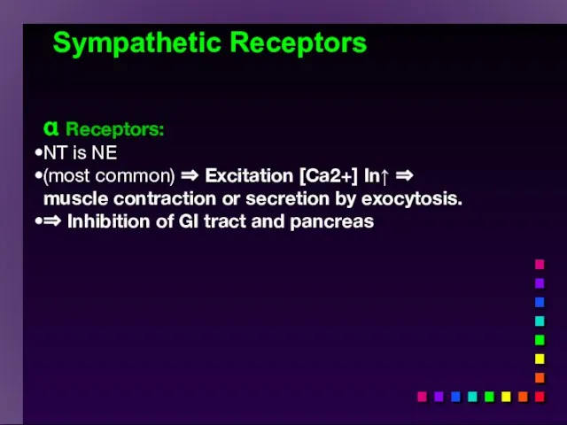
- 70. Sympathetic Receptors α Receptors: NT is NE (most common) ⇒ Excitation [Ca2+] In↑ ⇒ muscle contraction
- 71. β − Receptors Clinically more important β1 ⇒ Excitation heart ([E] = [NE]) “β - blockers”
- 72. Termination of NT Activity ACh: ACh esterase Catecholamine reuptake repackaging degradation (MAO) Blocked by cocaine Fig
- 73. Somatic Motor Division Pathway consists of single neuron from CNS to target Neuromuscular junction: nicotinic cholinergic
- 74. Myasthenia gravis MG: Antibodies block, alter, or destroy the receptors for acetylcholine at the neuromuscular junction
- 75. Direct (Ant)agonist = mimic or block the NT receptor (Ant)agonist = mimic or block secretion, reuptake
- 76. Strychnos Toxifera (Curare) from Koehler's Medicinal-Plants 1887 Direct Antagonists Atropine → muscarinic Curare → nicotinic Propranolol
- 77. Indirect (Ant)agonists Botulinum toxin → inhibits ACh release Parathion, malathion organophosphate insecticides → inhibit AChE (anticholinesterases)
- 78. Important physiological and functional differences exist Comparison of the two divisions
- 79. Table 11-4 Overview: The ANS
- 80. Overview: The ANS Compare the somatic motor pathway to the parasympathetic and sympathetic motor pathways
- 81. A Comparison of Somatic and Autonomic Function
- 82. Summary of Efferent NS
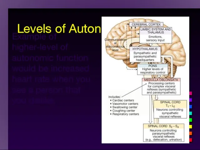
- 84. Activity in the ANS is controlled by centers in the brainstem that deal with visceral functioning
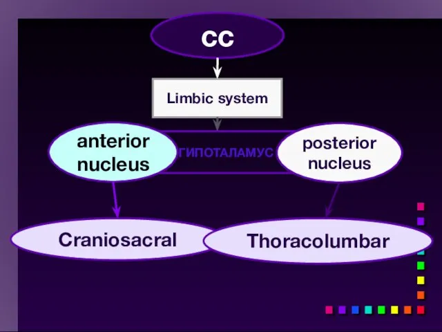
- 85. Levels of Autonomic Control Example of higher-level of autonomic function would be increased heart rate when
- 86. cc Limbic system ГИПОТАЛАМУС Craniosacral anterior nucleus posterior nucleus Thoracolumbar
- 88. Levels of Autonomic Control Example of higher-level of autonomic function would be increased heart rate when
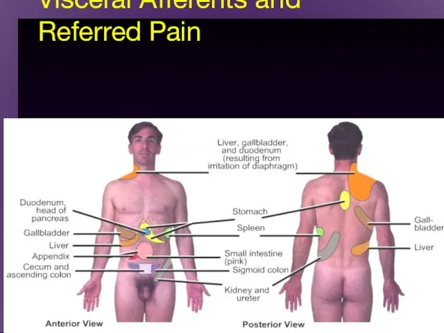
- 89. Visceral Afferents and Referred Pain
- 91. Скачать презентацию
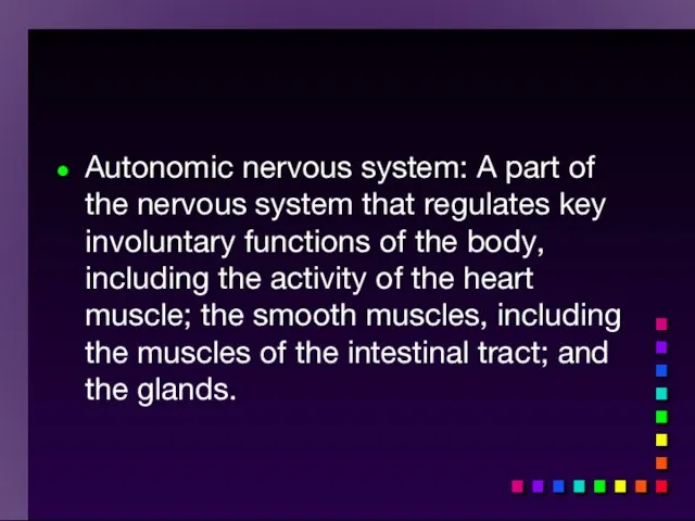
Autonomic nervous system: A part of the nervous system that regulates
Autonomic nervous system: A part of the nervous system that regulates
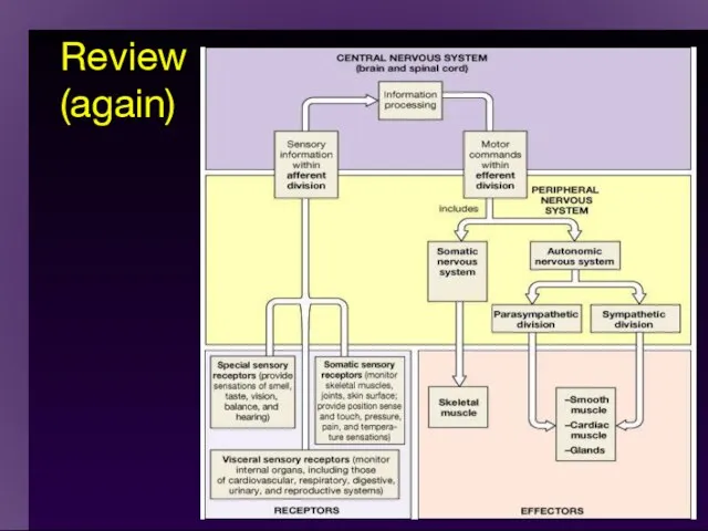
Review (again)
Review (again)
Autonomic Nervous System
Responsible for control of involuntary or visceral bodily
Autonomic Nervous System
Responsible for control of involuntary or visceral bodily
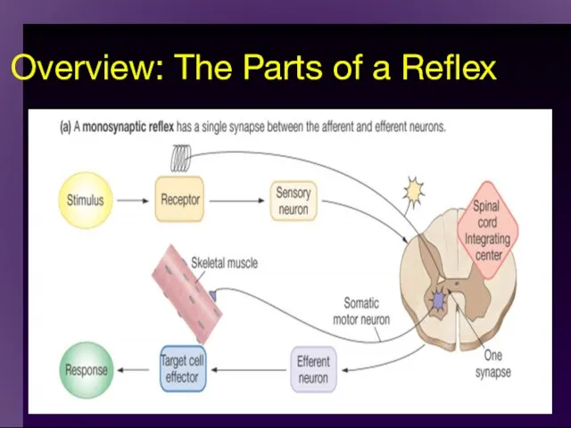
Overview: The Parts of a Reflex
Overview: The Parts of a Reflex
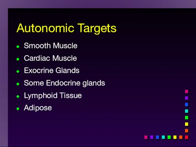
Autonomic Targets
Smooth Muscle
Cardiac Muscle
Exocrine Glands
Some Endocrine glands
Lymphoid Tissue
Adipose
Autonomic Targets
Smooth Muscle
Cardiac Muscle
Exocrine Glands
Some Endocrine glands
Lymphoid Tissue
Adipose
Divisions of ANS
Sympathetic
Parasympathetic
Metasympathetic
Divisions of ANS
Sympathetic
Parasympathetic
Metasympathetic
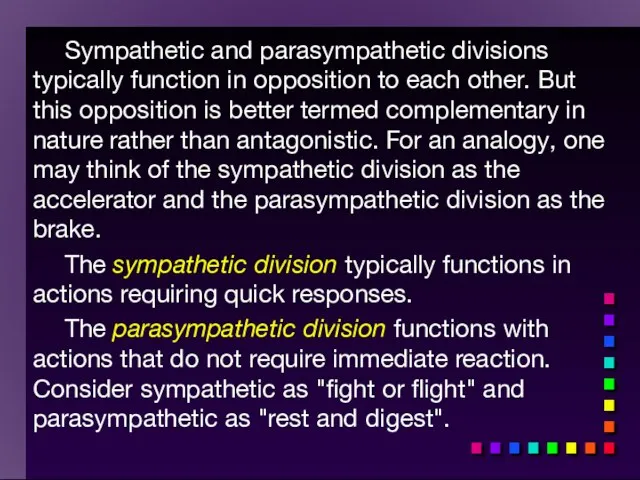
Sympathetic and parasympathetic divisions typically function in opposition to each other.
Sympathetic and parasympathetic divisions typically function in opposition to each other.
ANS
2 divisions:
Sympathetic
“Fight or flight”
“E” division
Exercise, excitement, emergency, and embarrassment
Parasympathetic
“Rest
ANS
2 divisions:
Sympathetic
“Fight or flight”
“E” division
Exercise, excitement, emergency, and embarrassment
Parasympathetic
“Rest
1. The autonomic nervous system (ANS) is an involuntary motor (efferent)
1. The autonomic nervous system (ANS) is an involuntary motor (efferent)
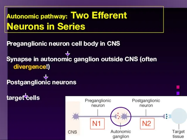
Autonomic pathway: Two Efferent Neurons in Series
Preganglionic neuron cell body in
Autonomic pathway: Two Efferent Neurons in Series
Preganglionic neuron cell body in

3. Although “involuntary”, the autonomic nervous system is regulated by higher
3. Although “involuntary”, the autonomic nervous system is regulated by higher
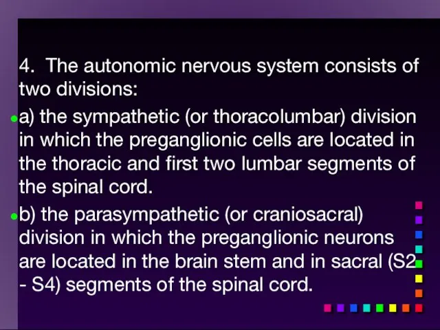
4. The autonomic nervous system consists of two divisions:
a) the sympathetic
4. The autonomic nervous system consists of two divisions:
a) the sympathetic
Sympathetic
“Fight or flight”
“E” division
Exercise, excitement, emergency, and embarrassment
Sympathetic
“Fight or flight”
“E” division
Exercise, excitement, emergency, and embarrassment
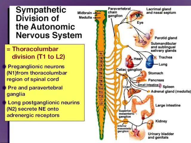
= Thoracolumbar
division (T1 to L2)
Preganglionic neurons (N1)from
= Thoracolumbar
division (T1 to L2)
Preganglionic neurons (N1)from
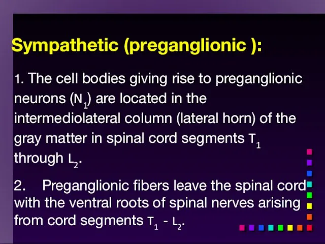
Sympathetic (preganglionic ):
1. The cell bodies giving rise to preganglionic neurons
Sympathetic (preganglionic ):
1. The cell bodies giving rise to preganglionic neurons
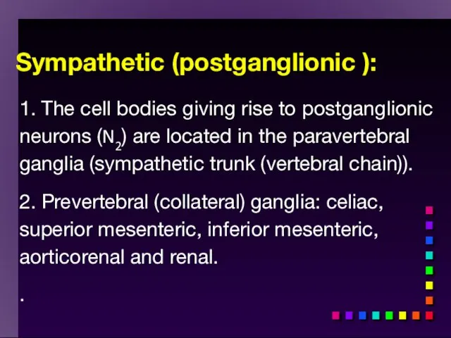
Sympathetic (postganglionic ):
1. The cell bodies giving rise to postganglionic neurons
Sympathetic (postganglionic ):
1. The cell bodies giving rise to postganglionic neurons
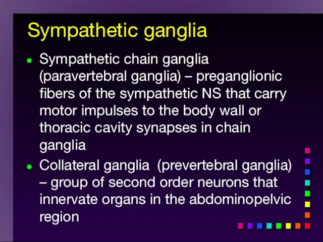
Sympathetic ganglia
Sympathetic chain ganglia (paravertebral ganglia) – preganglionic fibers of
Sympathetic ganglia
Sympathetic chain ganglia (paravertebral ganglia) – preganglionic fibers of
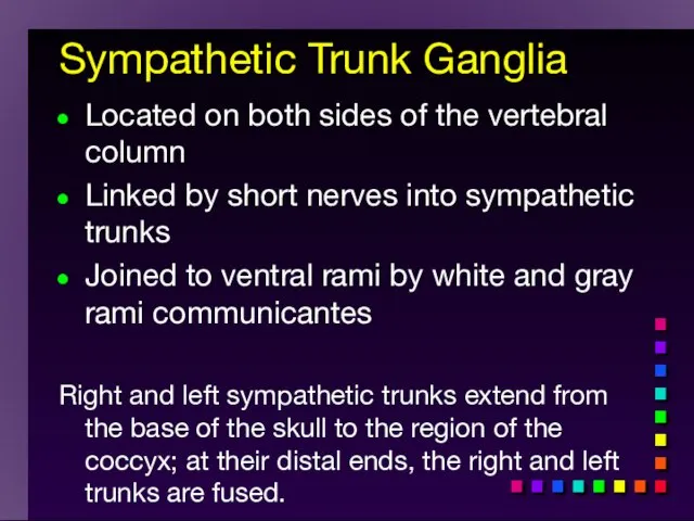
Sympathetic Trunk Ganglia
Located on both sides of the vertebral column
Linked by
Sympathetic Trunk Ganglia
Located on both sides of the vertebral column
Linked by
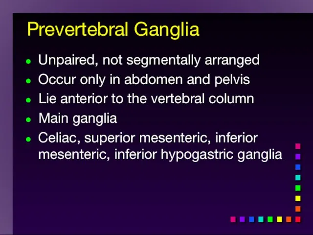
Prevertebral Ganglia
Unpaired, not segmentally arranged
Occur only in abdomen and pelvis
Lie anterior
Prevertebral Ganglia
Unpaired, not segmentally arranged
Occur only in abdomen and pelvis
Lie anterior
The Organization of the Sympathetic Division of the ANS
The Organization of the Sympathetic Division of the ANS
Copyright
Sympathetic Pathways to Periphery
Figure 15.9
Copyright
Sympathetic Pathways to Periphery
Figure 15.9
Rejoin spinal nerves and reach their destination by way of the
Rejoin spinal nerves and reach their destination by way of the
Copyright
Sympathetic Pathways to Thoracic Organs
Copyright
Sympathetic Pathways to Thoracic Organs
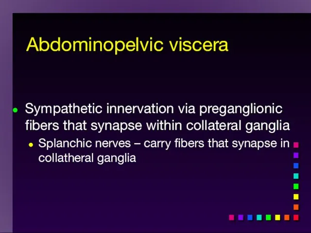
Sympathetic innervation via preganglionic fibers that synapse within collateral ganglia
Splanchic nerves
Sympathetic innervation via preganglionic fibers that synapse within collateral ganglia
Splanchic nerves
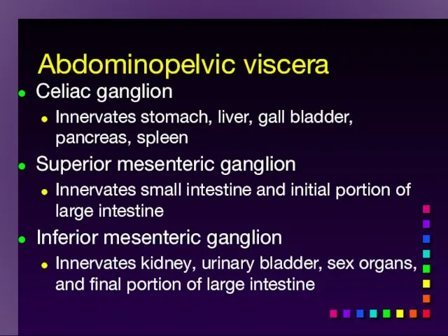
Celiac ganglion
Innervates stomach, liver, gall bladder, pancreas, spleen
Superior mesenteric ganglion
Innervates small
Celiac ganglion
Innervates stomach, liver, gall bladder, pancreas, spleen
Superior mesenteric ganglion
Innervates small
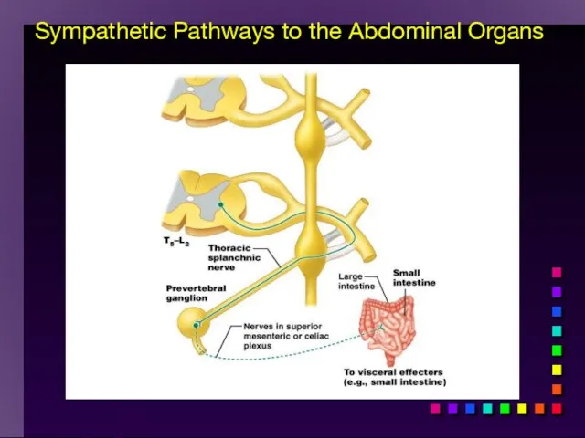
Copyright
Sympathetic Pathways to the Abdominal Organs
Copyright
Sympathetic Pathways to the Abdominal Organs
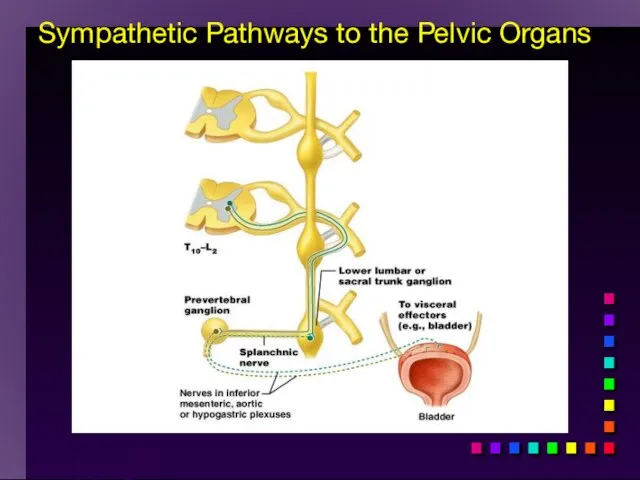
Copyright
Sympathetic Pathways to the Pelvic Organs
Copyright
Sympathetic Pathways to the Pelvic Organs
Other important considerations:
ganglion cells are usually located at some distance from
Other important considerations:
ganglion cells are usually located at some distance from
Sympathetic Division
A single sympathetic preganglionic fiber has many axon collaterals and
Sympathetic Division
A single sympathetic preganglionic fiber has many axon collaterals and
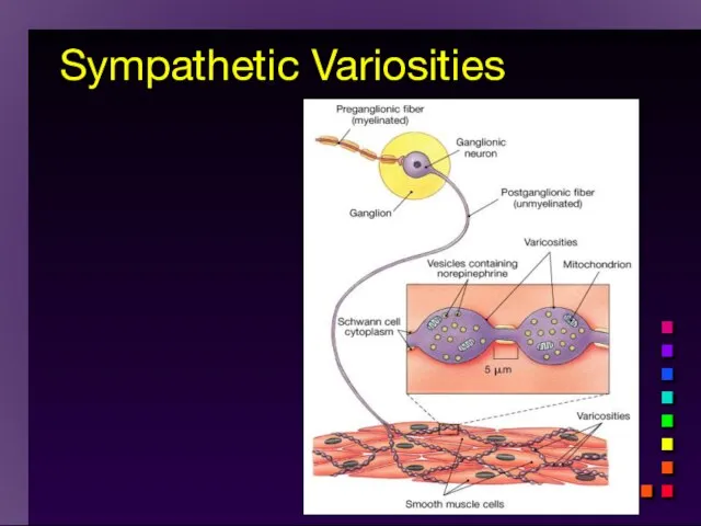
Sympathetic Variosities
Sympathetic Variosities
Effects of Sympathetic Division
cardiac output increases
SA node: heart rate (chronotropic) β1,
Effects of Sympathetic Division
cardiac output increases
SA node: heart rate (chronotropic) β1,
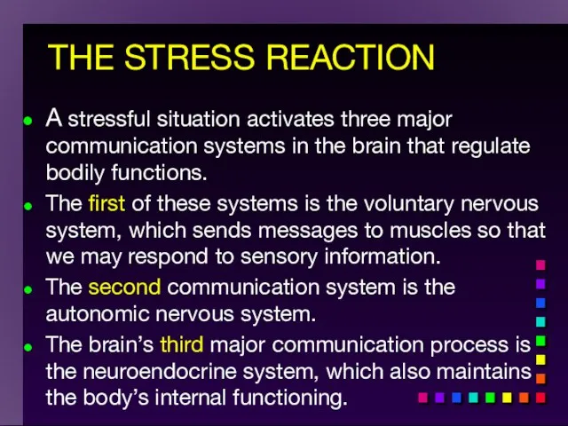
THE STRESS REACTION
A stressful situation activates three major communication systems in
THE STRESS REACTION
A stressful situation activates three major communication systems in
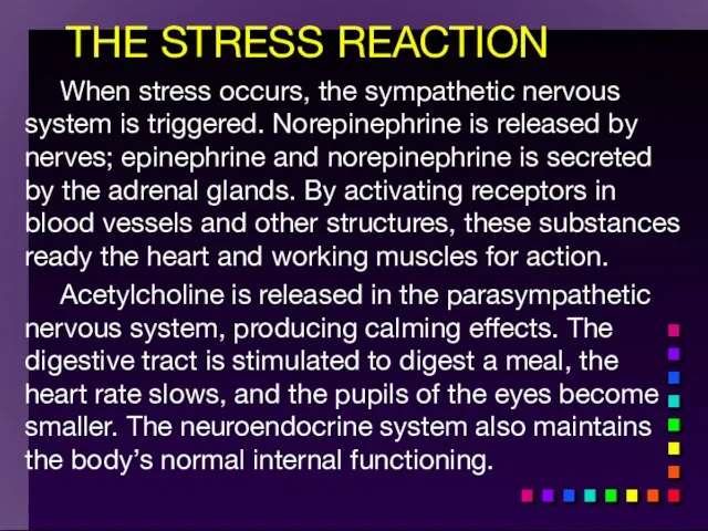
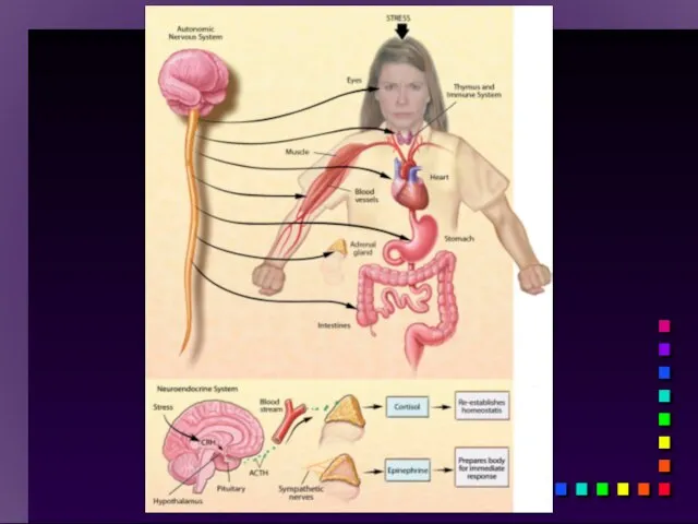
THE STRESS REACTION
When stress occurs, the sympathetic nervous system is triggered.
THE STRESS REACTION
When stress occurs, the sympathetic nervous system is triggered.
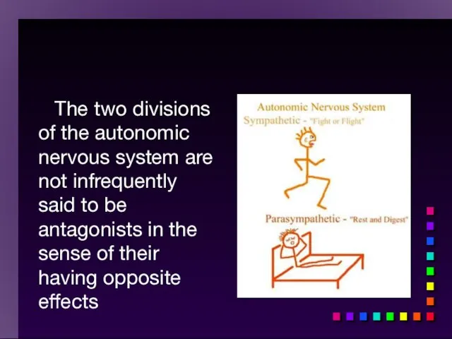
The two divisions of the autonomic nervous system are not
The two divisions of the autonomic nervous system are not
Homeostasis and the Autonomic Division
BP, HR, Resp., H2O balance, Temp. .
Homeostasis and the Autonomic Division
BP, HR, Resp., H2O balance, Temp. .
Other important considerations:
ganglion cells are usually located at some distance
Other important considerations:
ganglion cells are usually located at some distance
The terminations of most, but not all, sympathetic postganglionic fibers release
The terminations of most, but not all, sympathetic postganglionic fibers release
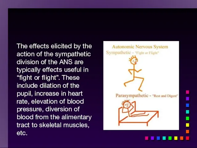
The effects elicited by the action of the sympathetic division of
The effects elicited by the action of the sympathetic division of
Parasympathetic
“Rest and digest”
“D” division
Digestion, defecation, and diuresis
Parasympathetic
“Rest and digest”
“D” division
Digestion, defecation, and diuresis
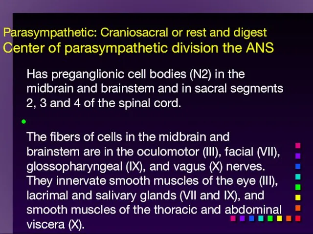
Parasympathetic: Craniosacral or rest and digest
Center of parasympathetic division the ANS
Parasympathetic: Craniosacral or rest and digest Center of parasympathetic division the ANS
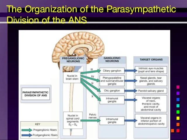
The Organization of the Parasympathetic Division of the ANS
The Organization of the Parasympathetic Division of the ANS
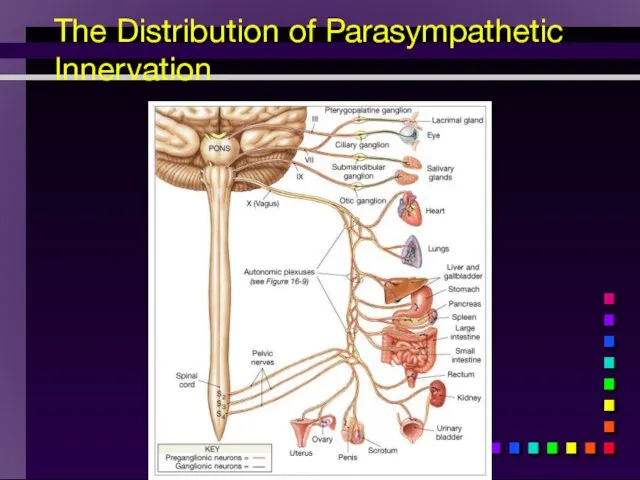
The Distribution of Parasympathetic Innervation
The Distribution of Parasympathetic Innervation
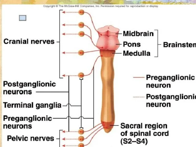
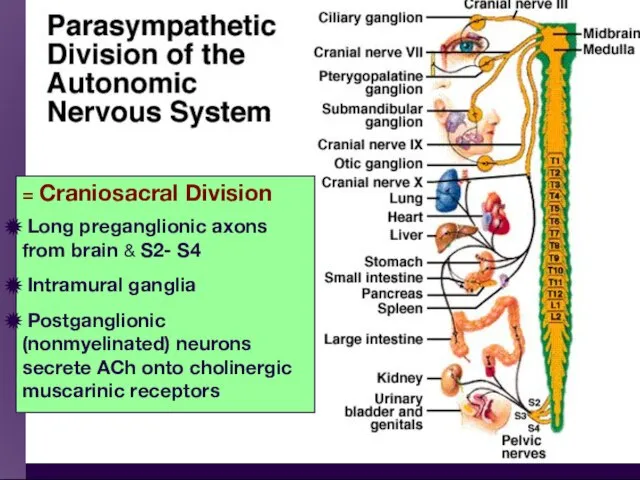
= Craniosacral Division
Long preganglionic axons from brain & S2- S4
= Craniosacral Division
Long preganglionic axons from brain & S2- S4
Parasympathetic: Craniosacral or rest and digest
Center of parasympathetic division the ANS
Parasympathetic: Craniosacral or rest and digest Center of parasympathetic division the ANS
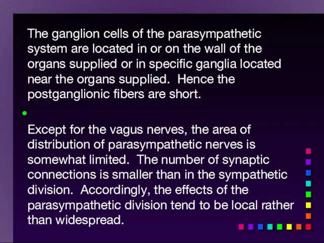
The ganglion cells of the parasympathetic system are located in or
The ganglion cells of the parasympathetic system are located in or

Most postganglionic parasympathetic fibers release acetylcholine at their terminations. These fibers
Most postganglionic parasympathetic fibers release acetylcholine at their terminations. These fibers

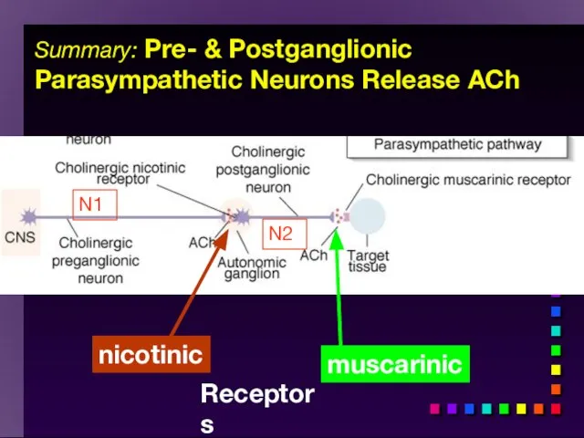
Summary: Pre- & Postganglionic Parasympathetic Neurons Release ACh
muscarinic
nicotinic
Receptors
N1
N2
Summary: Pre- & Postganglionic Parasympathetic Neurons Release ACh
muscarinic
nicotinic
Receptors
N1
N2
All parasympathetic fibers release ACh
Short-lived response as ACH is broken down
All parasympathetic fibers release ACh
Short-lived response as ACH is broken down
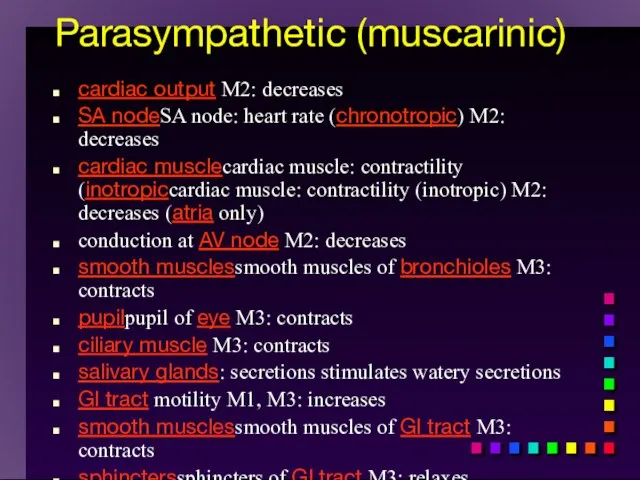
Parasympathetic (muscarinic)
cardiac output M2: decreases
SA nodeSA node: heart rate (chronotropic)
Parasympathetic (muscarinic)
cardiac output M2: decreases
SA nodeSA node: heart rate (chronotropic)
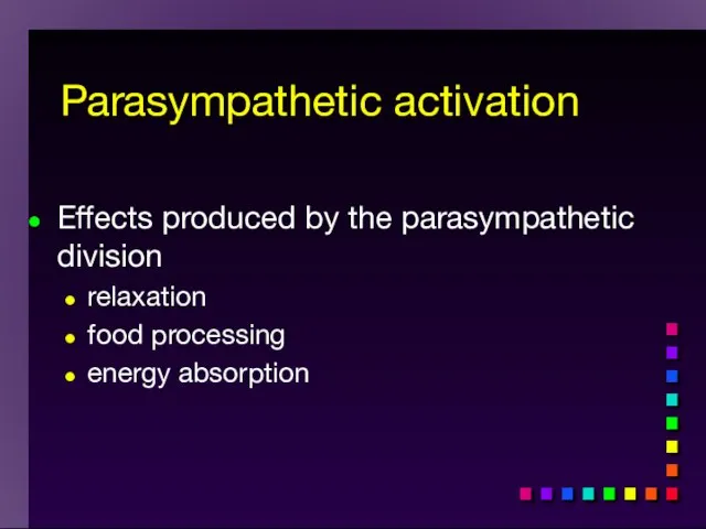
Effects produced by the parasympathetic division
relaxation
food processing
energy absorption
Parasympathetic activation
Effects produced by the parasympathetic division
relaxation
food processing
energy absorption
Parasympathetic activation
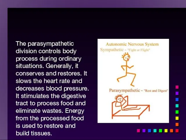
The parasympathetic division controls body process during ordinary situations. Generally, it
The parasympathetic division controls body process during ordinary situations. Generally, it
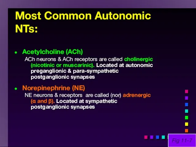
Most Common Autonomic NTs:
Acetylcholine (ACh)
ACh neurons & ACh receptors are called
Most Common Autonomic NTs:
Acetylcholine (ACh)
ACh neurons & ACh receptors are called
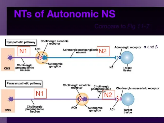
NTs of Autonomic NS
Compare to Fig 11-7
α and β
N1
N1
N2
N2
NTs of Autonomic NS
Compare to Fig 11-7
α and β
N1
N1
N2
N2
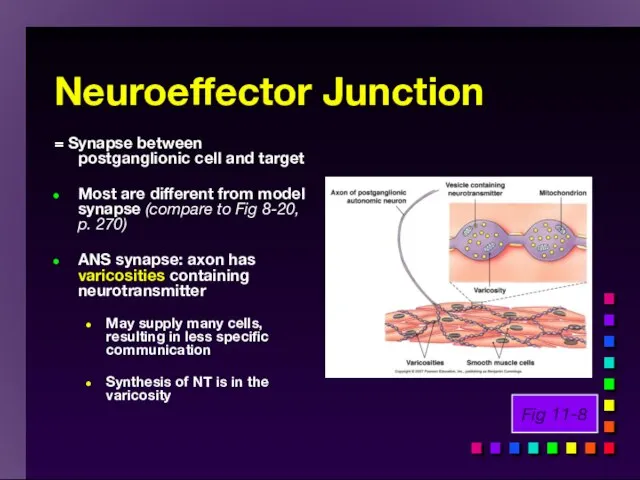
Neuroeffector Junction
= Synapse between postganglionic cell and target
Most are different from
Neuroeffector Junction
= Synapse between postganglionic cell and target
Most are different from
Summary: Pre- & Postganglionic Parasympathetic Neurons Release ACh
muscarinic
nicotinic
Receptors
N1
N2
Summary: Pre- & Postganglionic Parasympathetic Neurons Release ACh
muscarinic
nicotinic
Receptors
N1
N2
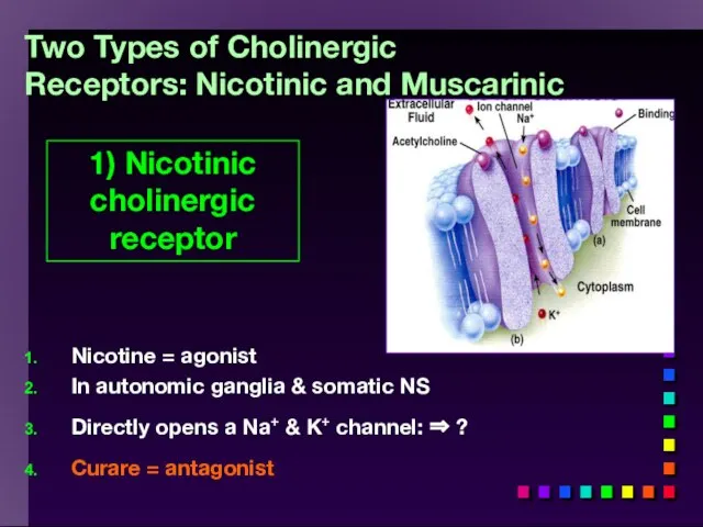
Two Types of Cholinergic Receptors: Nicotinic and Muscarinic
Nicotine = agonist
In autonomic
Two Types of Cholinergic Receptors: Nicotinic and Muscarinic
Nicotine = agonist
In autonomic
When the neurotransmitter, acetylcholine, attaches to the portion of the nicotinic
When the neurotransmitter, acetylcholine, attaches to the portion of the nicotinic
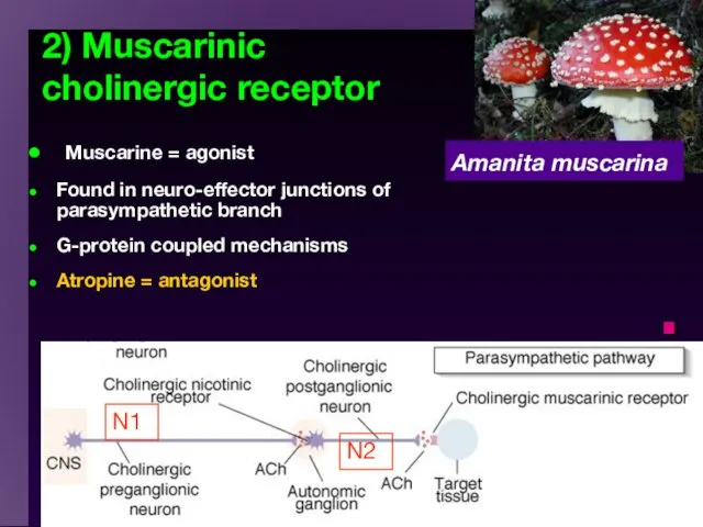
2) Muscarinic cholinergic receptor
Muscarine = agonist
Found in neuro-effector junctions of
2) Muscarinic cholinergic receptor
Muscarine = agonist
Found in neuro-effector junctions of
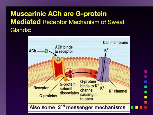
Muscarinic ACh are G-protein Mediated Receptor Mechanism of Sweat Glands:
Also some
Muscarinic ACh are G-protein Mediated Receptor Mechanism of Sweat Glands:
Also some
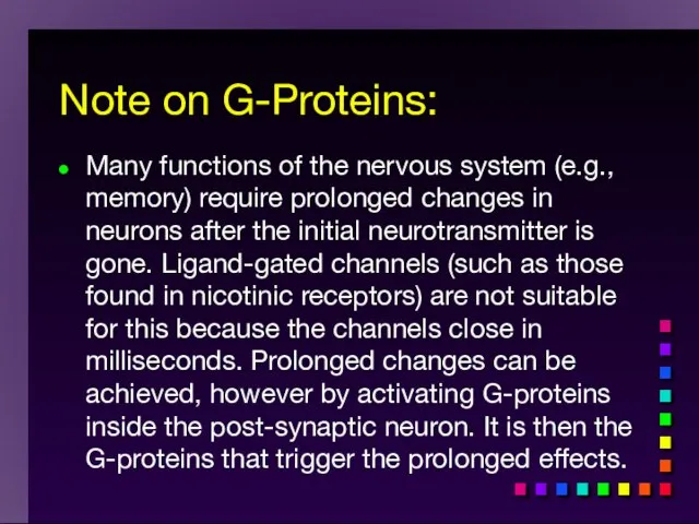
Note on G-Proteins:
Many functions of the nervous system (e.g., memory)
Note on G-Proteins:
Many functions of the nervous system (e.g., memory)
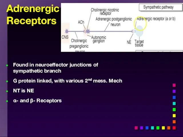
Adrenergic
Receptors
Found in neuroeffector junctions of sympathetic branch
G protein linked, with
Adrenergic
Receptors
Found in neuroeffector junctions of sympathetic branch
G protein linked, with
NE Action
NE Action
Sympathetic Receptors
α Receptors:
NT is NE
(most common) ⇒ Excitation [Ca2+]
Sympathetic Receptors
α Receptors:
NT is NE
(most common) ⇒ Excitation [Ca2+]
![β − Receptors Clinically more important β1 ⇒ Excitation heart ([E]](/_ipx/f_webp&q_80&fit_contain&s_1440x1080/imagesDir/jpg/543692/slide-70.jpg)
β − Receptors Clinically more important
β1 ⇒ Excitation heart ([E]
β − Receptors Clinically more important
β1 ⇒ Excitation heart ([E]
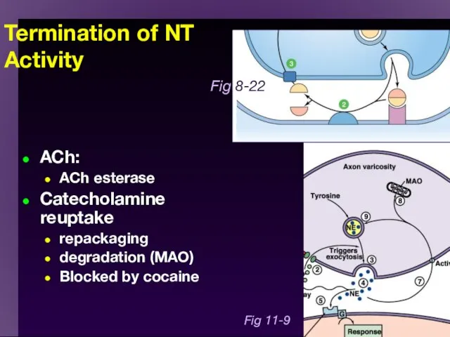
Termination of NT Activity
ACh:
ACh esterase
Catecholamine reuptake
repackaging
degradation (MAO)
Blocked by cocaine
Fig 11-9
Fig 8-22
Termination of NT Activity
ACh:
ACh esterase
Catecholamine reuptake
repackaging
degradation (MAO)
Blocked by cocaine
Fig 11-9
Fig 8-22
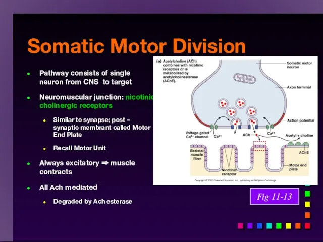
Somatic Motor Division
Pathway consists of single neuron from CNS to target
Neuromuscular
Somatic Motor Division
Pathway consists of single neuron from CNS to target
Neuromuscular
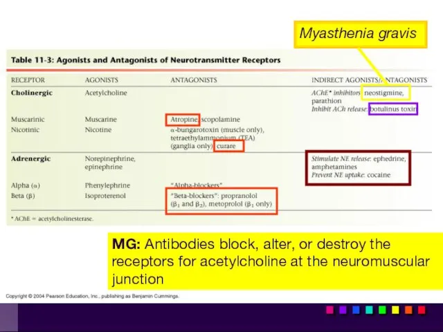
Myasthenia gravis
MG: Antibodies block, alter, or destroy the receptors for acetylcholine
Myasthenia gravis
MG: Antibodies block, alter, or destroy the receptors for acetylcholine
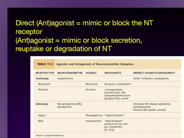
Direct (Ant)agonist = mimic or block the NT receptor
(Ant)agonist = mimic
Direct (Ant)agonist = mimic or block the NT receptor (Ant)agonist = mimic
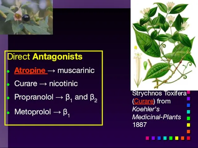
Strychnos Toxifera (Curare) from Koehler's Medicinal-Plants 1887
Direct Antagonists
Atropine → muscarinic
Curare
Strychnos Toxifera (Curare) from Koehler's Medicinal-Plants 1887
Direct Antagonists
Atropine → muscarinic
Curare
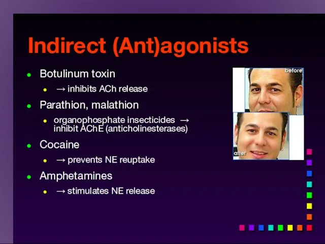
Indirect (Ant)agonists
Botulinum toxin
→ inhibits ACh release
Parathion, malathion
organophosphate insecticides → inhibit
Indirect (Ant)agonists
Botulinum toxin
→ inhibits ACh release
Parathion, malathion
organophosphate insecticides → inhibit
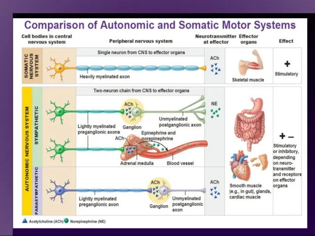
Important physiological and functional differences exist
Comparison of the two divisions
Important physiological and functional differences exist
Comparison of the two divisions
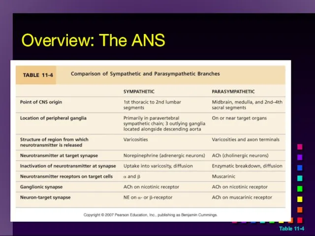
Table 11-4
Overview: The ANS
Table 11-4
Overview: The ANS
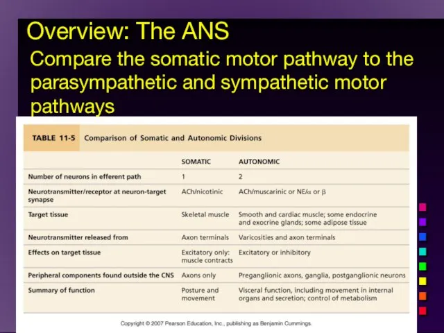
Overview: The ANS
Compare the somatic motor pathway to the parasympathetic
Overview: The ANS
Compare the somatic motor pathway to the parasympathetic
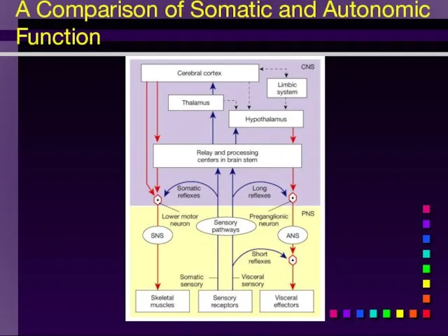
A Comparison of Somatic and Autonomic Function
A Comparison of Somatic and Autonomic Function
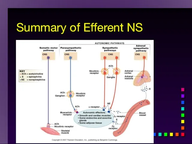
Summary of Efferent NS
Summary of Efferent NS
Activity in the ANS is controlled by centers in the brainstem
Activity in the ANS is controlled by centers in the brainstem
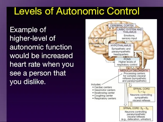
Levels of Autonomic Control
Example of higher-level of autonomic function would be
Levels of Autonomic Control
Example of higher-level of autonomic function would be
cc
Limbic system
ГИПОТАЛАМУС
Craniosacral
anterior
nucleus
posterior
nucleus
Thoracolumbar
cc
Limbic system
ГИПОТАЛАМУС
Craniosacral
anterior
nucleus
posterior
nucleus
Thoracolumbar

Levels of Autonomic Control
Example of higher-level of autonomic function would be
Levels of Autonomic Control
Example of higher-level of autonomic function would be
Visceral Afferents and Referred Pain
Visceral Afferents and Referred Pain
 Неочікувана радість
 Болезни системы крови
 ВНЧС. Заболевания височно-нижнечелюстного сустава
 Свойства вакцины Oxford-AstraZeneca (AZD1222)
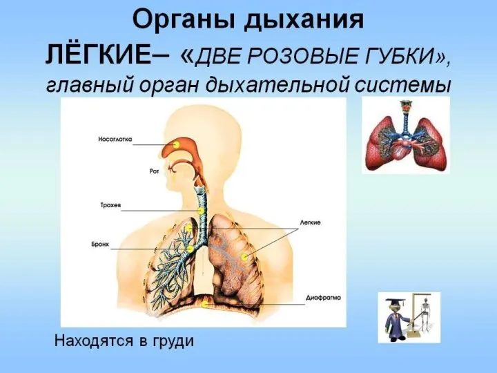 Органы дыхания
 Болезни органов дыхания
 Ветеринарная клиника
 Энцефалиты
 Жатырдан тыс жүктілікке жасалған хирургиялық емнен кейінгі науқастардың реабилитациясы
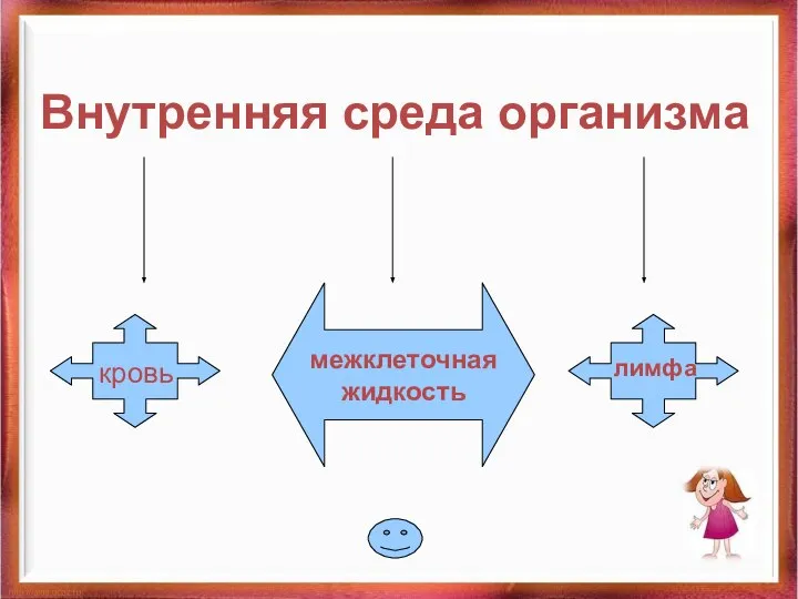 Внутренняя среда организма
 Диффузные заболевания соединительной ткани. Дерматологические аспекты
 Физико-химические свойства лекарственных препаратов железа
 Фармацевтическая биоэтика и биобезопасность
 Беседа о гипер и гипогликемических состояниях
 Covid-19. Выплаты стимулирующего характера
 Типы пациента в реабилитации зависимости
 Современное представление о патогенезе болезни Альцгеймера
 Диагностика типов привязанности
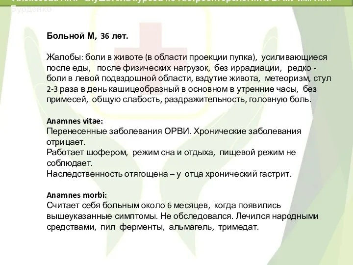 Эрозивный гастрит. Клинический случай
 Гигиена кожных покровов
 Туберкулез полости рта. (Лекция 5)
 Психология и педагогика высшей школы
 Регионарная анестезия. Спинномозговая. Эпидуральная
 Bronchitis
 Технологические особенности приготовления блюд из капусты для организации питания лиц страдающих заболеванием сердца и сосудов
 Вегетативна нервова система. Синдроми ураження різних відділів вегетативної нервової системи
 Ультразвуковая допплерография в стоматологии
 Практические аспекты применения ингибиторов PCSK-9 у пациентов высокого и очень высокого сердечно-сосудистого риска