- Surgical Neonatal Vomiting

Содержание
- 2. Interactive Case studies Summary of specific surgical conditions
- 3. What is a neonate? What is preterm? What is term?
- 4. Definitions Neonate – premature and term babies less that 44 weeks post-conceptional age Premature neonate Term
- 5. History and Symptoms
- 6. History and Symptoms Gestation Weight Antenatal history Colour of vomit Frequency of vomit Bowel opening Saliva?
- 7. Physical Findings
- 8. Physical findings Observations Erythema and bruising Distended Scaphoid abdomen Mass Anus – site, size and patency
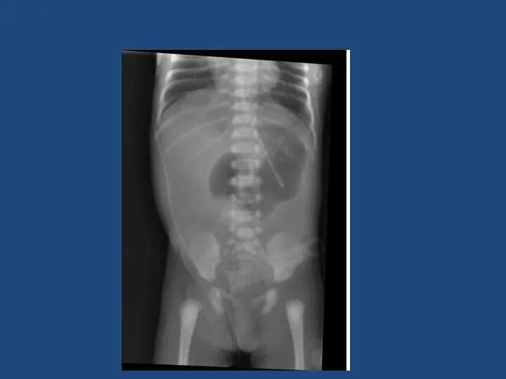
- 9. Investigations Plain AXR/CXR Upper/Lower GI contrast Abdominal USS
- 10. Case 1 Term neonate 1 day old Vomiting Relevant points in history Relevant examination findings Differential
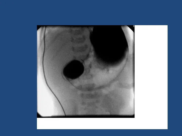
- 12. Oesophageal atresia and tracheo-oesophageal fistula 1 in 3500 liveborn births Antenatal Polyhydramnios, absent stomach, associated anomalies
- 13. Classification Type A: 8%, Type B: 1%, Type C: 86%, Type D 1%, Type E: 4%
- 14. Repair Right thoracotomy (usually) 4th or 5th intercoastal space Extrapleural approach +/- division of azygous vein
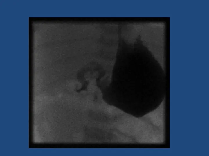
- 18. Duodenal Atresia 1 in 5000 Antenatal diagnosis – ‘double bubble’ Associated with Trisomy 21 - 30%,
- 20. Malrotation +/- volvulus
- 21. Malrotation 1 in 6000 present in babies 0.5% of autopsies show degree of malrotation Abnormal duodenal
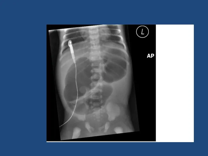
- 22. Malrotation + volvulus SURGICAL EMERGENCY Bilious vomiting in neonate Upper GI contrast to diagnose Emergency laparotomy
- 24. Jejunal/Ileal atresia Stenosis – 11% Type 1 – 23% Type 2 – 10% Type 3 –
- 25. Jejunal/ileal atresia 1 in 5000 births Aetiology – antenatal vascular compromise May have short bowel Resection
- 26. Meconium Ileus CF – 1 in 2500 births ~16% of babies with CF Inspissated sticky meconium
- 27. Microcolon in Meconium ileus
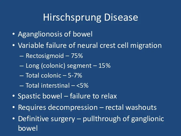
- 28. Hirschsprung Disease
- 29. Hirschsprung Disease 1 in 5000 births M:F 4:1 Associated with Trisomy 21 Delayed passage of meconium
- 30. Hirschsprung Disease Aganglionosis of bowel Variable failure of neural crest cell migration Rectosigmoid – 75% Long
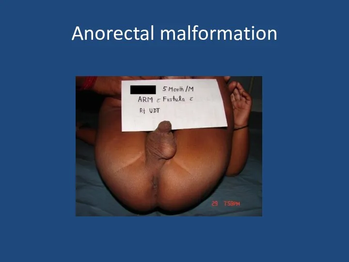
- 31. Anorectal malformation
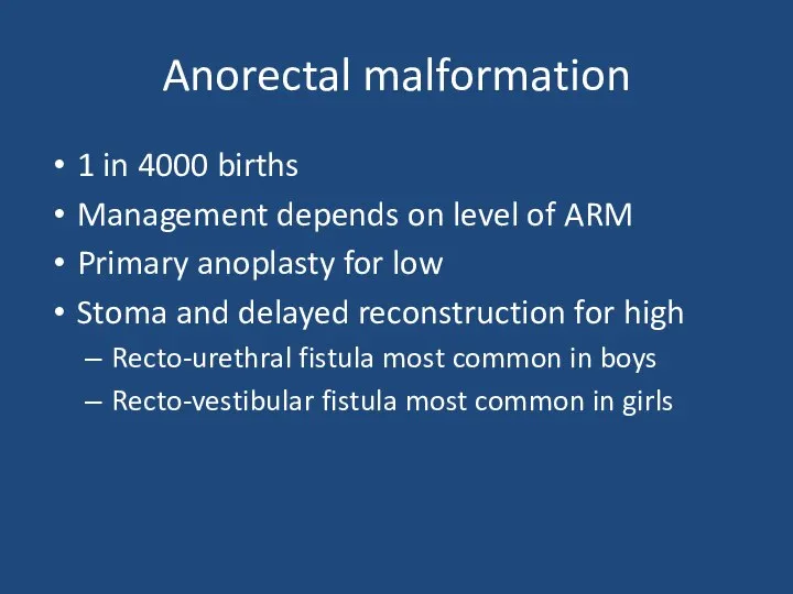
- 32. Anorectal malformation 1 in 4000 births Management depends on level of ARM Primary anoplasty for low
- 33. Case 2 3 week old term baby Relevant points in history Relevant examination findings Differential diagnosis
- 34. Infantile Hypertrophic Pyloric Stenosis 1-4:1000, M:F 4:1 Overgrowth of pyloric muscle Gastric outlet obstruction Increasing non-bilious
- 35. Pyloric stenosis
- 36. Infantile Hypertrophic Pyloric Stenosis Diagnosis – palpable mass on ‘test feed’ USS Pyloric length >16mm Single
- 37. Inguinal hernia
- 38. Inguinal hernia Usually can reduce If truly incacerated – emergency exploration Otherwise if premature baby or
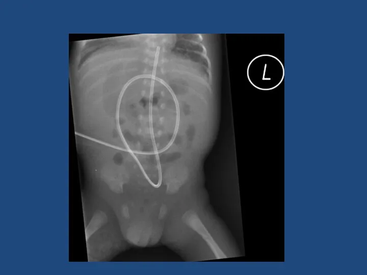
- 39. Case 3 Preterm neonate – bilious vomiting Born 27 weeks gestation Weight 1 kg 1 week
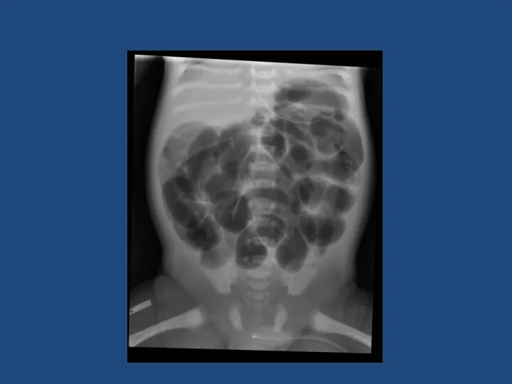
- 43. Necrotising Enterocolitis 90% in preterm 10% in term babies ~5% of all babies admitted to Neonatal
- 44. Necrotising Enterocolitis Surgery indicated for: Worsening clinical condition despite maximal supportive therapy Perforation Laparotomy Assess extent
- 46. Скачать презентацию
Interactive
Case studies
Summary of specific surgical conditions
Interactive
Case studies
Summary of specific surgical conditions
What is a neonate?
What is preterm?
What is term?
What is a neonate?
What is preterm?
What is term?
Definitions
Neonate – premature and term babies less that 44 weeks post-conceptional
Definitions
Neonate – premature and term babies less that 44 weeks post-conceptional
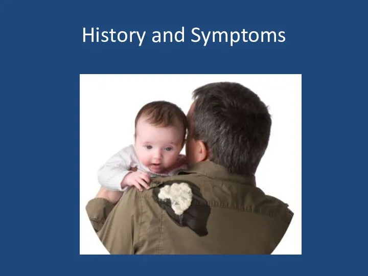
History and Symptoms
History and Symptoms
History and Symptoms
Gestation
Weight
Antenatal history
Colour of vomit
Frequency of vomit
Bowel opening
Saliva?
Associated co-morbidities
History and Symptoms
Gestation
Weight
Antenatal history
Colour of vomit
Frequency of vomit
Bowel opening
Saliva?
Associated co-morbidities
Physical Findings
Physical Findings
Physical findings
Observations
Erythema and bruising
Distended
Scaphoid abdomen
Mass
Anus – site, size and patency
Tenderness
External
Physical findings
Observations
Erythema and bruising
Distended
Scaphoid abdomen
Mass
Anus – site, size and patency
Tenderness
External
Investigations
Plain AXR/CXR
Upper/Lower GI contrast
Abdominal USS
Investigations
Plain AXR/CXR
Upper/Lower GI contrast
Abdominal USS
Case 1
Term neonate
1 day old
Vomiting
Relevant points in history
Relevant examination findings
Differential diagnosis
Case 1
Term neonate
1 day old
Vomiting
Relevant points in history
Relevant examination findings
Differential diagnosis
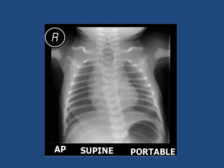
Oesophageal atresia and tracheo-oesophageal fistula
1 in 3500 liveborn births
Antenatal
Polyhydramnios, absent stomach,
Oesophageal atresia and tracheo-oesophageal fistula
1 in 3500 liveborn births
Antenatal
Polyhydramnios, absent stomach,
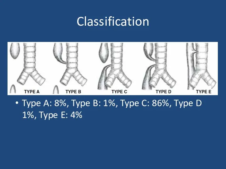
Classification
Type A: 8%, Type B: 1%, Type C: 86%, Type D
Classification
Type A: 8%, Type B: 1%, Type C: 86%, Type D
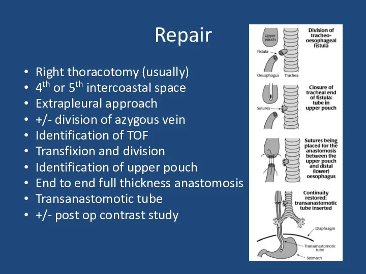
Repair
Right thoracotomy (usually)
4th or 5th intercoastal space
Extrapleural approach
+/- division of azygous
Repair
Right thoracotomy (usually)
4th or 5th intercoastal space
Extrapleural approach
+/- division of azygous
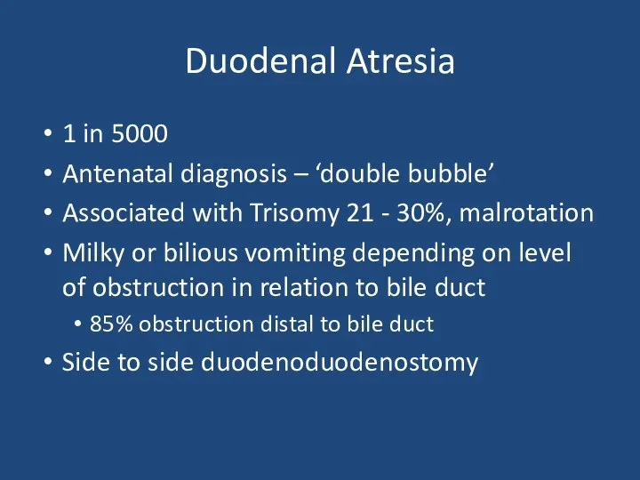
Duodenal Atresia
1 in 5000
Antenatal diagnosis – ‘double bubble’
Associated with Trisomy 21
Duodenal Atresia
1 in 5000
Antenatal diagnosis – ‘double bubble’
Associated with Trisomy 21
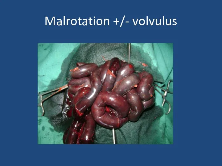
Malrotation +/- volvulus
Malrotation +/- volvulus
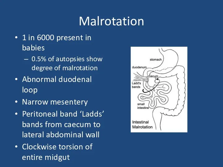
Malrotation
1 in 6000 present in babies
0.5% of autopsies show degree of
Malrotation
1 in 6000 present in babies
0.5% of autopsies show degree of
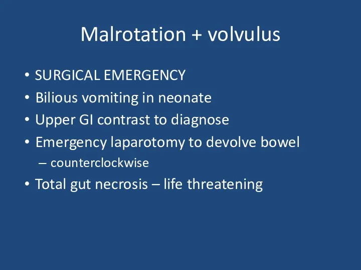
Malrotation + volvulus
SURGICAL EMERGENCY
Bilious vomiting in neonate
Upper GI contrast to
Malrotation + volvulus
SURGICAL EMERGENCY
Bilious vomiting in neonate
Upper GI contrast to
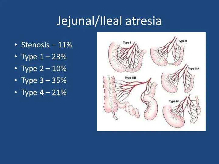
Jejunal/Ileal atresia
Stenosis – 11%
Type 1 – 23%
Type 2 – 10%
Type
Jejunal/Ileal atresia
Stenosis – 11%
Type 1 – 23%
Type 2 – 10%
Type
Jejunal/ileal atresia
1 in 5000 births
Aetiology – antenatal vascular compromise
May have short
Jejunal/ileal atresia
1 in 5000 births
Aetiology – antenatal vascular compromise
May have short
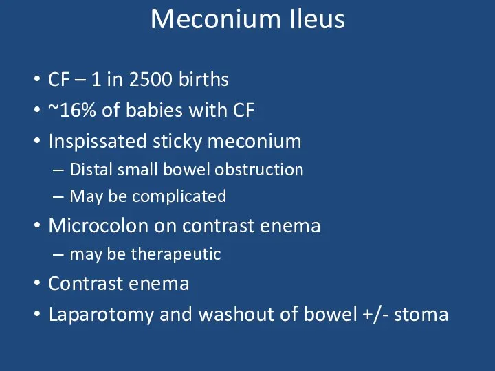
Meconium Ileus
CF – 1 in 2500 births
~16% of babies with CF
Inspissated
Meconium Ileus
CF – 1 in 2500 births
~16% of babies with CF
Inspissated
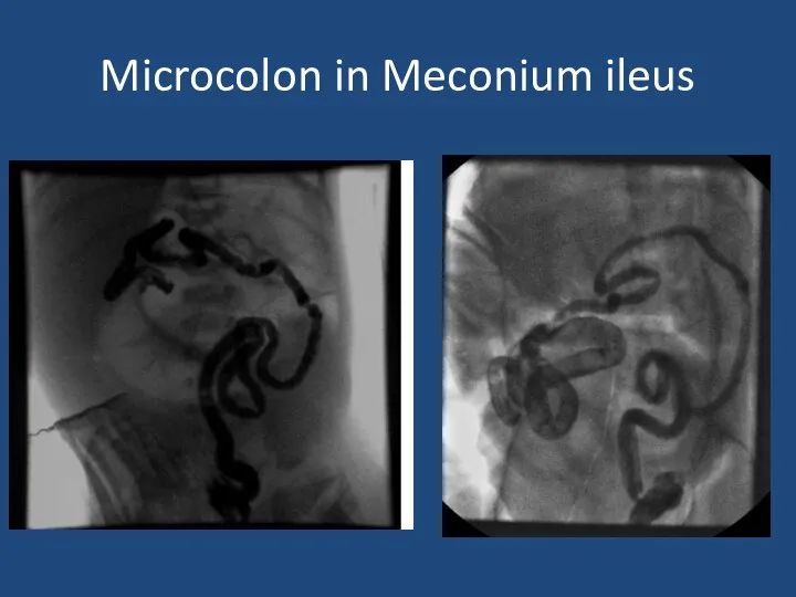
Microcolon in Meconium ileus
Microcolon in Meconium ileus
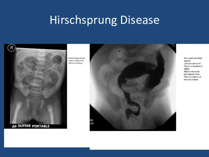
Hirschsprung Disease
Hirschsprung Disease
Hirschsprung Disease
1 in 5000 births
M:F 4:1
Associated with Trisomy 21
Delayed passage of
Hirschsprung Disease
1 in 5000 births
M:F 4:1
Associated with Trisomy 21
Delayed passage of
Hirschsprung Disease
Aganglionosis of bowel
Variable failure of neural crest cell migration
Rectosigmoid –
Hirschsprung Disease
Aganglionosis of bowel
Variable failure of neural crest cell migration
Rectosigmoid –
Anorectal malformation
Anorectal malformation
Anorectal malformation
1 in 4000 births
Management depends on level of ARM
Primary anoplasty
Anorectal malformation
1 in 4000 births
Management depends on level of ARM
Primary anoplasty
Case 2
3 week old term baby
Relevant points in history
Relevant examination findings
Differential
Case 2
3 week old term baby
Relevant points in history
Relevant examination findings
Differential
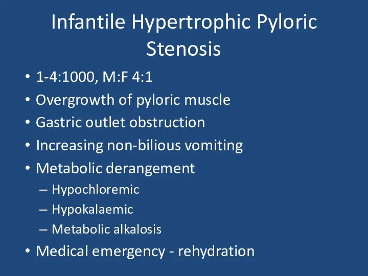
Infantile Hypertrophic Pyloric Stenosis
1-4:1000, M:F 4:1
Overgrowth of pyloric muscle
Gastric outlet obstruction
Increasing
Infantile Hypertrophic Pyloric Stenosis
1-4:1000, M:F 4:1
Overgrowth of pyloric muscle
Gastric outlet obstruction
Increasing
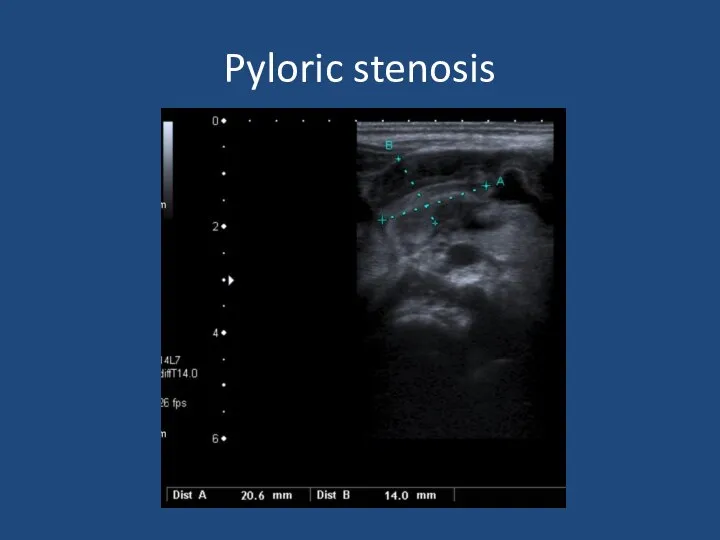
Pyloric stenosis
Pyloric stenosis
Infantile Hypertrophic Pyloric Stenosis
Diagnosis – palpable mass on ‘test feed’
USS
Pyloric length
Infantile Hypertrophic Pyloric Stenosis
Diagnosis – palpable mass on ‘test feed’
USS
Pyloric length
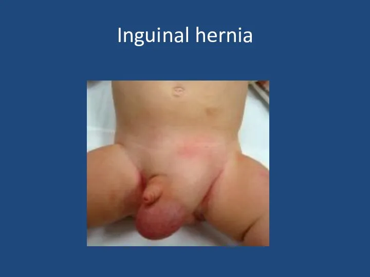
Inguinal hernia
Inguinal hernia
Inguinal hernia
Usually can reduce
If truly incacerated – emergency exploration
Otherwise if premature
Inguinal hernia
Usually can reduce
If truly incacerated – emergency exploration
Otherwise if premature
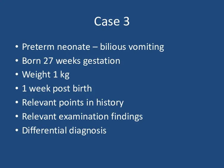
Case 3
Preterm neonate – bilious vomiting
Born 27 weeks gestation
Weight 1 kg
1
Case 3
Preterm neonate – bilious vomiting
Born 27 weeks gestation
Weight 1 kg
1
Necrotising Enterocolitis
90% in preterm 10% in term babies
~5% of all babies
Necrotising Enterocolitis
90% in preterm 10% in term babies
~5% of all babies
Necrotising Enterocolitis
Surgery indicated for:
Worsening clinical condition despite maximal supportive therapy
Perforation
Laparotomy
Assess extent
Necrotising Enterocolitis
Surgery indicated for:
Worsening clinical condition despite maximal supportive therapy
Perforation
Laparotomy
Assess extent
 Ультразвук в клинической диагностике глаз и орбиты
 Ортодонтия. Тістем дегеніміз орталық окклюзия жағдайында тістердің түйісуі
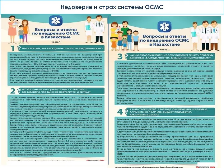 Недоверие и страх системы ОСМС
 Влияние антибиотиков на микрофлору
 Сүйек кемігі
 Хронические гепатиты, циррозы печени
 Хроническая сердечная недостаточность
 Сахар и сахарозаменители
 Вредное влияние алкоголя, курения и наркотиков в период эмбрионального развития
 Профилактика и ранняя диагностика онкологических заболеваний
 Уход за пациентами пожилого возраста в стационаре и дома
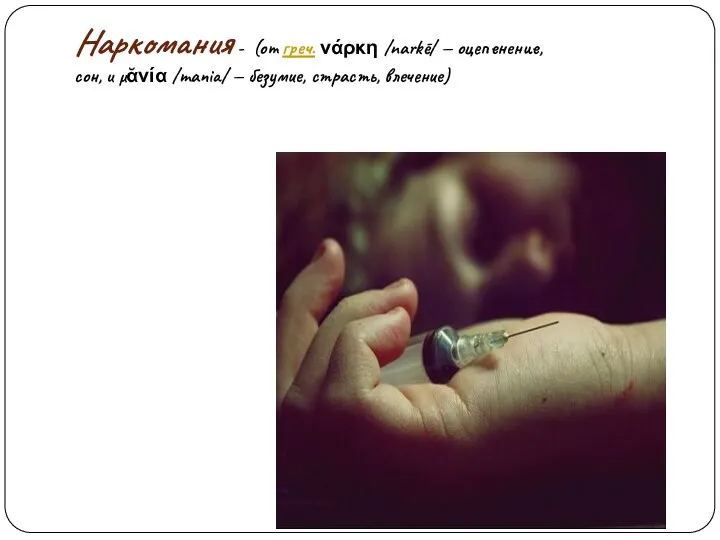 Наркомани. Наркотические средства
 Особенности первичной позитивной профилактики аддиктивного поведения в образовательных учреждениях
 Основные правила рационального питания
 Классный час на тему: Глаза - главные помощники человека. Гигиена зрения. Забота о глазах
 Правила оказания первой помощи при ранениях
 Сознание. Свойства сознания
 Методы обследования больного: субъективные и объективные
 Гистологиялық препараттардың электрондық нұсқасы
 Вірус Зіка
 Взгляд косметолога на акне и качество кожи лица
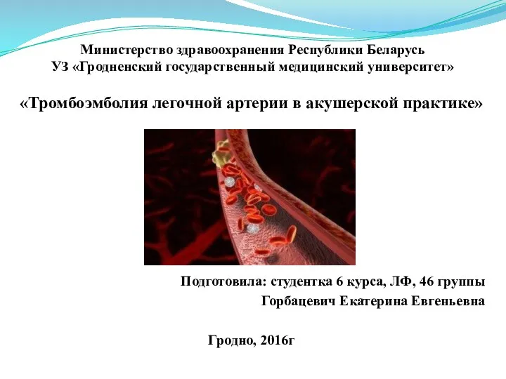 Тромбоэмболия легочной артерии в акушерской практике
 Профессиональное выгорание педагогов-психологов
 Детский церебральный паралич. Клинические симптомы
 Пуринергиялық синапстарда қозудың берілуіне әсер ететін дәрілер
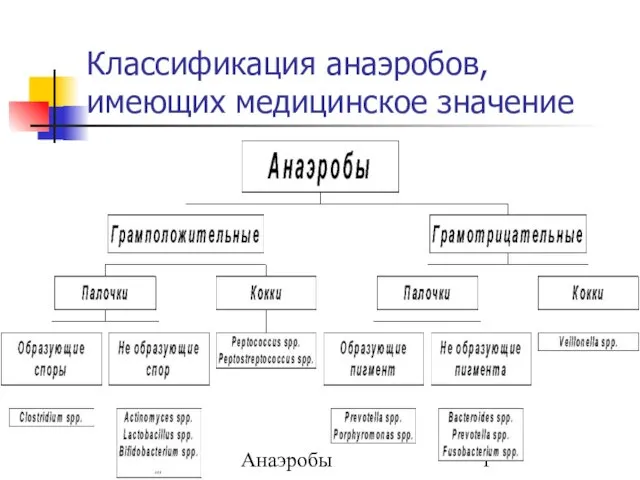 Классификация анаэробов, имеющих медицинское значение
 Виды медицинского страхования
 Когнитивная психология