- Anatomy, phtsiology & hystology of the skin

Содержание
- 2. The Skin Hypodermis (subcutaneous fatty tissue) Dermis the true skin Epidermis
- 3. Anatomy, phtsiology & hystology of the skin. I – Epidermis II- Dermis III - Hypodermis
- 4. Epidermis Stratum basale - Germinatinne layer Stratum spinosum - Prikle – cell layer Stratum granulosum -
- 5. Epidermis 1.Stratum basale (germinative layer) Keratinoblastis (1 layer, like a polisade). Melanoblastis (their ratio is 1:11
- 6. Epidermis 2. Stratum spinosum (pricle-cell layer) Dendritic epidermocytis (5-7 layers) Langhan’s cells Hrenstayin’s cells 3. Stratum
- 7. Epidermis 4. Stratum lucidum (lucid layer) These cells contain eleidin. Str. lucidum contains glycogens, lipoids, fatty
- 8. Dermis (the true skin) Structural amorphous interstitial substance: collagenous fibres elastics fibres argyrophile fibres vessels nerves
- 9. Dermis (the true skin) True homogeneous membrane Lipoids Mucopolysaccharides (mainly, hyaluronic and chondroitin – sulfuric acids)
- 10. Dermis (the true skin) Cells structure Fibroblasts Histiocytes Lymphocytes Mast cells Plasma cells Melanophages Epithelial appendages
- 11. Protective (barrier) functions of the skin Protects the organism from the damaging effect of sun rays
- 12. Protective (barrier) functions of the skin An acid (pH5.0-6,0) water-lipid mantle which attenuatus or neutralizis the
- 13. Hystology of the skin
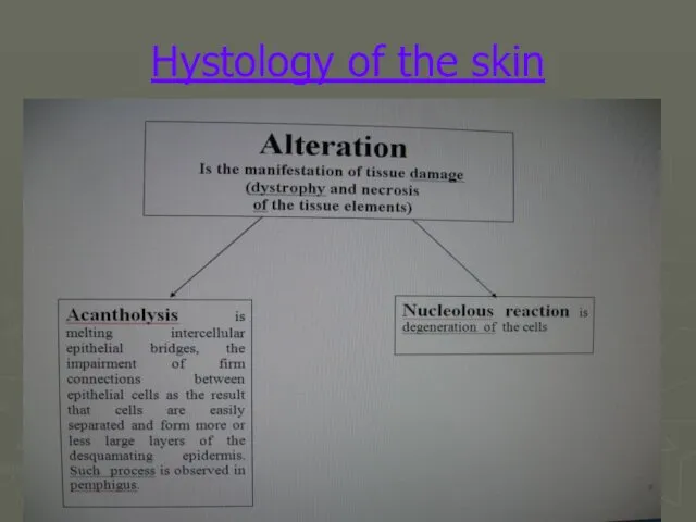
- 14. Hystology of the skin
- 15. Hystology of the skin
- 16. Hystology of the skin
- 19. spot spot spot Tubercle (tuberculum) Wheal (urtica) Primary morphological lesion papula
- 20. Primary morphological lesion Pustule (pustula) Phlyctena Staphyloccal impetigo Nodule (nodus) Vesicle (vesicula) Blister (bulla)
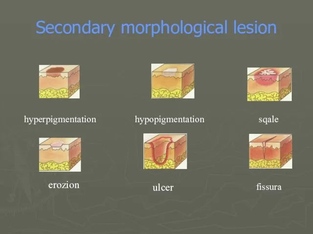
- 21. Secondary morphological lesion hyperpigmentation hypopigmentation sqale erozion ulcer fissura
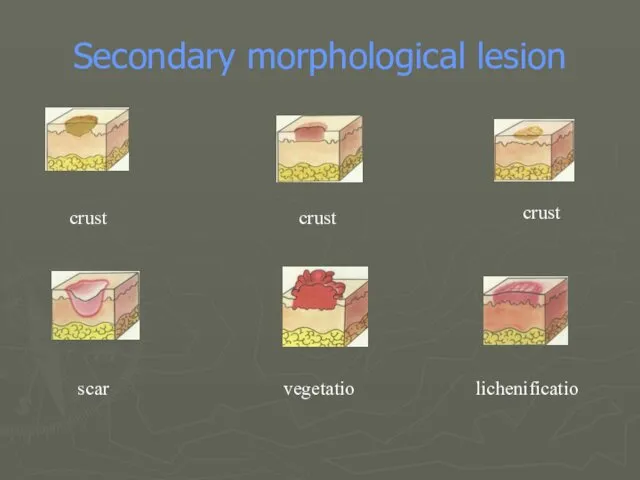
- 22. Secondary morphological lesion crust crust crust scar vegetatio lichenificatio
- 24. Скачать презентацию
The Skin
Hypodermis (subcutaneous fatty tissue)
Dermis the true skin
Epidermis
The Skin
Hypodermis (subcutaneous fatty tissue)
Dermis the true skin
Epidermis
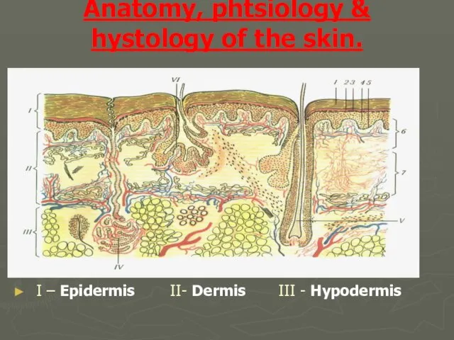
Anatomy, phtsiology & hystology of the skin.
I – Epidermis II- Dermis
Anatomy, phtsiology & hystology of the skin.
I – Epidermis II- Dermis
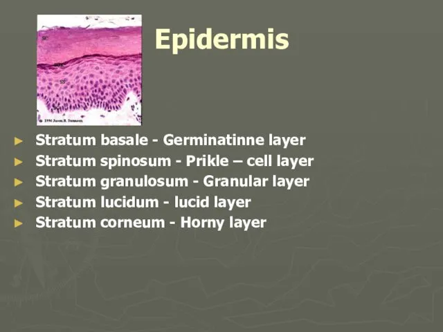
Epidermis
Stratum basale - Germinatinne layer
Stratum spinosum - Prikle – cell layer
Stratum
Epidermis
Stratum basale - Germinatinne layer
Stratum spinosum - Prikle – cell layer
Stratum
Epidermis
1.Stratum basale (germinative layer)
Keratinoblastis (1 layer, like a polisade).
Melanoblastis (their ratio
Epidermis
1.Stratum basale (germinative layer)
Keratinoblastis (1 layer, like a polisade).
Melanoblastis (their ratio
Epidermis
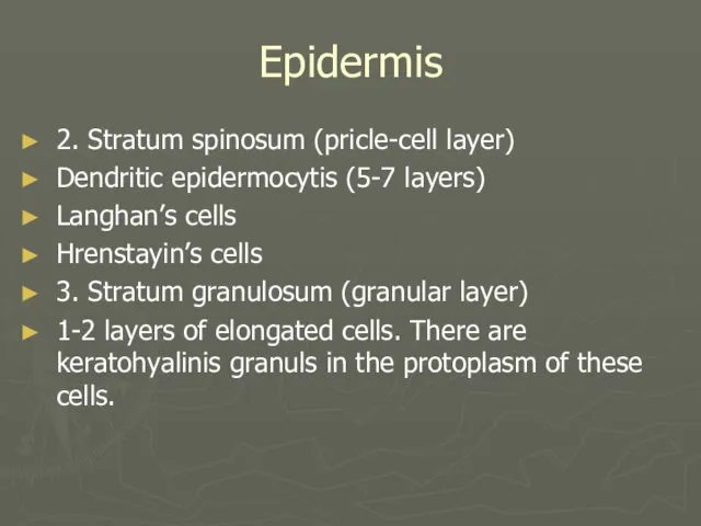
2. Stratum spinosum (pricle-cell layer)
Dendritic epidermocytis (5-7 layers)
Langhan’s cells
Hrenstayin’s cells
3. Stratum
Epidermis
2. Stratum spinosum (pricle-cell layer)
Dendritic epidermocytis (5-7 layers)
Langhan’s cells
Hrenstayin’s cells
3. Stratum
Epidermis
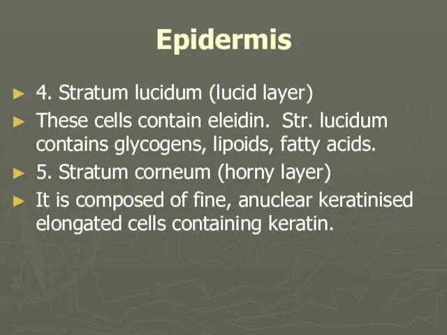
4. Stratum lucidum (lucid layer)
These cells contain eleidin. Str. lucidum
Epidermis
4. Stratum lucidum (lucid layer)
These cells contain eleidin. Str. lucidum
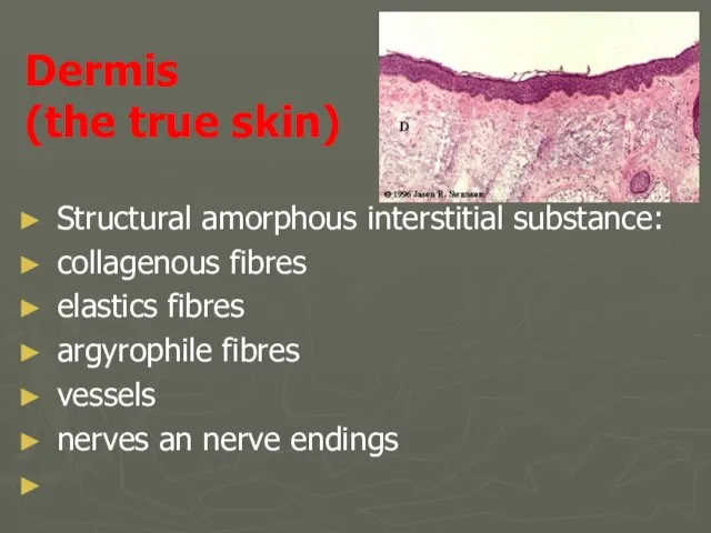
Dermis
(the true skin)
Structural amorphous interstitial substance:
collagenous fibres
elastics fibres
argyrophile fibres
vessels
nerves an
Dermis
(the true skin)
Structural amorphous interstitial substance:
collagenous fibres
elastics fibres
argyrophile fibres
vessels
nerves an
Dermis
(the true skin)
True homogeneous membrane
Lipoids
Mucopolysaccharides (mainly, hyaluronic and chondroitin
Dermis
(the true skin)
True homogeneous membrane
Lipoids
Mucopolysaccharides (mainly, hyaluronic and chondroitin
Dermis
(the true skin)
Cells structure
Fibroblasts
Histiocytes
Lymphocytes
Mast cells
Plasma cells
Melanophages
Epithelial appendages of the skin
Dermis
(the true skin)
Cells structure
Fibroblasts
Histiocytes
Lymphocytes
Mast cells
Plasma cells
Melanophages
Epithelial appendages of the skin

Protective (barrier) functions of the skin
Protects the organism from the
Protective (barrier) functions of the skin
Protects the organism from the
Protective (barrier) functions of the skin
An acid (pH5.0-6,0) water-lipid mantle
Protective (barrier) functions of the skin
An acid (pH5.0-6,0) water-lipid mantle
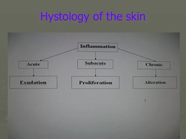
Hystology of the skin
Hystology of the skin

Hystology of the skin
Hystology of the skin

Hystology of the skin
Hystology of the skin
Hystology of the skin
Hystology of the skin


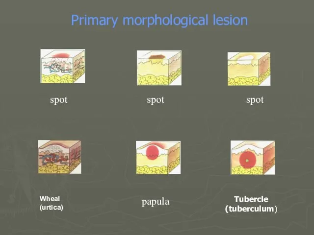
spot
spot
spot
Tubercle
(tuberculum)
Wheal
(urtica)
Primary morphological lesion
papula
spot
spot
spot
Tubercle
(tuberculum)
Wheal
(urtica)
Primary morphological lesion
papula

Primary morphological lesion
Pustule
(pustula)
Phlyctena
Staphyloccal impetigo
Nodule
(nodus)
Vesicle (vesicula)
Blister
(bulla)
Primary morphological lesion
Pustule
(pustula)
Phlyctena
Staphyloccal impetigo
Nodule
(nodus)
Vesicle (vesicula)
Blister
(bulla)
Secondary morphological lesion
hyperpigmentation
hypopigmentation
sqale
erozion
ulcer
fissura
Secondary morphological lesion
hyperpigmentation
hypopigmentation
sqale
erozion
ulcer
fissura
Secondary morphological lesion
crust
crust
crust
scar
vegetatio
lichenificatio
Secondary morphological lesion
crust
crust
crust
scar
vegetatio
lichenificatio
 Предмет, объект и задачи психологии как науки
 Лекарственные средства, влияющие на вегетативную нервную систему
 Международный день объятий
 Гонорея
 Клуб будущих и молодых мам Саратова. Роды
 Иммунодефицитный или инфекционно-воспалительный синдром
 Гидроторакс
 Административно-принудительные меры медицинского характера
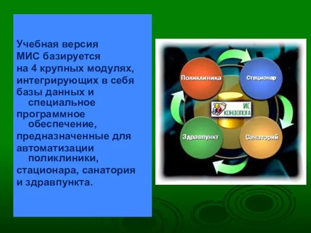 Учебная версия медицинской информационной системы (МИС)
 Легочные инфекции. Пневмонии
 Фармакотерапия в акушерстве
 ВКР: Профессиональная карта фельдшера при анемии у детей школьного возраста
 Болезнь Помпе
 Шұғыл және төтенше жағдайдағы жедел жәрдем беру қызметін ұйымдастыру
 Коронавирус! Обновлённая информация по состоянию на 15 сентября 2020 года
 Мочевая система
 Наследственные болезни человека
 Причины дизонтогенеза. Общие и частные закономерности аномального развития
 Опухоли: условие, причина
 ВКР: Методика физической реабилитации людей пожилого возраста с остеохондрозом шейного отдела позвоночника
 Панкреонекроз
 Презентация Свойства степени с натуральным показателем
 Практически нормальное зрение
 Ингарон. Лекарственная форма
 Амбулаторлық карта
 Амилоидоз. Классификация
 Внутричерепное давление
 Акселерация. Теории акселерации