- Cell injury. (Subject 2)

Содержание
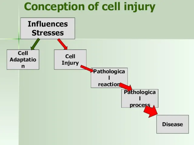
- 2. Conception of cell injury Cell Adaptation Pathological process Disease Pathological reaction Influences Stresses Cell Injury
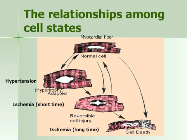
- 3. The relationships among cell states (Hypertrophy) Myocardial fiber Hypertension Ischemia (short time) Ischemia (long time)
- 4. Injury From Physical Agents Causes: Mechanical forces - trauma. Extremes of temperature – burns, heat stroke,
- 5. Other causes of cell damage Chemicals – substances or their metabolites Hypoxia – as a result
- 6. Types of Cell Injury Acute (strong irritants) Chronic (moderate irritants) Reversible (angina pectoris) Irreversible (myocardial infarction)
- 7. Signs of Cell Injury Morphological Functional changes of shape and color swelling or shrinking the disturbance
- 8. General Principles of Cell Injury Factors, which determine cell response Kind, severity, and duration of injury.
- 9. Major Processes of Cell Injury Decreased ATP production Injury by toxic oxygen radicals Disturbances of Ca
- 10. Example test Chose the example of specific cell injury from listed below: myocardial ischemia intestinal epithelial
- 11. Example test Which factors determine the type of cell’s response to injuring stimuli? kind of injuring
- 12. Example test Patient was made blood biochemical test in order to confirm hepatitis. Increased level of
- 13. Major Types of Cell Injury Hypoxia Chemicals Free radicals
- 14. Reversible Hypoxic Injury Lack of oxygen Decreased ATP formation failure of ATP dependent Na/K pumps and
- 15. Irreversible Hypoxic Injury ↑ membranes permeability Irreversible mytochondrial dysfunction ↓ intracellular pH loss of proteins, essential
- 16. Mechanisms of membranes damage Progressive loss of membrane phospholipids Cytoskeletal abnormalities Toxic oxygen radicals Lipid breakdown
- 17. Reperfusion injury Neutrophiles Calcium ions Blood stream Toxic oxygen radicals Cell damage Cytokines Enzymes activation Cell
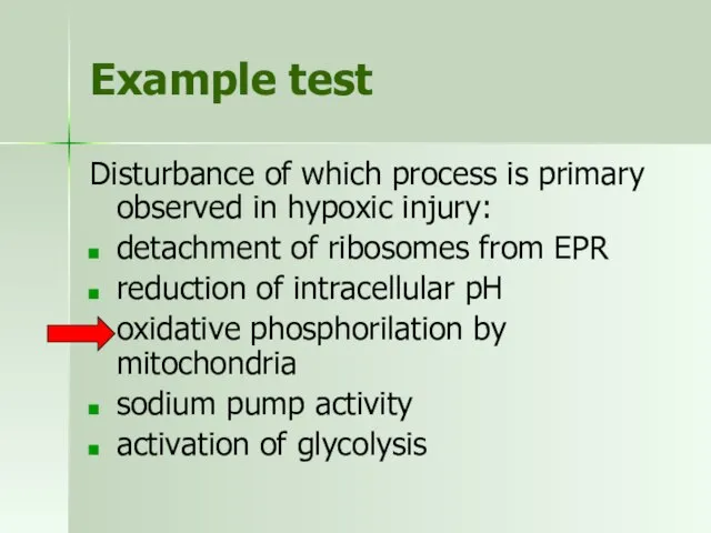
- 18. Example test Disturbance of which process is primary observed in hypoxic injury: detachment of ribosomes from
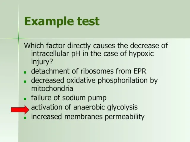
- 19. Example test Which factor directly causes the decrease of intracellular pH in the case of hypoxic
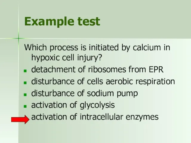
- 20. Example test Which process is initiated by calcium in hypoxic cell injury? detachment of ribosomes from
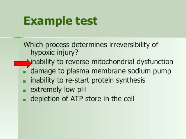
- 21. Example test Which process determines irreversibility of hypoxic injury? inability to reverse mitochondrial dysfunction damage to
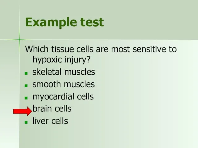
- 22. Example test Which tissue cells are most sensitive to hypoxic injury? skeletal muscles smooth muscles myocardial
- 23. Sources of free radicals
- 24. Reactive oxygen species Superoxide O2- Hydroxyl radical OH- Hydrogen peroxide H2O2
- 25. The effects of free radicals Positive: phagocytosis, energy production Negative: Lipid peroxidation of membranes Nonperoxidative mitochondrial
- 26. Antioxidative substances Enzymatic antioxidants Thioredoxin system Glutathione system Superoxide dismutase Catalase Non-enzymatic antioxidants Vitamins A, C,
- 27. Example test Choose the effect which IS NOT directly caused by free radicals: lipid peroxidation of
- 28. Chemical injury mechanisms Direct cytotoxic effect mercury damages GIT and kidneys cyanide breaks oxidative phosphorilation. Conversion
- 29. Outcomes of cell injury
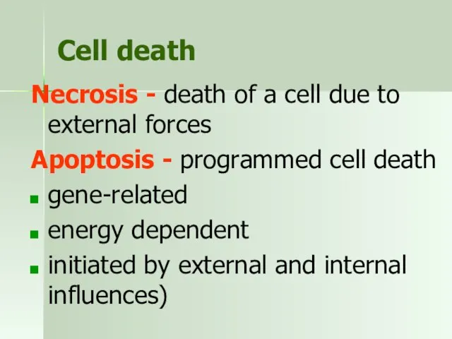
- 30. Cell death Necrosis - death of a cell due to external forces Apoptosis - programmed cell
- 31. Physiological apoptosis Frog plants amphibia human
- 32. Morphological signs of apoptosis Shrinkage of the cell. Condensation of chromatin around the nucleus. Formation of
- 33. Necrosis and apoptosis
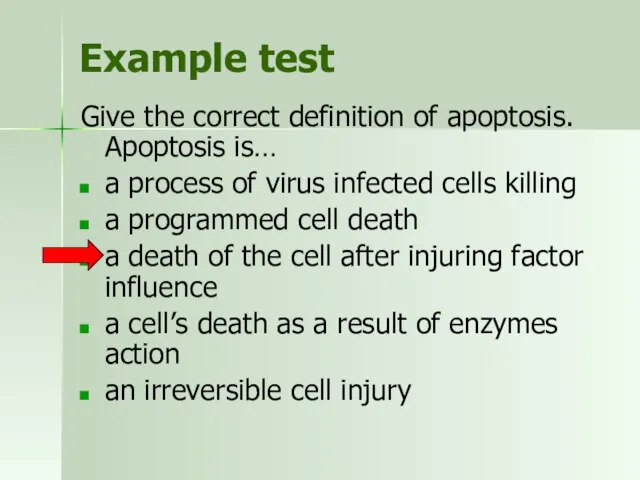
- 34. Example test Give the correct definition of apoptosis. Apoptosis is… a process of virus infected cells
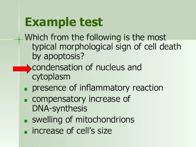
- 35. Example test Which from the following is the most typical morphological sign of cell death by
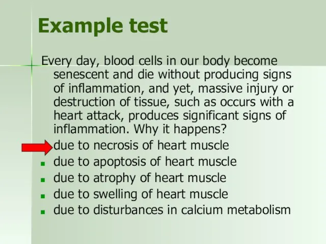
- 36. Example test Every day, blood cells in our body become senescent and die without producing signs
- 37. Cell Adaptation to Injury compensation of energy metabolism disturbance protection of cells membranes compensation of water-ion
- 38. Compensation of energy metabolism disturbance increased ATP formation, transport and effectiveness of ATP use increase of
- 39. Protection of cells membranes activation of antioxidants action activation of cells buffer system activation of EPR
- 40. Compensation of water-ion disbalance activation of ion “pumps” energy supply increase of ion-transporting enzymes activation of
- 41. Mechanisms of cell genome repair revealing and elimination of damaged DNA fragment replacement of damaged DNA
- 42. The types of cellular adaptations
- 43. Causes of atrophy decreased workload, loss of innervation, diminished blood supply, inadequate nutrition, loss of endocrine
- 44. Hypertrophy increase in the size of cells REASONS: increased functional demand specific hormonal stimulation occurs under
- 45. Hyperlasia increase in the number of cells It occurs in tissues where cells are capable of
- 46. Metaplasia one adult cell type (epithelial or mesenchymal) is replaced by another adult cell type Squamous
- 47. Dysplasia Deranged cell growth of a specific tissue that results in cells that vary in size,
- 48. Example test Cells may adapt to external and internal stimuli by undergoing changes in their size,
- 49. Example test Cells may adapt to external and internal stimuli by undergoing changes in their size,
- 51. Скачать презентацию
Conception of cell injury
Cell
Adaptation
Pathological
process
Disease
Pathological
reaction
Influences
Stresses
Cell Injury
Conception of cell injury
Cell
Adaptation
Pathological
process
Disease
Pathological
reaction
Influences
Stresses
Cell Injury
The relationships among cell states
(Hypertrophy)
Myocardial fiber
Hypertension
Ischemia (short time)
Ischemia (long time)
The relationships among cell states
(Hypertrophy)
Myocardial fiber
Hypertension
Ischemia (short time)
Ischemia (long time)
Injury From Physical Agents
Causes:
Mechanical forces - trauma.
Extremes of temperature –
Injury From Physical Agents
Causes:
Mechanical forces - trauma.
Extremes of temperature –
Other causes of cell damage
Chemicals – substances or their metabolites
Hypoxia –
Other causes of cell damage
Chemicals – substances or their metabolites
Hypoxia –
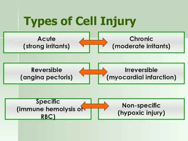
Types of Cell Injury
Acute
(strong irritants)
Chronic
(moderate irritants)
Reversible
(angina pectoris)
Irreversible
(myocardial infarction)
Non-specific
(hypoxic injury)
Specific
(immune
Types of Cell Injury
Acute
(strong irritants)
Chronic
(moderate irritants)
Reversible
(angina pectoris)
Irreversible
(myocardial infarction)
Non-specific
(hypoxic injury)
Specific
(immune
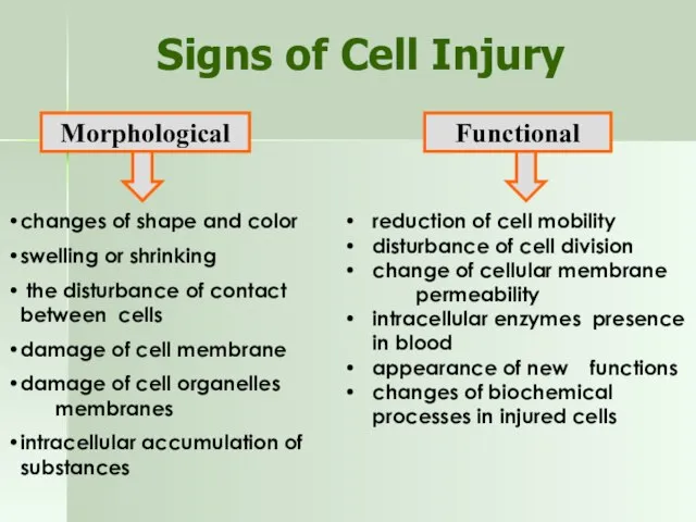
Signs of Cell Injury
Morphological
Functional
changes of shape and color
swelling or shrinking
Signs of Cell Injury
Morphological
Functional
changes of shape and color
swelling or shrinking
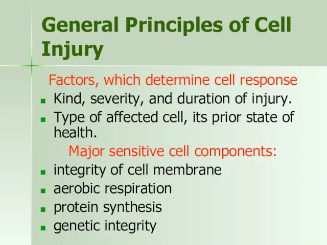
General Principles of Cell Injury
Factors, which determine cell response
Kind, severity, and
General Principles of Cell Injury
Factors, which determine cell response
Kind, severity, and
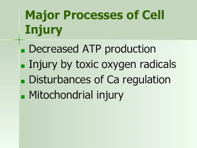
Major Processes of Cell Injury
Decreased ATP production
Injury by toxic oxygen radicals
Disturbances
Major Processes of Cell Injury
Decreased ATP production
Injury by toxic oxygen radicals
Disturbances
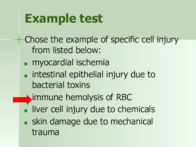
Example test
Chose the example of specific cell injury from listed below:
myocardial
Example test
Chose the example of specific cell injury from listed below:
myocardial
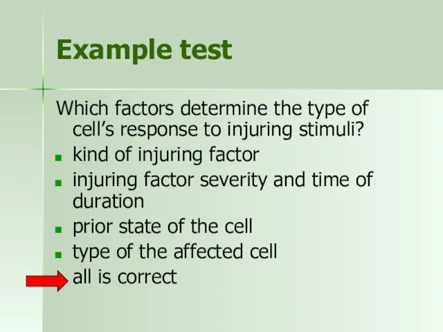
Example test
Which factors determine the type of cell’s response to injuring
Example test
Which factors determine the type of cell’s response to injuring
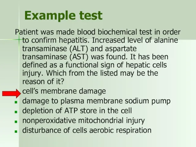
Example test
Patient was made blood biochemical test in order to confirm
Example test
Patient was made blood biochemical test in order to confirm
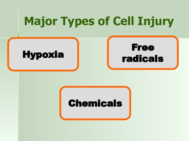
Major Types of Cell Injury
Hypoxia
Chemicals
Free radicals
Major Types of Cell Injury
Hypoxia
Chemicals
Free radicals
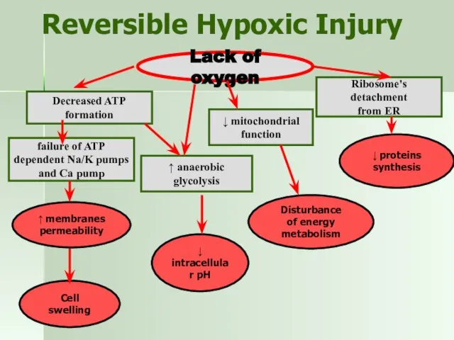
Reversible Hypoxic Injury
Lack of oxygen
Decreased ATP formation
failure of ATP
dependent Na/K
Reversible Hypoxic Injury
Lack of oxygen
Decreased ATP formation
failure of ATP
dependent Na/K
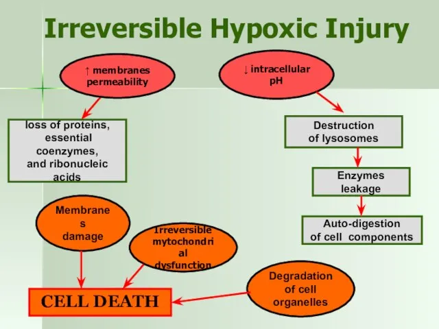
Irreversible Hypoxic Injury
↑ membranes
permeability
Irreversible
mytochondrial
dysfunction
↓ intracellular pH
loss of proteins,
essential
Irreversible Hypoxic Injury
↑ membranes
permeability
Irreversible
mytochondrial
dysfunction
↓ intracellular pH
loss of proteins,
essential
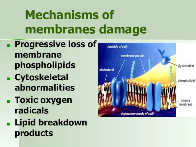
Mechanisms of membranes damage
Progressive loss of membrane phospholipids
Cytoskeletal abnormalities
Mechanisms of membranes damage
Progressive loss of membrane phospholipids
Cytoskeletal abnormalities
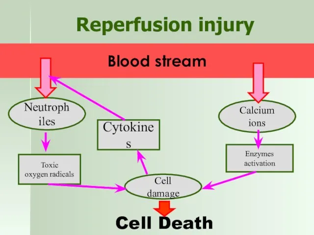
Reperfusion injury
Neutrophiles
Calcium ions
Blood stream
Toxic
oxygen radicals
Cell damage
Cytokines
Enzymes activation
Cell Death
Reperfusion injury
Neutrophiles
Calcium ions
Blood stream
Toxic
oxygen radicals
Cell damage
Cytokines
Enzymes activation
Cell Death
Example test
Disturbance of which process is primary observed in hypoxic injury:
detachment
Example test
Disturbance of which process is primary observed in hypoxic injury:
detachment
Example test
Which factor directly causes the decrease of intracellular pH in
Example test
Which factor directly causes the decrease of intracellular pH in
Example test
Which process is initiated by calcium in hypoxic cell injury?
detachment
Example test
Which process is initiated by calcium in hypoxic cell injury?
detachment
Example test
Which process determines irreversibility of hypoxic injury?
inability to reverse mitochondrial
Example test
Which process determines irreversibility of hypoxic injury?
inability to reverse mitochondrial
Example test
Which tissue cells are most sensitive to hypoxic injury?
skeletal muscles
smooth
Example test
Which tissue cells are most sensitive to hypoxic injury?
skeletal muscles
smooth
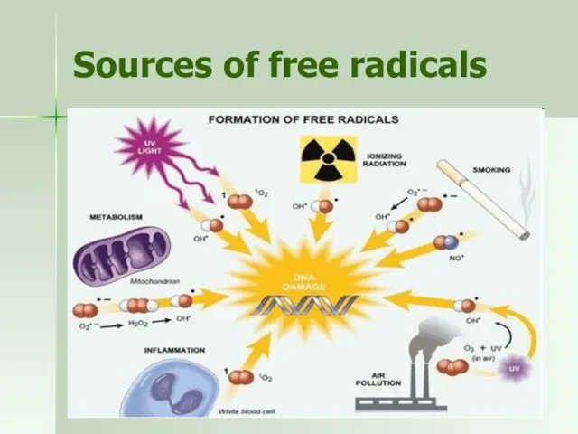
Sources of free radicals
Sources of free radicals
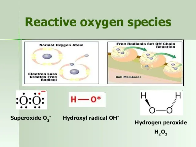
Reactive oxygen species
Superoxide O2-
Hydroxyl radical OH-
Hydrogen peroxide
H2O2
Reactive oxygen species
Superoxide O2-
Hydroxyl radical OH-
Hydrogen peroxide
H2O2
The effects of free radicals
Positive: phagocytosis, energy production
Negative:
Lipid peroxidation of membranes
Nonperoxidative
The effects of free radicals
Positive: phagocytosis, energy production
Negative:
Lipid peroxidation of membranes
Nonperoxidative
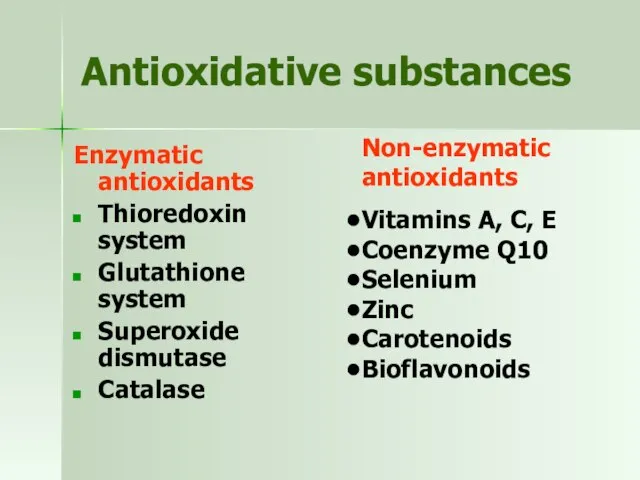
Antioxidative substances
Enzymatic antioxidants
Thioredoxin system
Glutathione system
Superoxide dismutase
Catalase
Non-enzymatic
Antioxidative substances
Enzymatic antioxidants
Thioredoxin system
Glutathione system
Superoxide dismutase
Catalase
Non-enzymatic
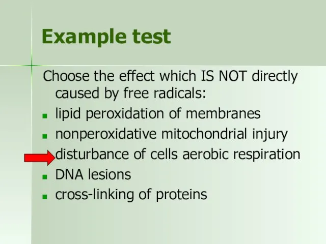
Example test
Choose the effect which IS NOT directly caused by free
Example test
Choose the effect which IS NOT directly caused by free
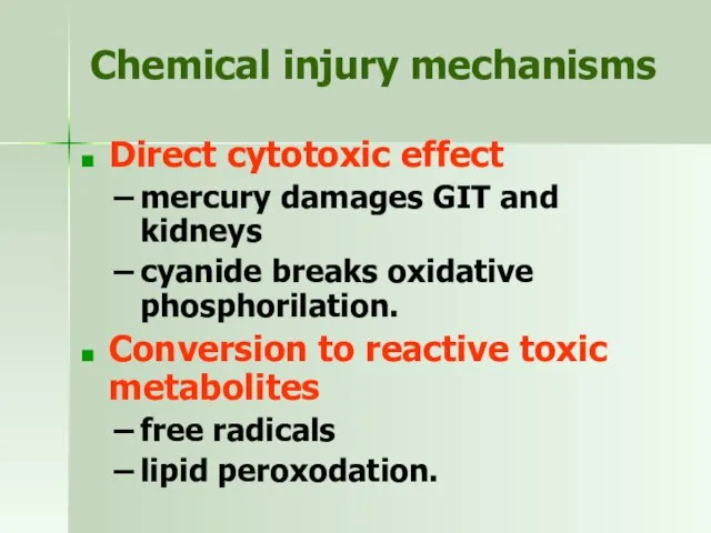
Chemical injury mechanisms
Direct cytotoxic effect
mercury damages GIT and kidneys
cyanide breaks
Chemical injury mechanisms
Direct cytotoxic effect
mercury damages GIT and kidneys
cyanide breaks
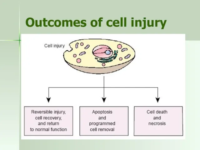
Outcomes of cell injury
Outcomes of cell injury
Cell death
Necrosis - death of a cell due to external forces
Apoptosis
Cell death
Necrosis - death of a cell due to external forces
Apoptosis
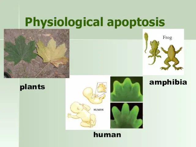
Physiological apoptosis
Frog
plants
amphibia
human
Physiological apoptosis
Frog
plants
amphibia
human
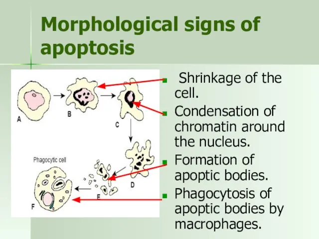
Morphological signs of apoptosis
Shrinkage of the cell.
Condensation of chromatin around
Morphological signs of apoptosis
Shrinkage of the cell.
Condensation of chromatin around
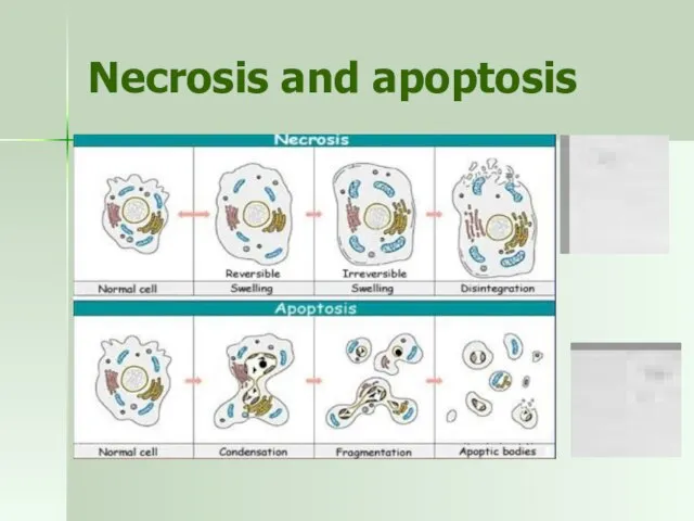
Necrosis and apoptosis
Necrosis and apoptosis
Example test
Give the correct definition of apoptosis. Apoptosis is…
a process of
Example test
Give the correct definition of apoptosis. Apoptosis is…
a process of
Example test
Which from the following is the most typical morphological sign
Example test
Which from the following is the most typical morphological sign
Example test
Every day, blood cells in our body become senescent and
Example test
Every day, blood cells in our body become senescent and
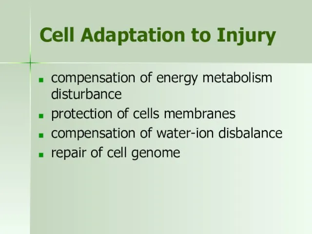
Cell Adaptation to Injury
compensation of energy metabolism disturbance
protection of cells membranes
compensation
Cell Adaptation to Injury
compensation of energy metabolism disturbance
protection of cells membranes
compensation
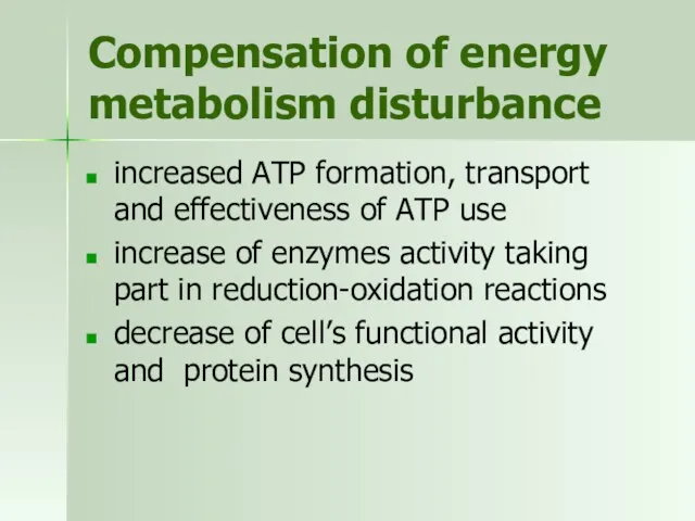
Compensation of energy metabolism disturbance
increased ATP formation, transport and effectiveness of
Compensation of energy metabolism disturbance
increased ATP formation, transport and effectiveness of
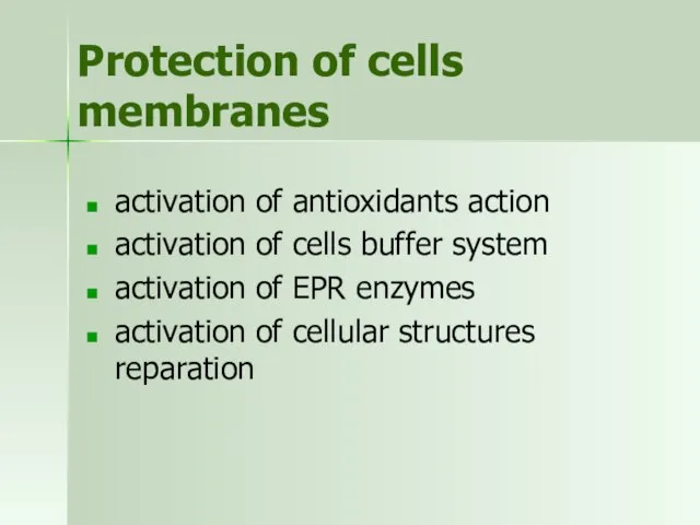
Protection of cells membranes
activation of antioxidants action
activation of cells buffer system
activation
Protection of cells membranes
activation of antioxidants action
activation of cells buffer system
activation
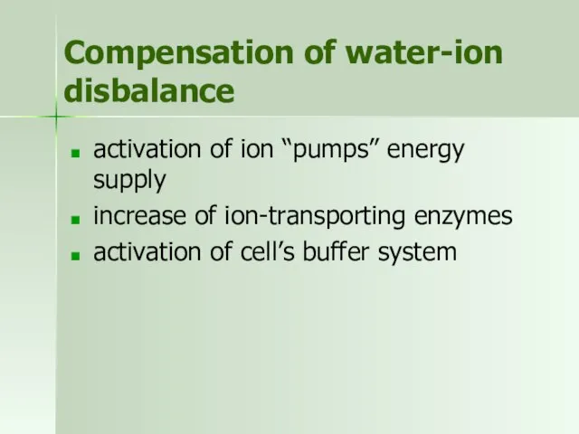
Compensation of water-ion disbalance
activation of ion “pumps” energy supply
increase of ion-transporting
Compensation of water-ion disbalance
activation of ion “pumps” energy supply
increase of ion-transporting
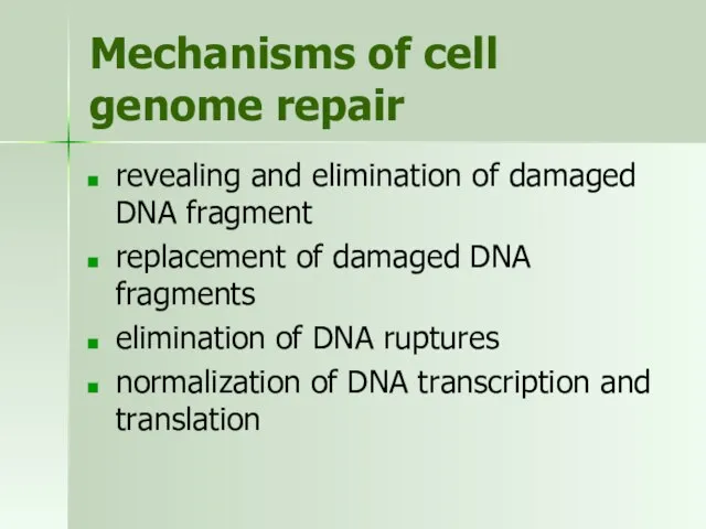
Mechanisms of cell genome repair
revealing and elimination of damaged DNA fragment
replacement
Mechanisms of cell genome repair
revealing and elimination of damaged DNA fragment
replacement
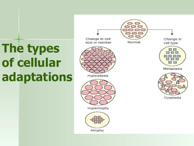
The types of cellular adaptations
The types of cellular adaptations
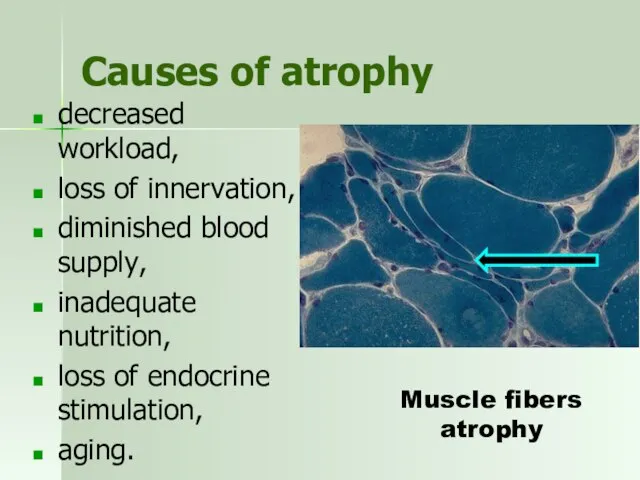
Causes of atrophy
decreased workload,
loss of innervation,
diminished blood supply,
inadequate
Causes of atrophy
decreased workload,
loss of innervation,
diminished blood supply,
inadequate
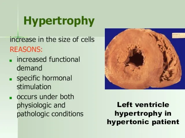
Hypertrophy
increase in the size of cells
REASONS:
increased functional demand
specific hormonal
Hypertrophy
increase in the size of cells
REASONS:
increased functional demand
specific hormonal
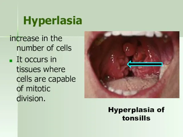
Hyperlasia
increase in the number of cells
It occurs in tissues where
Hyperlasia
increase in the number of cells
It occurs in tissues where
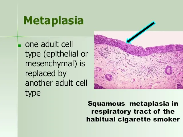
Metaplasia
one adult cell type (epithelial or mesenchymal) is replaced by
Metaplasia
one adult cell type (epithelial or mesenchymal) is replaced by
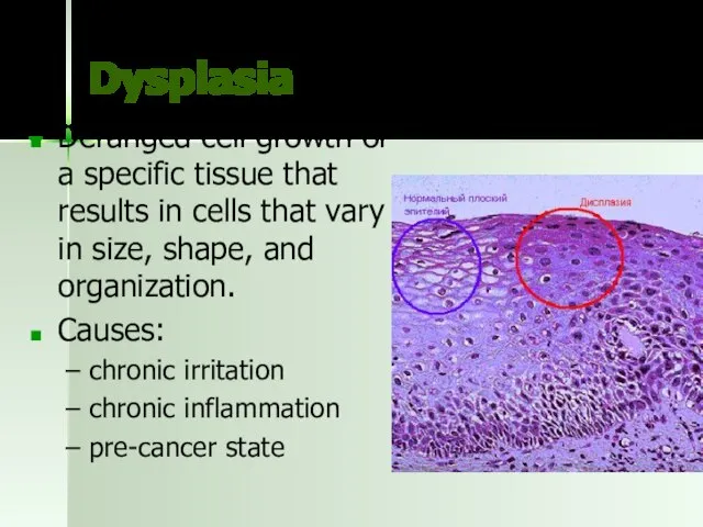
Dysplasia
Deranged cell growth of a specific tissue that results in cells
Dysplasia
Deranged cell growth of a specific tissue that results in cells
Example test
Cells may adapt to external and internal stimuli by undergoing
Example test
Cells may adapt to external and internal stimuli by undergoing
Example test
Cells may adapt to external and internal stimuli by undergoing
Example test
Cells may adapt to external and internal stimuli by undergoing
 Атом энергиясын бейбіт мақсатта пайдалану. Радиоактивті изотоптар мен иондаушы сәулелер көздерін медицинада қолдану
 Инфекциялық емес аурулармен
 Переломы костей конечностей
 Eurotab-Нефко. Проект Таблетки
 Заболевания щитовидной железы
 Этапы постановки ПВВК
 Hygiene of children
 Студенттің өзіндік жұмысы
 Этапы сестринского процесса
 Всегда – вместе, всегда – рядом
 ООО Научно - производственное предприятие Колловита
 Көкіріңді таяқша
 Добровольное медицинское страхование иностранных студентов. Программа Студент
 Сестринский процесс при аллергодерматозах. Синдром Лайелла, отёк Квинке
 История развития психопатологии в России
 СП при бронхиальной астме
 Патология клеток крови и костного мозга. Гемобластозы . Анемии
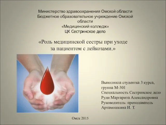 Роль медицинской сестры при уходе за пациентом с лейкозами
 Венерические заболевания
 Абразивные материалы
 Рациональное питание. Вскармливание детей первого года жизни. Виды вскармливания
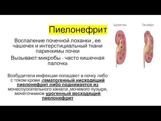 Пиелонефрит
 Функциональная анатомия мышц конечностей
 Регуляция сердечной деятельности. Лекция 2
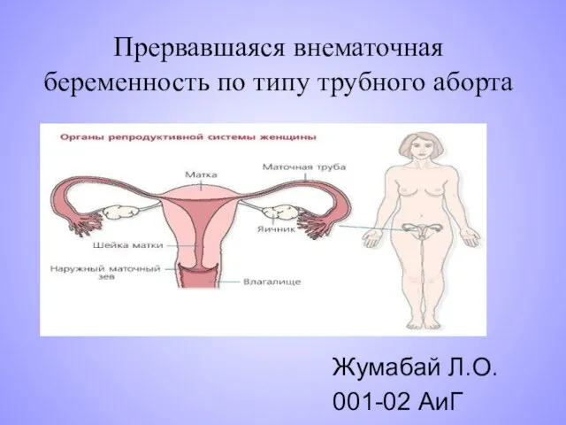 Прервавшаяся внематочная беременность по типу трубного аборта
 Возможности магнитнорезонансной томографии в диагностике патологических состояний локтевого сустава
 Неингаляционные виды наркоза
 Общие особенности инфекционных болезней