- Tissue processing

Содержание
- 2. Contents Introduction Specimen Accessioning Gross Examination Tissue Processing steps The paraffin Technique and its alternatives The
- 3. Introduction There are 3 main techniques which are used in preparing microscopical sections from tissues: The
- 4. …Introduction Tissues from the body taken for diagnosis of disease processes must be processed in the
- 5. Specimen Accessioning Tissue specimens received in the surgical pathology laboratory have a request form that lists
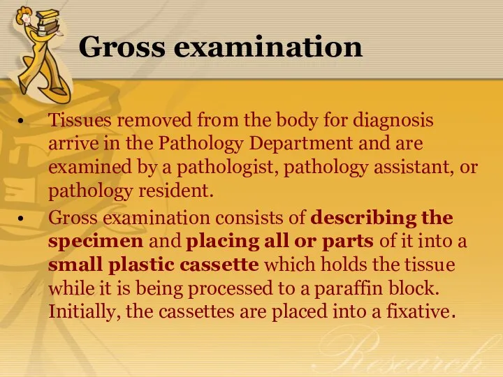
- 8. Gross examination Tissues removed from the body for diagnosis arrive in the Pathology Department and are
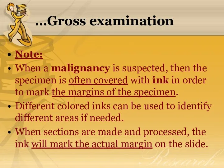
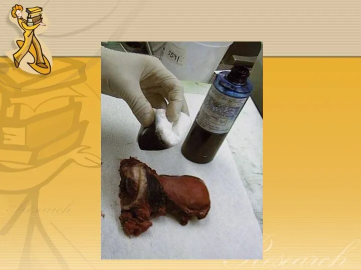
- 10. …Gross examination Note: When a malignancy is suspected, then the specimen is often covered with ink
- 12. Tissue Processing steps Biological tissues are generally rather soft, making it quite difficult to cut acceptably
- 13. …Tissue Processing steps The usual way this is done is with paraffin. Tissues embedded in paraffin,
- 14. The paraffin Technique Washing Following fixation, the tissues should be washed from 15 to 30 minutes.
- 15. ...The paraffin Technique Dehydration Wet fixed tissues (in aqueous solutions) cannot be directly infiltrated with paraffin.
- 16. …The paraffin technique Clearing The next step is called "clearing" and consists of removal of the
- 17. …The paraffin technique Impregnation in paraffin The tissues are put from 6 – 24 hours in
- 18. …The paraffin technique Embedding Finally, the tissue is infiltrated with the embedding agent, almost always paraffin.
- 19. …The paraffin technique Infiltration must be carried out at only a few degrees above the melting
- 20. …The paraffin technique Paraffin can be purchased that differ in melting point, for various hardnesses, depending
- 21. …The paraffin technique Using a completely different rationale for dehydration, Prento (1978) was able to reduce
- 22. …The paraffin technique Upon solidifying, paraffin shrinks 16.5 percent in volume. Paraplast is supposed to shrink
- 23. …The paraffin technique The above processes are almost always automated for the large volumes of routine
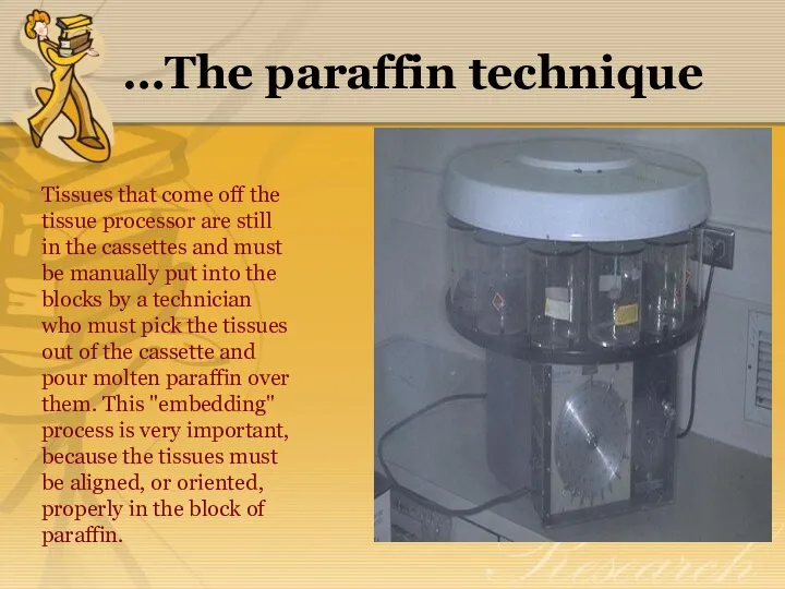
- 24. …The paraffin technique Tissues that come off the tissue processor are still in the cassettes and
- 26. Alternatives to paraffin embedding Alternatives to paraffin embedding include various plastics that allow thinner sections. Such
- 27. Note: Plastics require special reagents for dehydration and clearing that are expensive. For this reason, and
- 28. The freezing Technique In this method, the fresh or fixed tissues are frozen hardened and cut
- 29. Problems in Tissue Processing "Floaters" are small pieces of tissue that appear on a slide that
- 30. Problems in Tissue Processing If reusable cassettes are employed, you must be aware that tissue may
- 31. Problems in Tissue Processing Always be sure that you properly identify the tissue! This means that
- 32. Problems in Tissue Processing You must never submit a cassette of tissue with the wrong label.
- 33. Sectioning Frozen Sections Staining H & E staining Coverslipping Decalcification Artefacts in Histologic Sections
- 35. Скачать презентацию
Contents
Introduction
Specimen Accessioning
Gross Examination
Tissue Processing steps
The paraffin
Contents
Introduction
Specimen Accessioning
Gross Examination
Tissue Processing steps
The paraffin
Introduction
There are 3 main techniques which are used in preparing
Introduction
There are 3 main techniques which are used in preparing
…Introduction
Tissues from the body taken for diagnosis of disease processes
…Introduction
Tissues from the body taken for diagnosis of disease processes
Specimen Accessioning
Tissue specimens received in the surgical pathology laboratory have a
Specimen Accessioning
Tissue specimens received in the surgical pathology laboratory have a


Gross examination
Tissues removed from the body for diagnosis arrive in
Gross examination
Tissues removed from the body for diagnosis arrive in

…Gross examination
Note:
When a malignancy is suspected, then the specimen is
…Gross examination
Note:
When a malignancy is suspected, then the specimen is
Tissue Processing steps
Biological tissues are generally rather soft, making it
Tissue Processing steps
Biological tissues are generally rather soft, making it
…Tissue Processing steps
The usual way this is done is with
…Tissue Processing steps
The usual way this is done is with
The paraffin Technique
Washing
Following fixation, the tissues should be washed from 15
The paraffin Technique
Washing
Following fixation, the tissues should be washed from 15

...The paraffin Technique
Dehydration
Wet fixed tissues (in aqueous solutions) cannot be directly
...The paraffin Technique
Dehydration
Wet fixed tissues (in aqueous solutions) cannot be directly
…The paraffin technique
Clearing
The next step is called "clearing" and consists
…The paraffin technique
Clearing
The next step is called "clearing" and consists
…The paraffin technique
Impregnation in paraffin
The tissues are put from 6 –
…The paraffin technique
Impregnation in paraffin
The tissues are put from 6 –
…The paraffin technique
Embedding
Finally, the tissue is infiltrated with the embedding agent,
…The paraffin technique
Embedding
Finally, the tissue is infiltrated with the embedding agent,
…The paraffin technique
Infiltration must be carried out at only a few
…The paraffin technique
Infiltration must be carried out at only a few
…The paraffin technique
Paraffin can be purchased that differ in melting point,
…The paraffin technique
Paraffin can be purchased that differ in melting point,
…The paraffin technique
Using a completely different rationale for dehydration, Prento (1978)
…The paraffin technique
Using a completely different rationale for dehydration, Prento (1978)
…The paraffin technique
Upon solidifying, paraffin shrinks 16.5 percent in volume. Paraplast
…The paraffin technique
Upon solidifying, paraffin shrinks 16.5 percent in volume. Paraplast
…The paraffin technique
The above processes are almost always automated for the
…The paraffin technique
The above processes are almost always automated for the
…The paraffin technique
Tissues that come off the tissue processor are still
…The paraffin technique
Tissues that come off the tissue processor are still


Alternatives to paraffin embedding
Alternatives to paraffin embedding include various plastics that
Alternatives to paraffin embedding
Alternatives to paraffin embedding include various plastics that
Note:
Plastics require special reagents for dehydration and clearing that are expensive.
Note:
Plastics require special reagents for dehydration and clearing that are expensive.
The freezing Technique
In this method, the fresh or fixed tissues are
The freezing Technique
In this method, the fresh or fixed tissues are
Problems in Tissue Processing
"Floaters" are small pieces of tissue that appear
Problems in Tissue Processing
"Floaters" are small pieces of tissue that appear
Problems in Tissue Processing
If reusable cassettes are employed, you must be
Problems in Tissue Processing
If reusable cassettes are employed, you must be
Problems in Tissue Processing
Always be sure that you properly identify the
Problems in Tissue Processing
Always be sure that you properly identify the
Problems in Tissue Processing
You must never submit a cassette of tissue
Problems in Tissue Processing
You must never submit a cassette of tissue
Sectioning
Frozen Sections
Staining
H & E staining
Coverslipping
Decalcification
Artefacts in Histologic Sections
Sectioning
Frozen Sections
Staining
H & E staining
Coverslipping
Decalcification
Artefacts in Histologic Sections
 Знакомство с ротовой полостью
 Общие вопросы обследования больных с заболеваниями сердечно-сосудистой системы. Лекция 4 (модуль 1)
 Бронхиальная астма
 Студенттің өзіндік жұмысы
 Тератома
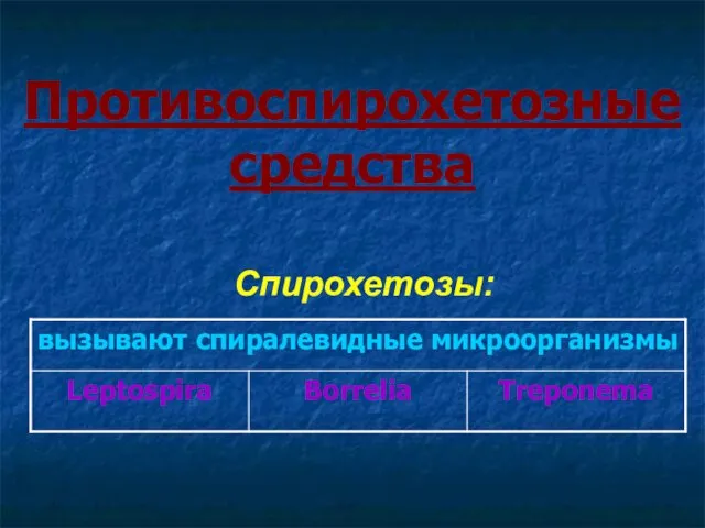 Противоспирохетозные средства
 Сульфаниламиды. Механизм действия
 Ожоги
Ожоги Влияние стресса на развитие сердечно-сосудистых заболеваний
 Физиология и методы исследования слухового анализатора
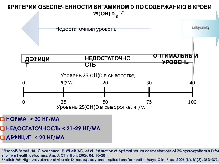 Критерии обеспеченности витамином d по содержанию в крови
 Курить – здоровью вредить!
 Законы рационального питания и их практическая значимость
 Эндокринология. Синдром Иценко Кушинга. (Лекция 6)
 Клиническая и биологическая смерть. Правила констатации смерти. Проблематика эвтаназии
 Перекрестный прикус
 Патология иммунной системы. Аллергия. Лекция №6
 Особенности течения и терапии септических послеродовых заболеваний в современных условиях (маститы, перитониты)
 Правила ухода за волосами
 Анатомия и физиология полости рта. Анатомия зубов, гистология твердых тканей зуба. Рентгеноанатомия зубов. (Лекция 2)
 Технология изготовления металлокерамических коронок коронки
 Асептика. Асептика анықтамасы, міндеттері. Қысқаша тарихи мағлұматтар
 Первая помощь при ожогах
 Хронические расстройства питания у детей. Гипотрофии. ЗВУР. Специальность: Сестринское дело
 Гигиена рук в клинической практике
 Позитивные и негативные последствия иммунной реакции
 Черепно–мозговая травма
 Психологическая коррекция самооценки младших школьников с нарушениями зрения нетрадиционными методами