- Objective physical examination in cardiovascular diseases: visual examination

Содержание
- 2. The cardiovascular examination is a portion of the physical examination that involves evaluation of the cardiovascular
- 3. The cardiac examination is based on the different methods of evaluation, comprising the following sections: measurement
- 4. Measurement of Vital Signs A good cardiac examination starts as soon you can lay eyes on
- 6. Many clues to the cardiac condition can be detected with a simple visual inspection. In the
- 7. Complete examination of all systems is essential to detect peripheral and systemic effects of cardiac disorders
- 8. Palpation Before auscultation, inspection of the precordium can be a useful indicator of previous surgery –
- 9. Auscultation Held by many as the key to physical examination, the importance of auscultation remains, but
- 10. Percussion There was a time when cardiac percussion was considered a useful addition in the clinical
- 11. Triglycerides Lower blood triglycerides by: Not overeating Limiting alcohol and simple sugars Spreading meals throughout the
- 13. Скачать презентацию
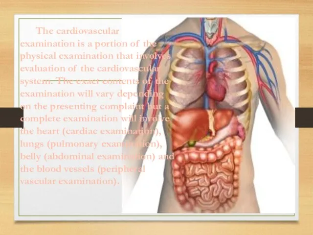
The cardiovascular examination is a portion of the physical examination that
The cardiovascular examination is a portion of the physical examination that

The cardiac examination is based on the different methods of evaluation,
The cardiac examination is based on the different methods of evaluation,
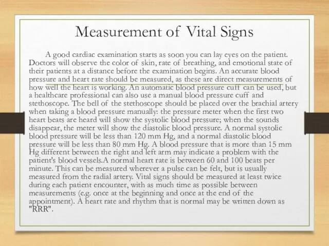
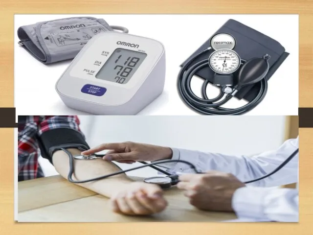
Measurement of Vital Signs
A good cardiac examination starts as soon you
Measurement of Vital Signs
A good cardiac examination starts as soon you
Many clues to the cardiac condition can be detected with a
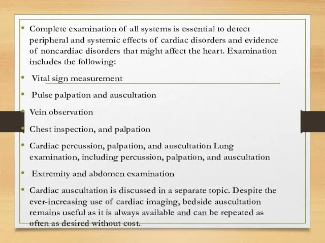
Complete examination of all systems is essential to detect peripheral and
Complete examination of all systems is essential to detect peripheral and
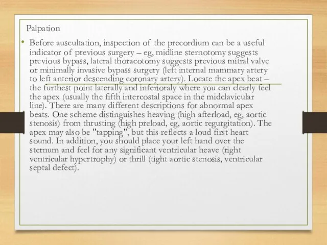
Palpation
Before auscultation, inspection of the precordium can be a useful
Palpation
Before auscultation, inspection of the precordium can be a useful
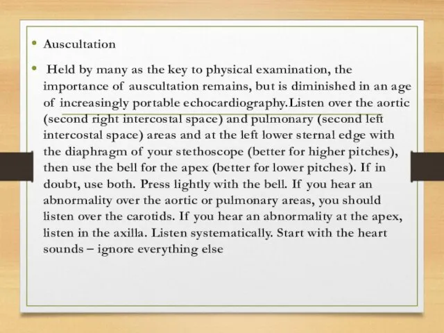
Auscultation
Held by many as the key to physical examination, the importance
Auscultation
Held by many as the key to physical examination, the importance
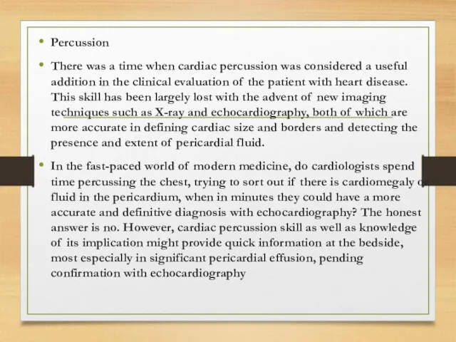
Percussion
There was a time when cardiac percussion was considered a useful
Percussion
There was a time when cardiac percussion was considered a useful
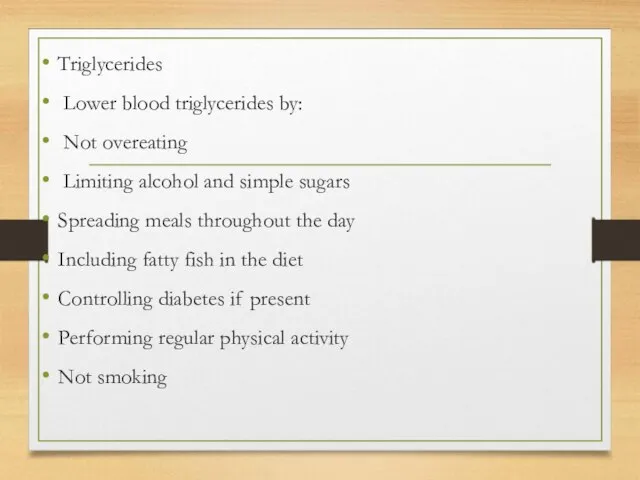
Triglycerides
Lower blood triglycerides by:
Not overeating
Limiting alcohol and simple sugars
Spreading meals throughout
Triglycerides
Lower blood triglycerides by:
Not overeating
Limiting alcohol and simple sugars
Spreading meals throughout
 Плюсы и минусы сыроедения
 Действия очевидцев при оказании первой помощи
 Тромбастения Гланцманна
 Онкология. Вводная
 Функциональная анатомия мочевых органов
 Особенности организации и оказания помощи пострадавшим с поражением ЧЛО на этапах медицинской эвакуации
 Ауру тарихы
 Диагностика инфекционных заболеваний у детей
 Информация по компании Интертич для ТОО Болашак Атырау
 Новые санитарные правила и нормативы
 Основы управления в здравоохранении
 Корреляции в психологическом исследовании
 Ауыз қуысының кілегей қабығының жүйелік ауруларында болатын зақымданулары
 Эффекты, возникающие при повторном введении лекарственных веществ. Взаимодействие лекарств
 Чему учат компьютерные игры
 Аллергия. Различия аллергии и иммунитета
 Предоперационная подготовка и послеоперационный уход за гинекологическими больными
 Противоэпилептические средства
 Внутренняя среда организма. Кровь
 1 декабря – день борьбы со СПИДом
 Кәсіби бағдарланған тұлғаның психодиагностикалық мәселелері
 Портальная гипертензия
 Балалардағы холецистит
 Несахарный диабет
 Улучшение деятельности по лекарственному обслуживанию населения и лечебно-профилактических учреждений
 Частное учреждение здравоохранения: больница РЖД-Медицина города Мичуринск
 Лечение тромбозов у детей
 Психические, физические, психологические и личностные особенности пожилых людей. Этапы старения