- Disorders of Skeletal Muscle

Содержание
- 2. Spinal Muscular Atrophy
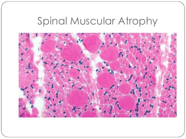
- 3. Spinal Muscular Atrophy
- 4. Disorders of Neuromuscular Junction Myasthenia gravis is an autoimmune disease with fluctuating muscle weakness that is
- 5. Disorders of Neuromuscular Junction Lambert-Eaton syndrome is caused by autoantibodies that inhibit the function of presynaptic
- 6. Miscellaneous Neuromuscular Junction Disorders Congenital myasthenic syndromes comprise a heterogeneous group of diseases that result from
- 7. Muscle Fiber Atrophy Neuropathic changes. Loss of innervation causes atrophy of myofibers. The two main morphologic
- 8. Patters of skeletal muscle injury
- 9. Inherited Disorders of Skeletal Muscle Muscular dystrophies are associated with progressive muscle injury in patients who
- 10. Dystrophinopathies: Duchenne and Becker Muscular Dystrophy The most common muscular dystrophies are X-linked and are caused
- 13. Other X-Linked and Autosomal Muscular Dystrophies Myotonic dystrophy. Myotonia, the sustained involuntary contraction of a group
- 14. Other X-Linked and Autosomal Muscular Dystrophies Emery-Dreifuss muscular dystrophy (EMD) is a genetically heterogeneous disorder caused
- 15. Channelopathies Ion channel myopathies are a group of familial disorders caused by inherited defects in ion
- 16. Metabolic Myopathies Myopathies due to inborn errors of metabolism include disorders of glycogen synthesis and degradation
- 17. Mitochondrial Myopathies Mitochondrial myopathies can stem from mutations in either the mitochondrial or nuclear genomes because
- 18. Acquired Disorders of Skeletal Muscle
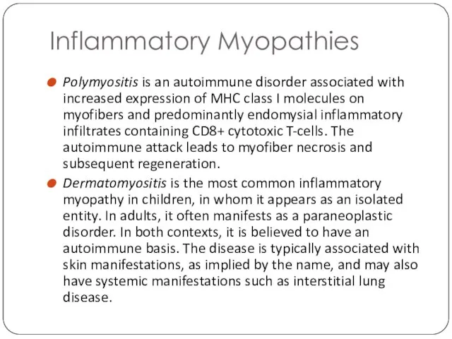
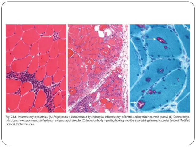
- 19. Inflammatory Myopathies Polymyositis is an autoimmune disorder associated with increased expression of MHC class I molecules
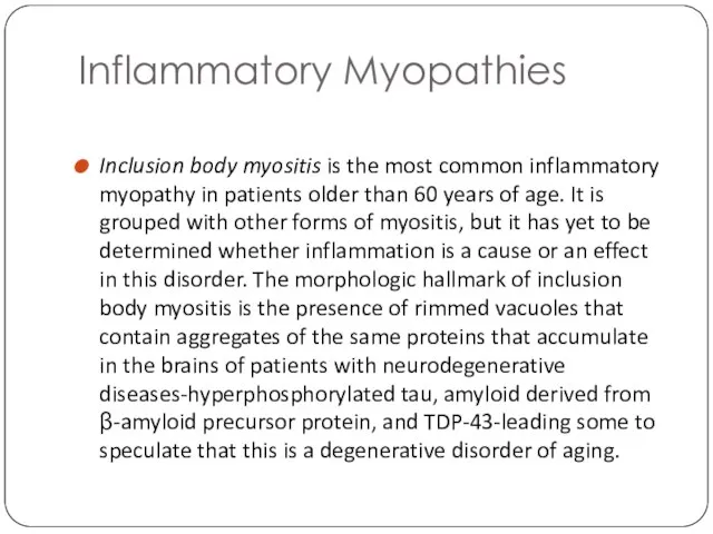
- 20. Inflammatory Myopathies Inclusion body myositis is the most common inflammatory myopathy in patients older than 60
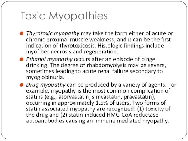
- 22. Toxic Myopathies Thyrotoxic myopathy may take the form either of acute or chronic proximal muscle weakness,
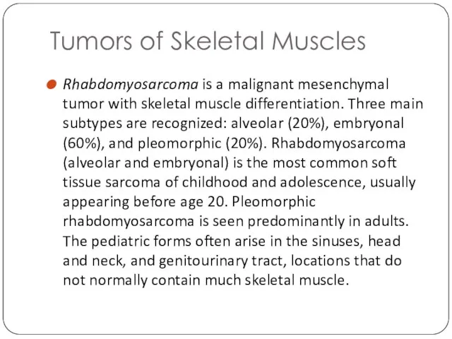
- 23. Tumors of Skeletal Muscles Rhabdomyosarcoma is a malignant mesenchymal tumor with skeletal muscle differentiation. Three main
- 24. A – embryonal subtype B – alveolar subtype
- 25. Tumors of Skeletal Muscles Leiomyoma, a benign tumor of smooth muscle, is most common in the
- 26. Tumors of Skeletal Muscles Soft tissue leiomyosarcoma accounts for 10% to 20% of soft tissue sarcomas.
- 27. Summary Skeletal muscle function can be impaired by a primary (inherited or acquired) myopathy or secondarily
- 29. Скачать презентацию
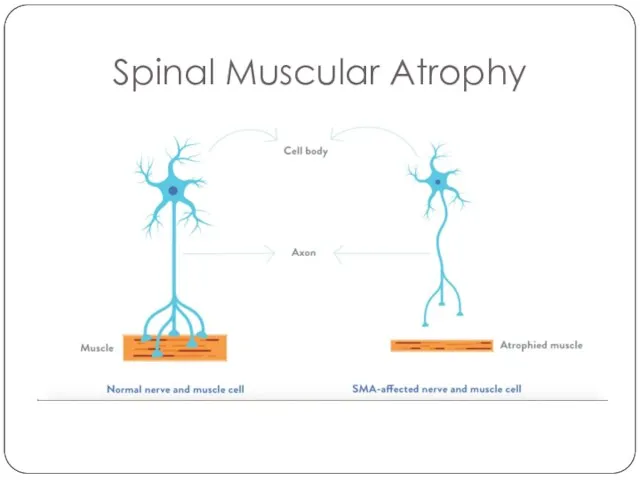
Spinal Muscular Atrophy
Spinal Muscular Atrophy
Spinal Muscular Atrophy
Spinal Muscular Atrophy
Disorders of Neuromuscular Junction
Myasthenia gravis is an autoimmune disease with fluctuating
Disorders of Neuromuscular Junction
Myasthenia gravis is an autoimmune disease with fluctuating
Disorders of Neuromuscular Junction
Lambert-Eaton syndrome is caused by autoantibodies that inhibit
Disorders of Neuromuscular Junction
Lambert-Eaton syndrome is caused by autoantibodies that inhibit
Miscellaneous Neuromuscular Junction Disorders
Congenital myasthenic syndromes comprise a heterogeneous group of
Miscellaneous Neuromuscular Junction Disorders
Congenital myasthenic syndromes comprise a heterogeneous group of
Muscle Fiber Atrophy
Neuropathic changes. Loss of innervation causes atrophy of myofibers.
Muscle Fiber Atrophy
Neuropathic changes. Loss of innervation causes atrophy of myofibers.
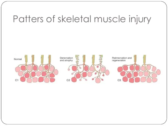
Patters of skeletal muscle injury
Patters of skeletal muscle injury
Inherited Disorders of Skeletal Muscle
Muscular dystrophies are associated with progressive muscle
Inherited Disorders of Skeletal Muscle
Muscular dystrophies are associated with progressive muscle
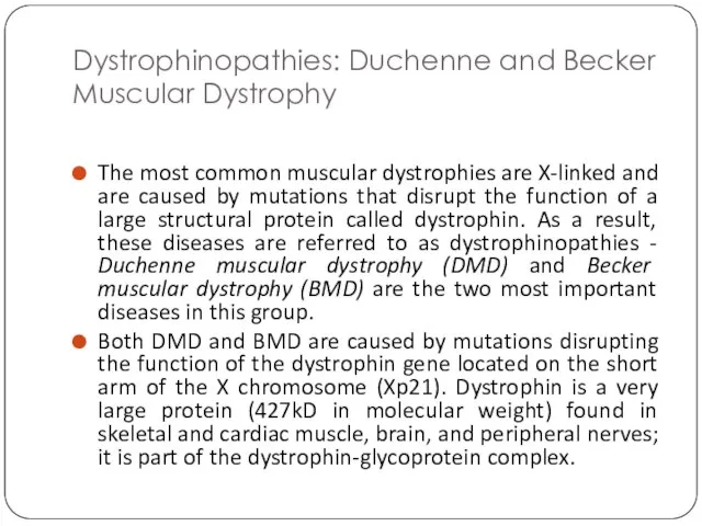
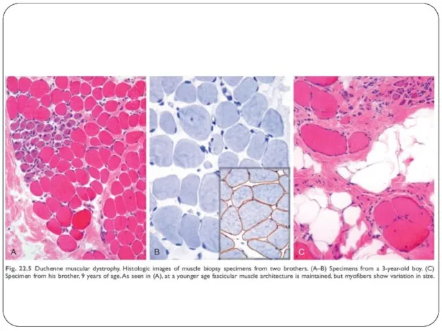
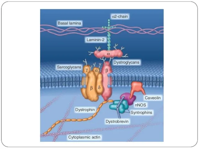
Dystrophinopathies: Duchenne and Becker Muscular Dystrophy
The most common muscular dystrophies are
Dystrophinopathies: Duchenne and Becker Muscular Dystrophy
The most common muscular dystrophies are
Other X-Linked and Autosomal Muscular Dystrophies
Myotonic dystrophy. Myotonia, the sustained involuntary
Other X-Linked and Autosomal Muscular Dystrophies
Myotonic dystrophy. Myotonia, the sustained involuntary
Other X-Linked and Autosomal Muscular Dystrophies
Emery-Dreifuss muscular dystrophy (EMD) is a
Other X-Linked and Autosomal Muscular Dystrophies
Emery-Dreifuss muscular dystrophy (EMD) is a
Channelopathies
Ion channel myopathies are a group of familial disorders caused by
Channelopathies
Ion channel myopathies are a group of familial disorders caused by
Metabolic Myopathies
Myopathies due to inborn errors of metabolism include disorders of
Metabolic Myopathies
Myopathies due to inborn errors of metabolism include disorders of
Mitochondrial Myopathies
Mitochondrial myopathies can stem from mutations in either the mitochondrial
Mitochondrial Myopathies
Mitochondrial myopathies can stem from mutations in either the mitochondrial
Acquired Disorders of Skeletal Muscle
Acquired Disorders of Skeletal Muscle
Inflammatory Myopathies
Polymyositis is an autoimmune disorder associated with increased expression of
Inflammatory Myopathies
Polymyositis is an autoimmune disorder associated with increased expression of
Inflammatory Myopathies
Inclusion body myositis is the most common inflammatory myopathy in
Inflammatory Myopathies
Inclusion body myositis is the most common inflammatory myopathy in
Toxic Myopathies
Thyrotoxic myopathy may take the form either of acute or
Toxic Myopathies
Thyrotoxic myopathy may take the form either of acute or
Tumors of Skeletal Muscles
Rhabdomyosarcoma is a malignant mesenchymal tumor with skeletal
Tumors of Skeletal Muscles
Rhabdomyosarcoma is a malignant mesenchymal tumor with skeletal
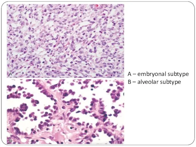
A – embryonal subtype
B – alveolar subtype
A – embryonal subtype
B – alveolar subtype
Tumors of Skeletal Muscles
Leiomyoma, a benign tumor of smooth muscle, is
Tumors of Skeletal Muscles
Leiomyoma, a benign tumor of smooth muscle, is
Tumors of Skeletal Muscles
Soft tissue leiomyosarcoma accounts for 10% to 20%
Tumors of Skeletal Muscles
Soft tissue leiomyosarcoma accounts for 10% to 20%
Summary
Skeletal muscle function can be impaired by a primary (inherited or
Summary
Skeletal muscle function can be impaired by a primary (inherited or
 Қант диабеті бар науқастарда ЗШЖИ
 Сестринский процесс при бронхитах, ХОБЛ и бронхиальной астме. Тема 4.5
 Ижтиомий фанлар кафедраси
 Эндопротезирование аортального клапана (TAVI)
 Повреждение менисков коленного сустава
 Специфика психологической работы с сотрудниками, вернувшимися из «горячих точек»
 Вплив наркотиків на статеве дозрівання та здоров’я підлітків
 Вербальное и невербальное общение: ключи к вашему успеху
 Малассезиоз. Разноцветный отрубевидный лишай
 Сестринский уход при заболеваниях носа и придаточных пазух носа, при заболеваниях глотки
 Медицинская и социальная защита населения старших возрастных групп. Гериатрическая помощь
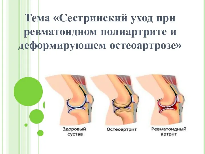 Сестринский уход при ревматоидном полиартрите и деформирующем остеоартрозе
 Лекарственные средства для лечения заболеваний бронхолегочной системы
 Микрофлора влагалища
 10 секретов Аюрведы для здоровья и долголетия
 СКА ВК ФЕСТ - Сувениры
 Порядок надання невідкладної медичної допомоги постраждалим та хворим на догоспітальному етапі. (Лекція № 2)
 Острый медиастенит
 Этиопатогенез дизартрии и её симптомы
 Диагностические тесты в психиатрии
 Жедел асқазан ішек, ауа тамшылы, зоонозды, трансмиссивті және аса қатерлі жұқпалы аурулар
 Основы традиционной китайской медицины с позиции прикладной кинезиологии
 Дерматоздардың ауыз қуысындағы көрінісі. Қызыл жалпақ теміреткі (КПЛ), жүйелі қызыл волчанка. Пузырчатка. Этиологиясы, клиникасы
 ЦВЗ
ЦВЗ Политравма
 Гормоны, принимающие участие в процессе адаптации. Стресс и адаптация
 Комплексная система очищения организма от компании Сибирское здоровье
 Менопаузальная гормонотерапия